

Introduction
The grapevine (Vitis spp.) is one of the most widely grown fruit crops, whose fruits can be processed into juice or wine or used for fresh as well as dried consumption. Grapevine is also one of the oldest and most economically important crops in the South Caucasus region, and it ranks in the leading places among the cultivated fruit crops in Azerbaijan. Many regions of Azerbaijan are abundant in precious local grapevine varieties that have not been explored yet. Grapevine production in Azerbaijan is intensive in two central viticulture regions, Salyan and Calilabad, both located in the south [1]. Multiple virus infections of grapevines are the major reason for yield losses and reductions in fruit quality. Such mixed infections with two or more viruses are the main problem in grapevine production worldwide and can cause up to 60% yield losses in vineyards [2]. To date, over 86 described viruses belonging to 17 different virus families and 34 genera can infect grapevines, some of them with very high prevalence [3]. Besides, these new virus strain(s) or isolate(s) infecting grapevines will likely be reported soon. The infected plants show various symptoms, including leaf chlorosis, mosaic, leaf rolling, fan leaf, reddening, and the appearance of necrotic spots, whereas multiple virus infections can significantly increase the severity of these symptoms [4]. The rate of possible economic losses caused by viral infections depends on grapevine varieties. Numerous factors contribute to the gradual reduction of grapevine yield losses, with viruses playing a prominent role, that are frequently associated with declining grape juice quality, decreased levels of soluble solids, and an increase in phenolic substances, as well as titratable acidity [5]. Various significant aspects of plant metabolism can also be disrupted, as well as qualitative parameters such as the number of aromatic compounds in grapes. Such changes as disruption of photosynthesis, transfer of respiratory assimilators, and the life cycle of the vine, as well as suppression of plant growth and development, a decrease in such qualities as survival, a decrease in the drought resistance of the vine, etc., can also be observed during viral infections of the plant. The occurrence of single and mixed viral infections often leads to exacerbations of symptoms and crop losses [6]. This may be due to the fact that viruses from perennial plants have a great potential for molecular variability. Genetic variation plays a significant role in the adaptation processes of viruses under unsatisfied conditions and increases the level of competitiveness, tolerance, and winter survival. The virus infections are associated with the main complex viral diseases of vines, including leafroll (GLD), corky rugose wood (CRW), and infectious degeneration (fanleaf). These are the most widespread and threatening viruses of grapevine [7]. It has been reported that most viruses and grapevine diseases are also part of various pathogen complexes (bacteria, phytoplasma, fungi, etc.), depending on the variety and composition of the vineyard. The most widely distributed multiple virus infections of grapevine include double infections of Grapevine fleck virus (GFkV) and Grapevine fanleaf virus (GFLV) or mixed infections of Grapevine leafroll associated virus-1 (GLRaV-1), Grapevine leafroll associated virus-2 (GLRaV-2) and Grapevine leafroll associated virus-3 (GLRaV-3) as well as double infections of Grapevine virus A (GVA) and Arabis mosaic virus (ArMV) or Grapevine virus A (GVA) and Grapevine leafroll associated virus-3 (GLRaV-3) and may cause changes in external and internal features of vines [8]. Multiple virus infections can increase the degree of leaf curl, cause noticeable changes in leaf color, the appearance of double nodes, shortening of internodes, abnormal development of shoots, fascia of bundles and butts, a delay in the growth of bushes, the appearance of infertile shoots, etc. Consequently, a viral infection negatively affects the efficiency of vine cultivation, reduces the main parameters of vine quality and the presentation of table grapes, and the severity of various symptoms increases with repeated viral infection [9]. On the other hand, it is known that plant viruses that cause disease in the same host plant can be both synergists and antagonists. Synergistic interactions have a “facilitating effect” on both sides or on one side and facilitate the reproduction of the virus in the host plant. Various synergistic situations can occur where one pathogen contributes to the transmission of another pathogen. This phenomenon naturally occurs in some complex pathogenic infections and is called “adjuvant dependence”. In contrast, only one pathogen benefits from antagonistic-type interactions, and its fitness for participation and activity is lower than that of the second pathogen. In addition, more or less predictable biological and epidemiological dependences on synergistic and antagonistic interactions between different viruses occur in plants. The study of existing interactions between pathogens can play an important role in elucidating their pathogenesis and evolution and, therefore, in developing effective and stable strategies for treating diseases [10]. At the same time, extensive research in this area can be useful in organizing effective and sustainable management strategies against complex diseases. Therefore, for the sustainable development of agriculture, it is necessary to select methods for combating various viruses, the primary diagnosis of these pathogens and mixed infections, taxonomic characteristics, and clarification of the spectrum of plant hosts and transmission routes of vectors. On the other hand, diagnosis, identification of pathogens, the study of their molecular biodiversity, and the study of the ways of transmission of viruses are of great importance for the timely prevention of diseases caused by various viruses [11]. Previous studies conducted with grapevine viruses included only single infections of GLRaVs, and only GLRaV-3 has been reported in the Azerbaijani vineyards as a result. At this point, the main purpose of this research was to study the prevalence of single and mixed viral infections of grapevines in two main viticulture regions of Azerbaijan. It should be noted that despite the high economic importance of the vine in the southern part of Azerbaijan, there is no information on the incidence of any mixed viral infections, including GFLV, and their impact on vegetative productivity and yield [12, 13].
Material and Methods
Field Sampling
To investigate the presence of single and mixed infections in Azerbaijani vineyards during the summer and autumn seasons from 2022 to 2023, a random survey of seven fields in the Salyan and Calilabad regions located in the southern part of Azerbaijan was conducted. A total of 166 leaf samples from local grapevine varieties, including Ag Shani, Gara Shani, Agadayi, Medrese, and Tebrizi were collected. The samples were obtained from grapevines exhibiting grapevine leafroll disease-like symptoms such as leaf mosaic, leaf rolling, leaf yellowing or reddening, vein banding, and shortening of internodes. Additionally, around 30 samples with visible wide-open veins resembling fan leaf symptoms were collected. The primary symptoms of a grapevine virus infection also include stunted growth, mottling of leaves, reduced fruit quality, distorted or discolored fruit, and overall weakened vine vigor. It’s important to note that the symptoms of GVA can be subtle and may not always be easily distinguishable from those caused by other grapevine viruses or environmental stressors. Molecular diagnostic tests are often necessary to confirm GVA infection. The collected leaf samples were transported to the laboratory and kept at 4ºC until further processing.
Virus Detection and Identification
AgriStrip - the Rapid One-Step Assay
The initial screening for the detection of grapevine leafroll-associated viruses in the original samples was checked by the rapid one-step assay AgriStrip based on lateral flow immunochromatography using specific antibodies (Bioreba AG, Reinach, Switzerland) and Agdia Inc. ImmunoStrip (USA). The strips were dipped with the “sample” side into the plant extract to allow the liquid to migrate upwards and initiate an antigen-antibody reaction, after which visible lines were recorded. Both test and control lines became visible with positive plant extracts, while negative plant samples gave only the upper control line. The line formation developed after 3 minutes and reached its maximum intensity after 10 minutes [14]. Photographs were taken of the ImmunoStrips to record the results (data not shown), and the dried test strips were stored in a dry, cool place.
Double-Antibody Sandwich Enzyme-Linked Immunosorbent Assay (DAS-ELISA)
The possible presence of grapevine leafroll-associated viruses (GLRaVs -1, 2, 3, and 4) and grapevine fanleaf virus (GFLV) was also carried out by DAS-ELISA using polyclonal antisera against these viruses, which were developed by Bioreba AG (Reinach, Switzerland) according to [15]. All buffers were prepared according to the manufacturer’s instructions. Grapevine leaf samples were homogenized (1:5 W/V) in Tris extraction buffer using a sterile mortar and pestle, and 100 µl volumes of the extracts were incubated overnight at 4ºC in microtiter plate wells previously treated with 100 µl of 1:1000 dilution of IgG in carbonate coating buffer. Microtiter plate wells were rinsed with washing buffer and incubated with 100 µl of the alkaline phosphatase-conjugated IgG diluted 1:1000 in conjugate buffer (3 h at 37ºC). Wells were washed and incubated in 100 µl of substrate for 1 h at room temperature, and the absorbance was determined at 405 nm using a microtiter plate reader (Stat Fax Microplate, Awareness Technology, USA). All samples were assayed in two repeats, and results were assigned as positive if the mean absorbance was greater than or equal to three times the average reading of the negative (healthy) controls. Alternatively, the presence of viruses was confirmed by using molecular assays.
RNA Extraction
Total RNA was extracted from 200 mg of scraped bark tissue from basal nodes, petioles, and/or midribs according to the CTAB method described by [16] with minor modifications. 200 mg of leaf tissue in pre-warmed 2 ml of extraction buffer was added into a sterile extraction bag, and 4 μl of β-mercaptoethanol was added. The sample extracts were transferred into the microtubes, incubated at 60ºC for 15 min, and vortexed at 5 min intervals. An equal volume of chloroform: isoamyl alcohol (24:1) was added, vortexed for 10 min, and centrifuged at 8000 rpm for 10 min at 4ºC. RNA was precipitated by adding 1/3 volume of 7.5 M lithium chloride (LiCl). Samples were incubated on the ice at 4ºC overnight and centrifuged at 8000 rpm for 30 min at 4ºC after incubation. The pellet was resuspended in 100 μl DEPC-treated water, 0.2 volume 2M sodium acetate (pH 5.2), and 2 volume 100% ethanol. Tubes were incubated at -20ºC for 2h and then centrifuged at 8000 rpm for 10 min. The supernatant was thrown away, and the pellet was washed with 70% ethanol. The extracted RNA was stored at -20 or -80ºC for further use. The degree of RNA purity and concentration was measured by analyzing the samples’ spectrophotometric absorbance at 230, 260, and 280 nm wavelengths and calculating the ratios of A260/A280 and A260/A230. Furthermore, the quality of RNA was assessed through the use of 1% agarose gel electrophoresis, followed by visualization under UV light after staining with ethidium bromide.
Reverse Transcription-Polymerase Chain Reaction (RT-PCR)
RT-PCR was done using the virus-specific primers mentioned in Table 1. RT-PCR was performed in two steps. To synthesize the complementary strand (cDNA), 2 μl of extracted RNA was submitted to reverse transcription in a final volume of 20 μl, using 2 μl RT buffer 10x (0.5 M Tris-HCl, 0.7 M KCl, 0.1 M MgCl2, pH 8), 1μl DTT (100 mol/μl), 1μl dNTPs (10 mmol/ μl), 0.5 μl RNase inhibitor (10 mmol/μl) and 2 μl reverse primer (100 pmol/μl) for one hour at 47ºC with 0.5 μl MMuLV reverse transcriptase (200 U/μl). Five μl of the RT reactions were used for PCR. The PCR reaction was carried out in a final volume of 20 μl containing 20 ng cDNA, 10mM of each dNTP (Solis BioDyne, Estonia), 1.6 mM MgCl2, 1U of Taq DNA polymerase (Solis BioDyne, Estonia), 0.5 μl of each primer pair (10 pmol/μl), and 1X PCR buffer. PCRs were performed in a thermal cycler (SimpliAmp Thermal Cycler, applied biosystems, USA), and the PCR products were held at 4ºC or stored at -20ºC and analyzed by electrophoresis in 1% agarose gel using 1X TBE (Tris-Borate EDTA buffer) and stained with ethidium bromide. An ultraviolet (UV) transilluminator was used to visualize DNA bands. Amplified products were also visualized using the UV-Gel Doc system (UK). The molecular weight of the PCR products was estimated using a 2-log DNA Ladder (NEB, UK).
Table 1.
Primers used for testing GFLV, GVA, and GLRaVs (1,2,3,4) infection by the RT-PCR assay.
Immunocapture RT-PCR (IC-RT-PCR)
Each 0.2 ml PCR tube was incubated for 3 h at 37ºC with 100 µl of GLRaV-2 IgG (1/1000) antiserum diluted in carbonate buffer, as used in DAS-ELISA, then washed three times with 100 µl of PBST (washing buffer). Fresh leaf tissue from grapevine samples was homogenized in grapevine extraction buffer 1x, centrifuged at 10,000 rpm for 5 min, and 100 μl clarified extract was incubated overnight at 4ºC in each PCR tube. Then 25 μl of deionized water and 1.5 μl reverse primer (10 pmol) were added to the tubes after washing three times with 100 µl washing buffer. The tubes were incubated at 65ºC for 5 min, and then cooled on ice. IC-RT-PCR was done as described before [17]. The IC-RT-PCR reaction consists of one cycle at 42ºC for 55 min and a second cycle at 94ºC for 3 min followed by 35 cycles (denaturing at 94ºC for 30 s, annealing at 60ºC for 30 s, and extension at 72ºC for 50 s), and a final extension cycle at 72ºC for 10 min.
Results and Discussion
Detection of GFLV, GVA, and GLRaVs (1-4)
Throughout the survey period, typical symptoms of viral agents that include inter-venial discoloration, redness, chlorosis, and downward leaf rolling were observed on both red and white cultivars (Fig. 1). The occurrence of leaf curling symptoms did not show a significant difference between regions and fields as a visual assessment over a period of two years.
During the survey, disease symptoms in field plants were evaluated concurrently with camera imaging. Fan leaf symptoms were observed in three out of seven vineyards. It is worth noting that the symptoms were barely noticeable at the onset of the growing season, and they gradually became more apparent as the season progressed toward the end of the harvest period, which spans from July to August. The prominent symptoms of viral infection were observed mainly towards the end of the growing season from late August to October, particularly in red cultivars. On the other hand, the symptoms resembling those caused by viruses did not display a significant difference between the various dates of sample collection. GLD symptoms were more pronounced in red cultivars, whereas white-berried cultivars may show subtle symptoms or be completely symptomless. During the entire duration of the study, as a result of serological tests, 108/166 (65%) of the tested samples yielded a positive result for at least one of the viruses listed: GFLV, GLRaV-1, GLRaV-2, GLRaV-3, GLRaV-4, and GVA. The total number of tested and infected samples from each region is shown in Table 2. In some cases, when the ImmuneStrips did not receive positive responses, the samples obtained by DAS-ELISA were exposed to positive signals. The frequency of occurrence (percentage) of these viruses is shown in Fig. 2.
Fig. 1.
Mild chlorosis. a) shortening of internodes; b) different sizes of berries and reduction in fruit color due to the irregular fruit ripening; c) interveinal reddening; d) interveinal discoloration (reddening and chlorosis); e) downward leaf rolling; f) in virus-infected plants.
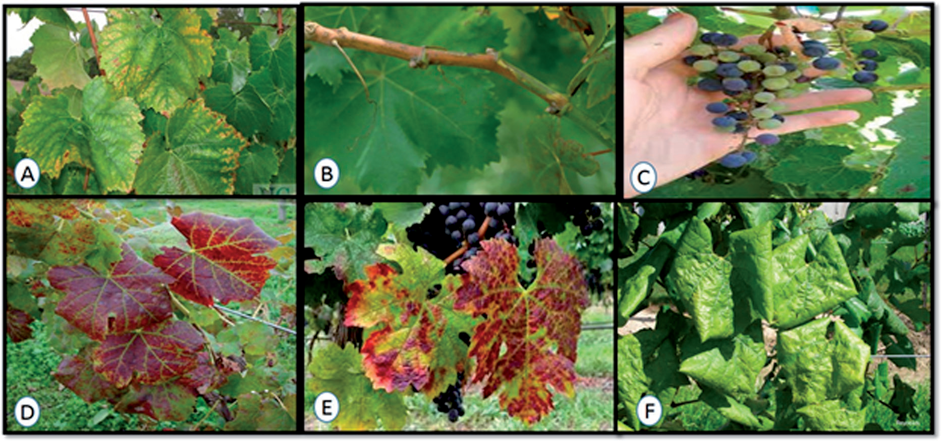
Table 2.
Results of AgriStrip (immune strips) and DAS-ELISA tests run on grapevine samples randomly taken from seven fields in two locations using polyclonal commercially available antibodies (Bioreba AG, Reinach, Switzerland).
Thus, a total of 166 samples were collected and analyzed by serological tests. It is widely recognized that grapevine is a crucial host for plant viruses, with the viruses linked to grapevine leafroll disease (GLD) being the most prevalent and economically significant worldwide [18]. The global impact of GLD has had a detrimental effect on both the quality and quantity of grape production worldwide. It has resulted in reduced grape yields and quality, leading to significant economic and agricultural challenges for the global wine and grape industry.
Over an extended period of grapevine cultivation, more than 90 viruses and virus-like agents have been identified [19, 20]. These agents exhibit diversity not only in their symptoms within the plant but also in their genomic structures [21]. The predominant grapevine viruses belong to various categories, including positive-sense single-stranded RNA ((+) ssRNA) genomes (comprising 74 species), negative-sense single-stranded RNA ((–) ssRNA–9 species), double-stranded RNA (dsRNA–2 species), and DNA viruses (8 species) [22, 23]. In the last 14 years, the application of high-throughput sequencing (HTS) techniques has led to the discovery of over 35 new grapevine viruses, including 21 species with (+) ssRNA genomes [20]. These (+) RNA viruses can act as causative agents of economically significant diseases, such as infectious degeneration and decline, grapevine leafroll disease, rugose wood disease, and fleck disease [21, 24]. In Azerbaijan, there is a shortage of contemporary research on plant viruses. So this study represents the initial documented occurrence of GFLV, GLRaV-1, GLRaV-2, GLRaV-1+3, GLRaV-4, and GVA infections in grapevines; the effects of these pathogens on grape production in the region remain unexplored and require further investigation. On the other hand, GLRaV-1+3 as mixed infections were identified and predominated.
In our survey, four distinct grapevine leafroll-associated viruses (GLRaVs), including GLRaV-1, GLRaV-2, GLRaV-3, and GLRaV-4, were identified (Table 2). These viruses belong to the family Closteroviridae and are classified into two genera: Ampelovirus (GLRaV-1, -3, -4) and Closterovirus (GLRaV-2). Additionally, within the Ampelovirus genus, GLRaV-1 and -3 were assigned to subgroup I, while GLRaV-4 was assigned to subgroup II [25]. According to [7], GLRaVs are primarily confined to phloem-associated cells and exhibit an uneven distribution within plants. As grapevines are often propagated vegetatively to preserve clonal consistency, infected planting material is a common source of virus introduction in vineyards [26]. Certain GLRaVs can also be transmitted locally by insect vectors such as mealybugs and soft-scale species [27]. However, there has been no investigation into the vector transmission of these viruses. GLRaVs were detected in six fields (70 samples from three different vineyards in Salyan and 96 samples from four different vineyards in Calilabad were analyzed). GFLV was found to be prevalent in a field in Salyan where GVA was not identified. In one field in Salyan where GVA was not detected and GLRaVs were rare, GFLV was prevalent. However, GVA was detected in the samples collected, only in the first year of the survey in Calilabad. The vineyards of Salyan had not yielded any detections of this virus. The presence of GVA was determined in two grapevine samples (1.20%) out of the mentioned 166 samples. Grapevine virus A (GVA), belonging to the genus Vitivirus in the family Betaflexiviridae, is known to infect phloem cells and can cause significant crop losses of 5-22% in grapevine cultivars. Its transmission occurs through various species of Pseudococcid mealybugs. Such an infection rate is low in comparison with the sanitary status of Italian cultivars – 8% [3], grapevines in Turkey – 53% [28, 29], or Tunisia – 63% [30]. Fig. 3 illustrates the viticulture area and locations where the viruses were detected based on the results of the DAS-ELISA and validated through molecular analysis using RT-PCR (Fig. 4). It should be noted that only IC-RT-PCR results for GLRaV-2 were presented in Fig. 5. The IC-RT-PCR and RT-PCR methods were used to detect grape viruses, also revealing that the most prevalent type of mixed viral infection was GLRaV-1+3 (10.84%). This combination of viruses is typically identified through symptoms such as interveinal reddening, leaf chlorosis, and the presence of necrotic spots. Out of a total of 166 plant samples, 17 (10.24%) of GLRaV-1, 18 (10.84%) of GLRaV-2, 32 (19.28%) of GLRaV-3, 11 (6.63%), 10 (6.02%) of GFLV, and 2 (1.20%) of GVA were detected. The IC-RTPCR technique, which omits the necessity for extracting viral or plant total RNA, effectively addresses concerns regarding RNase contamination, offers simplicity in execution, and yields dependable results when applied in the epidemiological study of viral diseases.
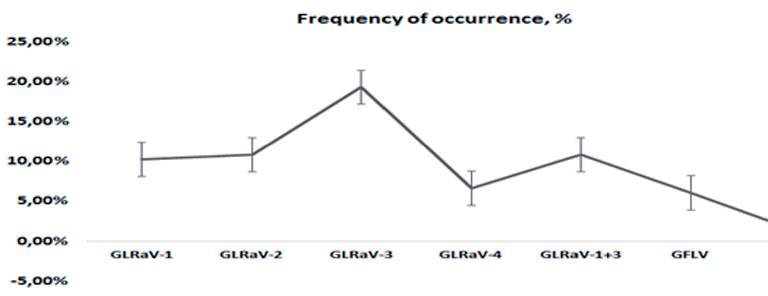
Fig. 3.
The viticulture area that was studied and the locations where the viruses were detected based on the results of the ELISA test were presented.
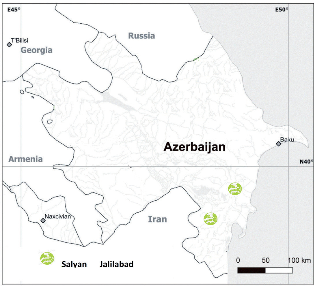
Fig. 4.
Agarose gel electrophoresis showing results of RT-PCR detection of GLRaV-3 using primer pair 8504v/9445c; (a), GFLV using primer pair GFLV-F/GFLV-R; (b), GLRaV-2 using primer pair GLR2CP1/GLR2CP2; (c), GLRaV-1 using primer pair LR1F/LR1rev; (d), GLRaV-4 using primer pair LR4F/LR4R; (e), GVA using primer pair GVA-F/GVA-R; (f). Total RNA was isolated from leaves obtained from distinct grapevine accessions from different geographical regions (lines 1-11), and the expected length of the amplified fragment was 942 bp; a) 1560 bp; b) 600 bp; c) 401 bp; d) 500 bp; e) and 272 bp; f) Line: M–marker (2-Log DNA Ladder (NEB, UK). RT-PCR products were separated in 1% agarose gel.
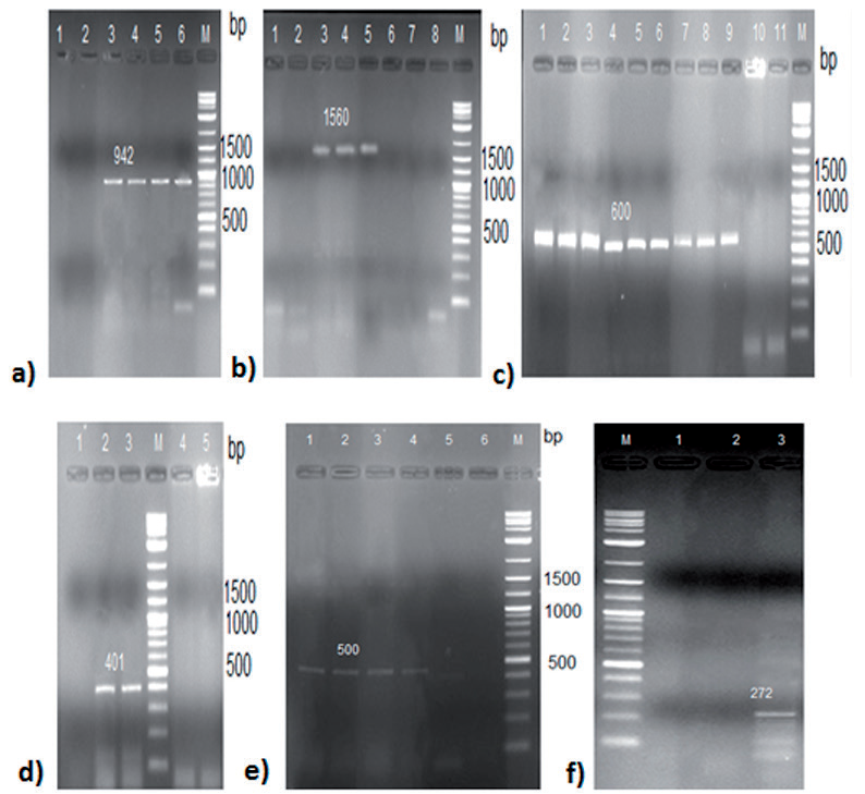
Fig. 5.
Amplification of the GLRaV-2 genomic fragment using virus-specific primers GLR2CP1/GLR2CP2. M – Molecular weight marker 2-Log DNA Ladder (NEB, UK). IC-RT-PCR was performed on extracts of infected grapevine plants with specific antibodies. The arrow indicates the position in the gel of the expected 600 bp fragment. IC-RT-PCR products were separated in 1% agarose gel.
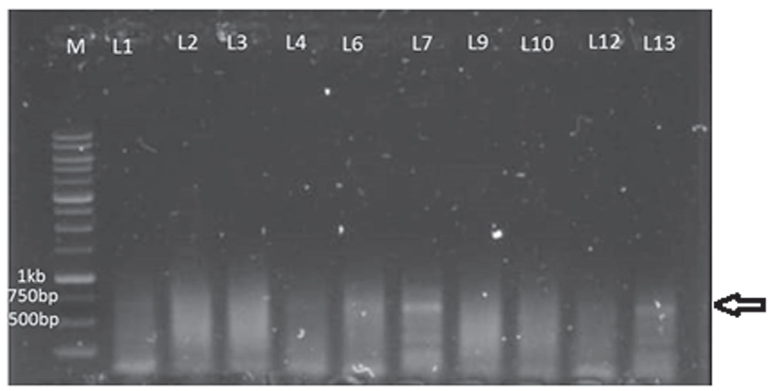
In addition to these specific fragments (Figs. 4 and 5), sometimes we observed the presence of other ‘non-specific’ amplification products, more marked in IC-RT-PCR than in RT-PCR. The presence of extra bands in the reaction could be attributed to the potential degenerations in the primers and the RT-PCR conditions utilized. The identification of infected plants plays a significant role in managing and controlling grapevine disease [31]. The factors that influence the manifestation of grapevine fanleaf or leafroll symptoms remain uncertain, but they may encompass the type of virus, scion variety, environmental factors, rootstock type, and the possibility of other viruses co-infecting the plant [32]. In the current study, there was no correlation between the expression of virus symptoms in the plant and its location, vineyard age, and varieties. Among the samples, only a few that tested negative for viruses using RT-PCR and IC-RT-PCR displayed visible yellow and red mosaic or decline symptoms. These samples might have been infected by other viruses or viroids with comparable symptoms, or other factors, such as mutations in primer sites, low levels of virus particles during the sampling period, or unusually high levels of plant substances that could potentially disrupt the reaction, could account for the inability to detect the virus through RT-PCR. Efficiently managing the spread of grapevine viruses requires the availability of diagnostic methods that are sensitive, rapid, and dependable. In this study, we assessed the sensitivity and specificity of four diagnostic techniques: RT-PCR, IC-RT-PCR, DAS-ELISA, and ImmunoStrips. The validation of serological test results was done by IC-RT-PCR and RT-PCR.
Conclusions
The present study was the first to mark the occurrence and current status of major RNA genome viruses infecting grapevine in Azerbaijan. The investigation revealed that grapevine leafroll-associated viruses (GLRaV 1-4) were the predominant viral strains, with GLRaV-3 emerging as a notable concern. These virus distributions were noticed in both surveyed regions and varied during the study period. Currently, no curative in-field treatment is available for GLD, and only prophylactic measures can be applied [33, 34]. Diagnostic methods based on serological and molecular analyses have been developed for reliable disease detection, but unknown viruses or new strains of known viruses may remain undetected. The existence of viral plant pathogens, particularly those resulting from combined infections, implies that the utilization of contaminated planting materials for vineyards and the absence of proper monitoring of their distribution may have a significant contribution to the spread of viruses. The accurate diagnosis of viruses is important for the application of the right management strategies for controlling plant viruses in agriculture.

