

Introduction
Nickel (Ni) pollution in aquatic ecosystems, primarily caused by industrial activities, poses significant risks to both organisms and the food web due to its persistence and toxicity [1-4]. Industrial activities such as mining, electroplating, battery manufacturing, and Ni alloy production are among the primary sources of Ni contamination in freshwater and coastal ecosystems. Once released into aquatic environments, Ni accumulates in sediments and water, where it is readily absorbed by aquatic organisms, leading to bioaccumulation and potential toxic effects [5-7]. Exposure to Ni has been associated with physiological stress, developmental abnormalities, and reduced survival rates in aquatic invertebrates, including gastropods [8-12]. Understanding the mechanisms of Ni accumulation, detoxification, and tissue-specific distribution in bioindicator species is crucial for assessing its environmental impact and mitigating its adverse effects [1, 2, 13-18].
Among freshwater snails, Pomacea insularum, a species of apple snail, is an effective biomonitoring organism due to its sedentary nature and frequent interactions with Ni-contaminated sediments [2, 19, 20]. This species is widely distributed in freshwater habitats and has demonstrated resilience to polluted environments, making it an ideal model for assessing heavy metal contamination [10, 11, 21-23]. Studies indicate that heavy metals often accumulate in specific tissues, with each tissue playing a distinct role in metal absorption and detoxification [18]. The shell, for instance, acts as a long-term storage site, reflecting cumulative exposure, while soft tissues manage initial metal absorption and detoxification processes [3, 19, 20]. However, despite existing knowledge of general heavy metal accumulation in gastropods, Ni’s specific pathways and detoxification mechanisms in P. insularum remain underexplored, highlighting a critical knowledge gap for effective biomonitoring [2-4, 23].
This study aims to address this gap by examining Ni’s tissue-specific accumulation, detoxification, and depuration in P. insularum under natural field conditions. Through a combination of field sampling and transplantation experiments, we investigate the extent of Ni bioaccumulation across different tissues and assess how this species responds to changes in environmental Ni exposure. The study employs correlation and factor analyses to elucidate the relationships between tissue-specific Ni accumulation patterns, revealing the coordinated role of different tissues in metal detoxification. Findings from this research contribute to a deeper understanding of Ni bioaccumulation in freshwater gastropods, reinforcing P. insularum as a valuable biomonitoring tool for Ni contamination in aquatic ecosystems. By examining both short-term responses in soft tissues and long-term sequestration in the shell, this study provides critical insights for environmental monitoring and pollution management strategies.
Materials and Methods
Study Area and Sample Collection
The freshwater snail Pomacea insularum and its habitat surface sediments (0-10 cm depth) were collected from 13 sampling locations across rivers, ponds, and lakes in Peninsular Malaysia (Fig. 1, Table 1). These sites represented a diverse range of freshwater environments with varying degrees of heavy metal exposure. Sampling locations were chosen based on their proximity to potential heavy metal contamination sources, including industrial effluents and agricultural runoff.
Fig. 1.
Sampling sites of Pomacea insularum and their habitat surface sediments in Peninsular Malaysia. Note: The sampling points are estimations only and follow the sampling sites indicated in Table 1.
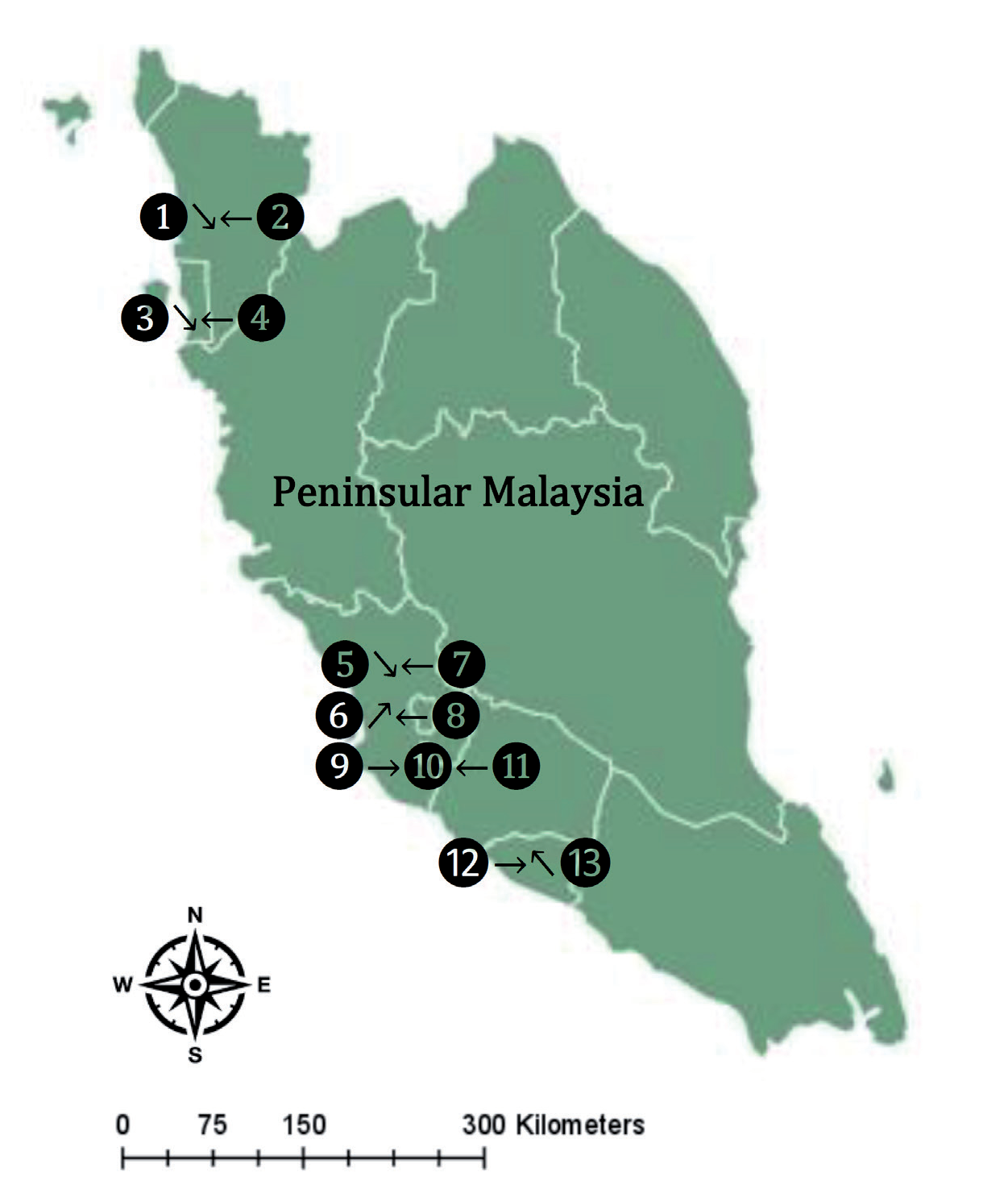
Table 1.
The description and GPS reading of the sampling locations for Pomacea insularum.
Snails were hand-picked and immediately placed in clean, labeled plastic bags. Surface sediments were collected using a stainless steel scoop. All samples were transported to the laboratory on ice and stored at -20ºC until further analysis.
Transplantation Experiment
A transplantation experiment was conducted using P. insularum at two selected sites: the Juru River and UPM Lake. The Juru River, classified as a polluted site, is influenced by anthropogenic activities, including industrial discharge and urban development [15, 16]. In contrast, UPM Lake was selected as a reference site due to its minimal anthropogenic influence.
A total of at least 100 P. insularum individuals (2.8-3.5 cm in shell length) were collected from UPM Lake and acclimatized in the laboratory for 24 hours. The snails were divided into five net cages (32 cm × 24 cm), with each cage containing 20 individuals. These cages were then transferred to the Juru River to initiate the exposure experiment. Concurrently, 100 native snails from the Juru River were collected and transplanted to UPM Lake. To assess Ni accumulation, 10-20 individuals were retrieved from the Juru River at weeks 1, 3, and 7 for metal analysis. Likewise, snails transplanted to UPM Lake were sampled at the same intervals for the depuration study to evaluate Ni elimination.
Sample Preparation and Ni Analysis
In the laboratory, collected snails were thawed, and individual specimens were carefully dissected to obtain eight distinct tissues: cephalic tentacle (CT), mantle, pineal sac (PS), remaining soft tissues (REM), digestive tract (DT), foot, operculum, and shell. Each tissue was rinsed with deionized water to remove external contaminants, blotted dry with filter paper, and weighed. Surface sediments and snail tissues were dried at 105ºC until a constant weight was reached. Sediment samples were then sieved through a 63-µm mesh for homogenization. Dried snail tissues and sediment samples were stored in acid-washed polyethylene containers until further analysis [15, 16, 18].
Triplicate samples of each tissue type and sieved sediment samples were analyzed for Ni content. Tissue samples were digested with concentrated nitric acid (HNO3; BDH grade, 69%), whereas sediment samples were digested using a combination of concentrated nitric acid (HNO3; BDH grade, 69%) and perchloric acid (HClO4; BDH grade, 60%). The digestion process was carried out at 40ºC for one hour, followed by heating at 140ºC for an additional three hours. Post-digestion, all samples were filtered and diluted with deionized water to a final volume of 40 mL [15, 16, 18].
Ni concentrations were determined using an Atomic Absorption Spectrophotometer (AAS) AAnalyst Model 800 with an air-acetylene flame. Calibration standards were prepared using certified Ni reference materials, and quality assurance was ensured by incorporating sediment reference materials with known concentrations alongside blank samples in each batch of analyses.
Statistical Analysis
Statistical analyses were conducted using NCSS software [24]. Descriptive statistics were calculated for Ni concentrations in each tissue. Pearson’s correlation coefficients were used to examine the relationships between Ni levels across tissues and surface sediments. Multiple regression analyses were performed to identify potential predictors of Ni accumulation in different tissues.
Factor analysis with Varimax rotation was applied to detect patterns of Ni accumulation and categorize tissues based on their metal uptake and retention characteristics. Graphical representations were generated using KaleidaGraph version 3.08 (November 1996). One-way analysis of variance (ANOVA) was employed to compare mean Ni concentrations, followed by the Newman-Keuls multiple comparison test to determine statistically significant differences.
Results
Spatial and Tissue-Specific Variations in Ni Accumulation in Pomacea insularum
The Ni concentrations in Pomacea insularum tissues and their habitat surface sediments exhibited notable spatial variations across different sampling sites in Peninsular Malaysia (Table 2, Fig. 2). The highest Ni concentrations in the shell were recorded at Kedah-2, UPM-2, and UPM-3, with values exceeding 30 mg/kgdry weight, indicating significant Ni deposition in the exoskeletal structure of the snails. In contrast, lower Ni concentrations, ranging from 20 to 25 mg/kg, were observed at sites such as Melaka-1 and Juru-1, suggesting site-specific differences in Ni bioavailability and environmental exposure.
Ni concentrations in REM showed a similar trend, with the highest values recorded in Malacca-2 and Kedah-2, exceeding 10 mg/kg dry weight, while other sites exhibited moderate to low Ni accumulation between 5 and 8 mg/kg. The CT displayed variability in Ni accumulation, with Kedah-2 and UPM-1 showing the highest levels, surpassing 8 mg/kg, while other locations had Ni concentrations ranging from 4 to 6 mg/kg dry weight.
The foot tissue exhibited the highest Ni concentration at Kedah-2 (~9.5 mg/kg), whereas Juru-2 and Melaka-1 recorded lower values (~4 mg/kg). Similarly, the highest Ni accumulation in the mantle was observed at Subang-2 (~15 mg/kg), while other sites had moderate concentrations between 5 and 10 mg/kg dry weight. The operculum also exhibited variability, with elevated Ni levels at Kedah-2 and UPM-1 (~9 mg/kg), while the lowest concentrations (~3-6 mg/kg) were found at other locations.
Table 2.
Overall statistics of Ni concentrations (mg/kg dry weight) in the eight parts of Pomacea insularum and their habitat surface sediments (SED) collected from 13 sites in Peninsular Malaysia. N = 13.
Fig. 2.
Concentrations (mean±SE, mg/kg dry weight) of Ni in the different tissues of Pomacea insularum and their habitat surface sediments (SED), collected from Peninsular Malaysia. Note: PS = pineal sac; Oper = operculum; CT = cephalic tentacle; DT = digestive tract; REM = remaining soft tissues. Fig. 2. Concentrations (mean±SE, mg/kg dry weight) of Ni in the different tissues of Pomacea insularum and their habitat surface sediments (SED), collected from Peninsular Malaysia. Note: PS = pineal sac; Oper = operculum; CT = cephalic tentacle; DT = digestive tract; REM = remaining soft tissues.
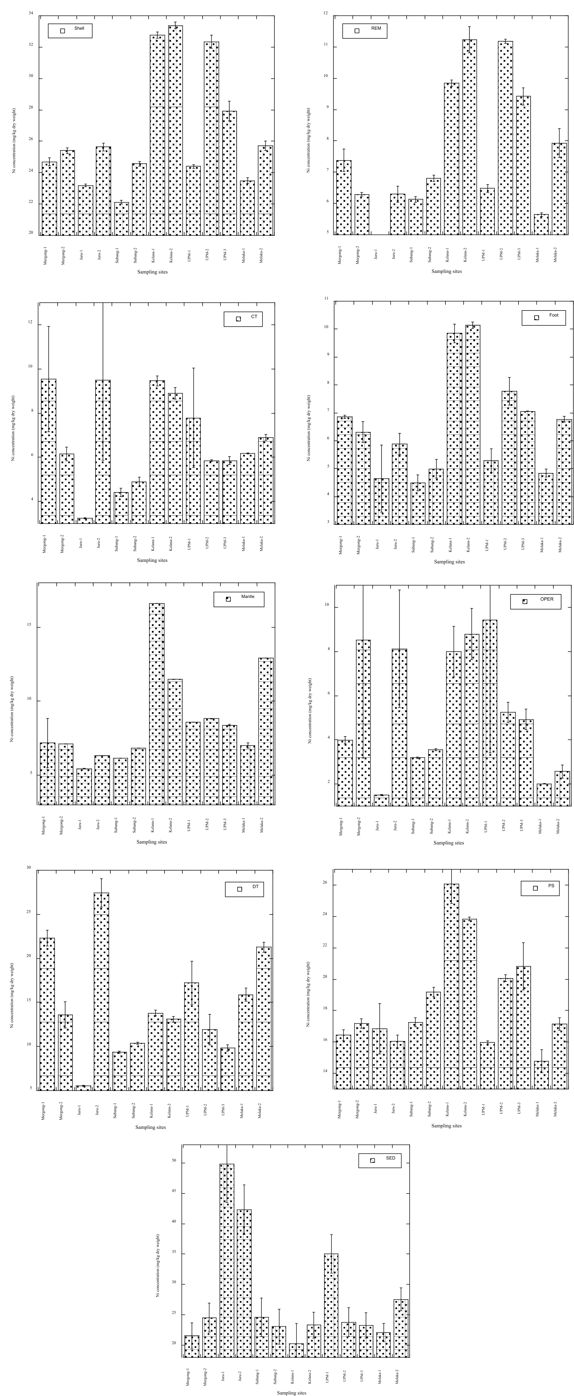
The DT exhibited the highest Ni accumulation at Juru-1 (~25 mg/kg), indicating significant Ni uptake through dietary exposure. Other locations had Ni concentrations between 10 and 18 mg/kg dry weight. Similarly, Ni accumulation in the PS was highest at Selangor-2 (~26 mg/kg), followed by Kedah-2 (~24 mg/kg), while lower concentrations (~15 mg/kg) were recorded at Melaka-1 and Juru-1.
Ni concentrations in surface sediments varied significantly among sampling sites, with the highest levels recorded in Melaka-2 (~50 mg/kg dry weight) and Juru-1 (~45 mg/kg). Moderate Ni concentrations (~20-30 mg/kg) were observed at sites such as Kedah-2 and UPM-2, while Malacca-2 exhibited the lowest Ni levels (~15 mg/kg). The elevated Ni concentrations in sediments at Melaka-2 and Juru-1 were reflected in higher Ni accumulation in soft tissues such as the DT, mantle, and PS, indicating the influence of environmental metal exposure on bioaccumulation patterns in P. insularum.
Table 2 shows the overall statistical analysis of Ni concentrations in different tissues of P. insularum and their corresponding surface sediments across 13 sites in Peninsular Malaysia, revealing substantial variations in bioaccumulation patterns. Among the tissues, the shell exhibited the highest mean Ni concentration (26.59 mg/kg dry weight), followed by the PS (18.59 mg/kg) and DT (14.74 mg/kg). The highest Ni accumulation across all samples was recorded in the surface sediments (mean: 27.79 mg/kg; max: 49.81 mg/kg).
In contrast, the operculum exhibited the lowest mean Ni concentration (5.37 mg/kg), reflecting its limited role in Ni accumulation. The CT, foot, and mantle showed moderate Ni levels, with mean concentrations of 6.82 mg/kg, 6.53 mg/kg, and 8.68 mg/kg, respectively, indicating tissue-specific variability in Ni uptake.
Correlation Coefficients of Ni Levels
The correlation analysis of Ni concentrations between different tissues of P. insularum and their habitat surface sediments revealed significant relationships, highlighting tissue-specific Ni accumulation patterns (Table 3). A strong positive correlation was observed between the shell and REM (r = 0.929, p<0.05), suggesting a similar Ni retention mechanism in these structures. Likewise, the foot exhibited strong correlations with the shell (r = 0.937), REM (r = 0.879), and CT (r = 0.616), indicating a common pathway for Ni uptake in external and locomotory tissues. The mantle also showed a significant correlation with the foot (r = 0.790) and REM (r = 0.649), reinforcing the role of these tissues in metal accumulation. The PS displayed significant correlations with both the foot (r = 0.848) and the mantle (r = 0.735), suggesting its involvement in Ni detoxification processes. The operculum exhibited a significant correlation with the CT (r = 0.628), implying a shared Ni retention mechanism in these external structures.
In contrast, Ni concentrations in the DT showed a moderate positive correlation with the CT (r = 0.753), indicating that ingestion may contribute to localized Ni accumulation in these regions. However, Ni levels in sediments showed weak and mostly negative correlations with all tissues, suggesting that direct sediment contact alone does not fully explain Ni bioaccumulation in P. insularum. The strongest negative correlation was observed between sediment Ni concentrations and REM (r = -0.542), followed by CT (r = -0.215), mantle (r = -0.400), and PS (r = -0.414).
Factor Structure Summary of Ni Levels in the Different Tissues
Table 4 presents the factor structure summary after Varimax rotation, providing insights into the patterns of Ni accumulation across different tissues of P. insularum. The high loading of the shell on Factor 1 (-0.930) confirms its function as the primary long-term repository for Ni, where the metal is effectively sequestered after being absorbed and processed by soft tissues. This strong negative loading suggests that once Ni is transferred to the shell, it is no longer bioavailable, emphasizing its role in detoxifying and isolating the metal from the organism’s metabolic processes.
The DC strongly loaded Factor 2 (0.961), indicating its central role in processing Ni absorbed from ingested material. The high loadings of the foot (0.870) and mantle (0.668) on Factor 1 suggest that these tissues play significant roles in the early Ni absorption and redistribution stages. These soft tissues are likely involved in the initial uptake of Ni from the environment, which is then transferred to the shell for long-term storage.
The CT showed moderate loadings on both Factor 2 (0.773) and Factor 3 (0.491), suggesting its involvement in multiple Ni detoxification aspects, including initial metal absorption and redistribution to other tissues. The REM and operculum exhibited weaker loadings on the various factors, indicating that these tissues are less involved in the active processing of Ni and more likely to serve as intermediary storage sites.
The factor analysis highlights a well-coordinated system in which soft tissues handle Ni’s initial absorption and redistribution, and the shell serves as the ultimate storage site. The findings underscore the importance of tissue-specific roles in Ni bioavailability, with soft tissues playing active roles in detoxification and redistribution, and the shell functioning as a detoxification sink where Ni is permanently sequestered.
Table 3.
Correlation coefficients of Ni levels between the eight parts of Pomacea insularum and their habitat surface sediments (SED) collected from 13 sites in Peninsular Malaysia.
Table 4.
Factor Structure Summary after Varimax Rotation based on Ni levels in the eight parts of Pomacea insularum collected from 13 sites in Peninsular Malaysia. N = 39. Values in bold are the tissues selected using the factor analysis.
Field Transplantation
The accumulation of Ni concentrations in Pomacea insularum tissues over a seven-week transplantation period from the unpolluted UPM Lake to the polluted Juru River showed significant temporal variations across different tissues (Table 5). The highest Ni accumulation was observed in the PS, which increased from 15.4 mg/kg at week 0 to 25.4 mg/kg at week 7, recording the highest accumulation percentage (AP) of 65.4%. Similarly, the REM showed a significant increase from 5.63 mg/kg to 8.01 mg/kg, with an AP of 42.3%, indicating a strong affinity for Ni accumulation over time. The DT also exhibited notable Ni retention, with concentrations rising from 12.2 mg/kg at week 0 to 15.5 mg/kg at week 7, corresponding to an AP of 27.0%. The CT showed moderate Ni accumulation, increasing from 4.54 mg/kg to 5.77 mg/kg, with an AP of 27.1%.
In contrast, tissues such as the shell, foot, mantle, and operculum exhibited lower Ni accumulation percentages, indicating a slower rate of metal incorporation. The shell Ni concentration increased from 22.9 mg/kg at week 0 to 27.9 mg/kg at week 7, with an AP of 21.9%, suggesting that Ni was gradually incorporated into the exoskeleton. The mantle showed a similar trend, with an AP of 21.0%, indicating moderate Ni retention. The foot and operculum had the lowest Ni accumulation, with AP values of 10.1% and 10.7%, respectively, suggesting that these tissues do not actively retain Ni.
Table 5.
Accumulation of Ni concentrations (mean±SE, mg/kg dry weight) in the different tissues of Pomacea insularum after 7 weeks transplanted from UPM Lake (unpolluted site) to Juru River (polluted site).
[i] Note: CT-Cephalic tentacle; DT-Digestive tract; PS-Pineal sac REM = Remaining soft tissues. Different alphabets indicate significant differences at P<0.05 based on the Student-Newman-Keuls test. Values in brackets = Rate of accumulation [Metal level at the end of accumulation – Metal level at 0 week/week of accumulation]. The native snails collected from UPM pond are 3.60±0.03 cm for shell heights and 2.84±0.02 cm for shell widths, while those from the Juru River are 3.22±0.05 cm for shell heights and 2.58±0.04 cm for shell widths. AP = accumulation percentage [(Week 7 – Week 0/Week 0) x 100%].
The depuration of Ni concentrations in P. insularum tissues over a seven-week period after transplantation from the polluted Juru River to the unpolluted UPM Lake exhibited significant reductions across all tissues (Table 6). The DT showed the highest depuration percentage (DP) of 70.0%, with Ni concentrations decreasing from 15.5 mg/kg at week 0 to 4.65 mg/kg at week 7. This substantial reduction indicates effective elimination of Ni from the digestive system, likely due to the absence of continuous exposure to contaminated sediments and water. Similarly, the CT demonstrated a high DP of 54.3%, with Ni levels decreasing from 5.77 mg/kg to 2.64 mg/kg, suggesting active excretion and physiological regulation of Ni in sensory tissues.
Other tissues also exhibited notable depuration trends. The PS showed a DP of 49.5%, with Ni concentrations reducing from 25.42±0.24 mg/kg to 12.9±0.31 mg/kg. The REM experienced a DP of 40.2%, highlighting a moderate decline in Ni levels from 8.01±0.04 mg/kg to 4.79±0.17 mg/kg. The shell and operculum had similar DPs of 31.2% and 31.5%, respectively, indicating a gradual release of Ni from these calcified structures. In contrast, the foot exhibited the lowest DP of 14.9%, reflecting limited depuration in muscular tissues. Overall, the results suggest that P. insularum effectively reduces Ni concentrations in most tissues when transferred to a cleaner environment, with the DT, CT, and PS showing the highest depuration rates.
Discussion
Spatial Ni Bioaccumulation and Tissue-Specific Distribution in Pomacea insularum
The results demonstrate significant variations in Ni accumulation across different tissues of P. insularum, emphasizing the influence of environmental Ni exposure and tissue-specific metal uptake mechanisms. The shell exhibited the highest Ni concentration (3.25-26.1 mg/kg), likely due to its role as a long-term storage site for metals through biomineralization processes [25-30]. Similarly, the PS and DT showed elevated Ni levels, reaching 25.4 mg/kg, suggesting that these tissues actively accumulate and regulate metal content. The REM, CT, and mantle exhibited moderate Ni levels, whereas the operculum had the lowest Ni concentration, indicating minimal Ni retention [31-35]. These findings highlight that Ni accumulation in P. insularum is tissue-dependent, with calcified structures and metabolic organs playing key roles in Ni retention [35-40].
Table 6.
Depuration of Ni concentrations (mean±SE, mg/kg dry weight) in the different tissues of Pomacea insularum after 7 weeks, transplanted from Juru River (polluted site) to UPM Lake (unpolluted site).
[i] Note: CT-Cephalic tentacle; DT-Digestive tract; PS-Pineal sac; REM = Remaining soft tissues. Different alphabets indicate significant differences at P<0.05 based on the Student-Newman-Keuls test. Values in brackets = Rate of depuration [Metal level at the end of the depuration – Metal level at 0 week)/Week of depuration]. The native snails collected from the Juru River are 3.22±0.05 cm for shell heights and 2.58±0.04 cm for shell widths, while those from UPM are 3.60±0.03 cm for shell heights and 2.84±0.02 cm for shell widths. DP = Depuration percentage [(Week 7 – Week 0/Week 0) x 100%].
The observed Ni accumulation patterns in P. insularum align with previous studies on other gastropod species (Table 7). Comparative analysis with other freshwater snails shows that Somatogyrus georgianus accumulated the highest Ni levels (101-253 mg/kg) under laboratory exposure conditions (7.80-810 µg/L Ni), followed by Elimia cahawbensis (8.64-12.6 mg/kg under 13.5-820 µg/L Ni) and Elimia spp. (0.20-6.73 mg/kg under 13.1-854 µg/L Ni) [41]. In contrast, field-collected P. canaliculata from Lake Dakong Napo, Philippines, exhibited a maximum Ni concentration of 28.3 mg/kg [25], which is comparable to the Ni levels in P. insularum from Peninsular Malaysia (3.25-26.1 mg/kg) [18].
The transplantation study further demonstrated dynamic Ni accumulation and depuration responses in P. insularum. After 49 days of exposure to polluted field conditions, Ni concentrations in soft tissues increased from 4.54 mg/kg to a maximum of 25.4 mg/kg, confirming active Ni bioaccumulation in contaminated environments. Conversely, after 49 days of depuration in unpolluted conditions, Ni concentrations declined, with a minimum recorded level of 2.64 mg/kg, reflecting the species’ capacity to regulate and excrete excess Ni over time.
The observed correlations further support the differential Ni retention capabilities of various tissues. Strong positive correlations between the shell and REM and between the foot and shell indicate similar Ni incorporation patterns, likely due to external exposure and physiological Ni deposition [18-21]. The DT exhibited a significant correlation with the CT, suggesting that dietary intake contributes to localized Ni accumulation. However, Ni levels in sediments did not correlate significantly with any tissues, and negative correlations were observed between sediments and REM, CT, and PS. These findings suggest that Ni accumulation in P. insularum is not solely dependent on sediment exposure but is influenced by physiological regulation and metal uptake pathways [14, 22]. The comprehensive comparison of Ni accumulation between P. insularum and other gastropods underscores its suitability as a biomonitor, providing valuable insights into metal contamination trends across freshwater ecosystems.
High Accumulation of Ni in PS and DC Due to Lack of (or without) Ni Excretion
Fig. 3 shows the overall conceptual model of Ni accumulation strategies in P. insularum. The high accumulation of Ni in the PS and DC of P. insularum can be explained by the absence of effective excretion mechanisms in these tissues. Referring to Fig. 3, the absorbed Ni enters the metabolically available pool, where it is utilized for essential processes. However, excess Ni that surpasses metabolic requirements in the PS and DC is not effectively excreted. Instead, it is detoxified and stored in an inactive form within the tissues [6, 14, 22-25]
Metals taken up from solution by crustaceans in general [27] appear to be added to the body storage without excretion by Palaemon elegans and the barnacle Elminius modestus [28]. Metallothionein in the cytosol of aquatic invertebrates’ main Ni storage organ binds most of it [29, 30]. Indirect data suggests that metallothionein may deposit insoluble non-essential metals in lysosomal residual bodies after severe metal exposure. If cells with lysosomal leftover bodies line a route with external access, metal-rich cell inclusions may be expelled, revealing the ultimate accumulation pattern.
Table 7.
Comparison of Ni concentrations (mg/kg dry weight) in the snails reported in the literature.
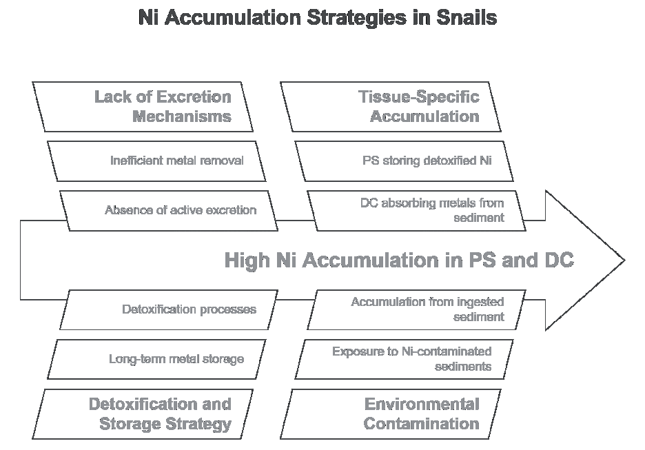
Pomacea insularum employs a distinctive strategy for managing Ni exposure through detoxification and storage rather than excretion. This strategy is evident in the PS and DC, which act as long-term storage sites for excess Ni [31, 32]. The role of these tissues in Ni accumulation is crucial for understanding the biomonitoring capabilities of Pomacea species. Research suggests that P. canaliculata and other apple snails exhibit differential abilities to bioconcentrate heavy metals, including Ni, with the midgut gland and kidney showing the highest bioconcentration factors for several metals [33, 34]. DC, involved in nutrient absorption and processing, plays a significant role in metal accumulation, particularly due to its exposure to Ni-contaminated sediments [35].
In P. insularum, the sensitivity to metal exposure underscores the importance of the DC and PS in Ni accumulation and detoxification processes [15, 16]. These findings highlight the importance of these anatomical features in assessing environmental contamination in freshwater ecosystems [36]. The DC, being a primary site for metal uptake, accumulates Ni over time, as it lacks an efficient excretion pathway [22]. This tissue absorbs metals from ingested sediment, and without a mechanism to remove excess Ni, it stores the metal in detoxified forms [32, 37-39].
Similarly, the PS accumulates Ni due to the absence of active excretion, functioning as another storage site for detoxified metals. The lack of an efficient excretion mechanism in both the DC and PS suggests that P. insularum has evolved a strategy focused on metal storage rather than excretion [22, 40]. This strategy ensures that Ni does not accumulate toxins in metabolically active tissues, although it leads to significant accumulation in these storage tissues [23, 32]. Consequently, this detoxification strategy protects vital functions, resulting in long-term metal storage in the PS and DC [22, 32].
Therefore, the high Ni accumulation in the PS and DC of P. insularum results from the lack of effective excretion mechanisms in these tissues. Instead of being excreted, excess Ni is detoxified and stored, leading to elevated metal concentrations in these tissues. This process is central to the organism’s metal regulation strategy and reflects the unique role of the PS and DC in managing Ni exposure in contaminated environments.
Ni Accumulation in the Shells as a Long-Term Storage Site
The shells of P. insularum play a pivotal role in the organism’s ability to manage and detoxify excessive Ni exposure, accumulating the highest Ni concentrations among the tissues studied. Unlike the model presented for the regulation and excretion of Ni in soft tissues, the shell functions as a long-term storage site for detoxified metals, allowing the organism to sequester potentially toxic elements in a biologically inert form [3, 13, 41]. This process is crucial for maintaining metabolic function while managing environmental metal contamination.
In the case of P. insularum, soft tissues, such as the DC and foot, follow the regulatory model depicted in Fig. 3, where Ni absorbed from environmental sources enters the metabolically available pool. Ni is utilized for metabolic processes from this pool or transferred to detoxified storage sites. However, while soft tissues utilize excretion as a mechanism for Ni regulation, this excretion pathway does not apply to the hard tissue of the shell. Instead, Ni that is detoxified and stored in soft tissues is ultimately transferred to the shell for permanent sequestration, making the shell a critical component in the overall metal regulation strategy of the organism.
The shells of various marine organisms, such as Telescopium telescopium and Mytilus edulis, play a crucial role in the sequestration of Ni, making them valuable for biomonitoring environmental pollution. Shells act as microconcentrators for Ni, with studies showing significant correlations between Ni concentrations in shells and sediment Ni levels, indicating their potential as biomonitoring materials, especially in tropical intertidal areas [3]. The development of ultrasound-assisted extraction methods has further enhanced the efficiency of Ni detection in mussel tissues, facilitating rapid bioaccumulation assessment in polluted coastal environments [42]. The mineral composition of shells, influenced by environmental factors, provides insights into historical pollution levels, as variations in elemental ratios reflect the water conditions during shell formation [43]. Consequently, the ability of shells to sequester Ni and their responsiveness to environmental changes underscores their importance in monitoring metal pollution [44].
The shell of aquatic organisms serves as a crucial immobilization site for Ni accumulation, effectively reducing its bioavailability and preventing interference with cellular processes in soft tissues [45]. High levels of Ni in shells are attributed to their role as endpoints in the detoxification pathway, where Ni that cannot be excreted is safely stored. This contrasts with soft tissues, which must balance uptake, metabolic use, and excretion to maintain low metal concentrations [46, 47]. The absence of an excretion pathway in the shell allows it to serve as a repository for Ni over the organism’s lifetime, providing a cumulative record of Ni exposure. This makes the shell an invaluable indicator for long-term biomonitoring, as it retains a historical record of metal accumulation. The shell’s function is particularly important in environments with fluctuating metal levels, where it serves as a stable sink for excess metals, preventing toxic effects in metabolically active tissues [48, 49].
From an ecotoxicological perspective, the shell’s high Ni accumulation highlights its utility as a biomonitor for assessing Ni pollution in aquatic ecosystems. Unlike soft tissues, which may exhibit variability in Ni concentrations due to dynamic regulatory and excretion processes, the shell provides a consistent and long-term record of Ni exposure [50, 51]. This stability makes it a reliable indicator for assessing environmental health in ecosystems contaminated with heavy metals [45]. Although laboratory experiments offer direct insights into contaminants’ effects, using bivalve shells as biomonitors presents a valuable approach to studying the long-term impacts of Ni pollution in the field [14, 41, 45]. This body of research demonstrates the significance of shell-based biomonitoring in understanding the broader implications of Ni contamination on aquatic ecosystems.
Therefore, while the regulatory model depicted in the image accurately describes Ni management in soft tissues, it does not apply to the shell of P. insularum. Instead, the shell acts as a permanent storage site for detoxified Ni, reflecting the organism’s strategy of sequestering toxic metals in an inert form to prevent damage to essential metabolic processes. The accumulation of Ni in the shell underscores its critical role in the organism’s overall metal regulation system and its importance as a tool for biomonitoring environmental Ni pollution.
Low Accumulation of Ni in CT and Foot Due to Ni Excretion
Fig. 4 shows the overall conceptual model for the Ni regulation strategies in Pomacea insularum. The low accumulation of Ni in the CT and foot of P. insularum can be attributed to efficient excretion mechanisms in these tissues. Ni, absorbed into the body from environmental sources, enters the metabolically available pool, where it is utilized for essential metabolic functions [26]. However, unlike tissues such as the PS and DC, the CT and foot have developed mechanisms to excrete excess Ni, preventing its accumulation in the body.
Amphipods ingesting non-essential metals from their food seem to manage it by detoxifying them inside the cells of the ventral caeca. Corophium volutator processes dietary cadmium in this manner [52]. The regulation of Ni in the freshwater gastropod P. lineata involves several physiological mechanisms aimed at mitigating toxicity. Ni exposure disrupts ion homeostasis, particularly affecting magnesium (Mg²⁺) levels, which are crucial for cellular functions. Research shows that Ni antagonizes Mg²⁺ uptake, leading to significant reductions in whole-body Mg²⁺ concentrations, impairing essential physiological processes in gastropods [5]. Additionally, Ni exposure induces oxidative stress, promoting the production of reactive oxygen species (ROS), which can damage cellular components [53]. Chronic exposure to Ni has been linked to adverse effects on growth and reproduction, further highlighting its impact on physiological health [54]. The regulation of Ni toxicity in P. insularum is thus a complex interplay of regulatory mechanisms, oxidative stress responses, and potential alterations in energy metabolism, requiring further research for a more comprehensive understanding [46, 55].
Tissues like the central organs [51] and the foot of P. insularum are in direct contact with the aquatic environment and can absorb metals like Ni from the water. However, these tissues exhibit efficient excretory mechanisms that prevent the accumulation of Ni to toxic levels [38, 45]. The rapid excretion of Ni from the central tissues and foot ensures that they function as initial interfaces for Ni absorption without becoming long-term storage sites. These efficient excretion pathways in the foot enable the tissue to eliminate excess Ni ingested from sediments, ensuring minimal Ni accumulation within the organism [32, 45, 56].
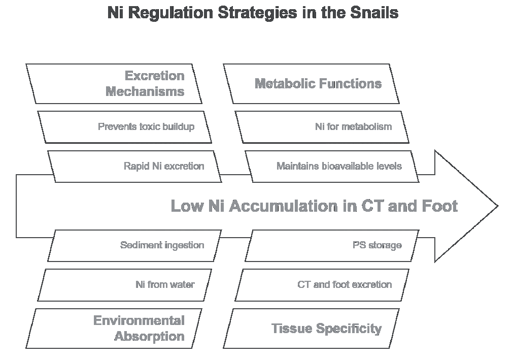
This excretion-driven mechanism is vital in preventing Ni overaccumulation, which could become toxic at high concentrations [45]. By quickly excreting Ni from these tissues, P. insularum maintains Ni concentrations at bioavailable levels for essential physiological processes while preventing harmful buildup. In contrast, tissues such as the PS and DC rely more on metal storage than excretion to manage Ni levels. This explains why Ni concentrations remain relatively low in excretory tissues compared to organs that utilize storage as their primary regulation strategy [32, 39, 45]. Understanding these differential strategies – excretion versus storage – highlights how P. insularum effectively manages metal exposure and mitigates the toxic effects of Ni in its aquatic environment.
Hence, the low Ni levels in the CT and foot result from their efficient excretion mechanisms, which enable P. insularum to regulate and manage Ni exposure effectively. This contrasts with other tissues, such as the PS, which accumulate Ni due to the absence of excretion. The CT and foot, therefore, serve as important sites for metal regulation through excretion, ensuring that P. insularum can thrive in environments with varying levels of Ni contamination.
Tissue-Specific Bioavailability and Redistribution of Ni
Ni bioavailability and redistribution in P. insularum can be understood through its sensitivity to heavy metals, including Ni, as shown in toxicity studies. Research highlights that juvenile snails are more sensitive to Ni than adults, with median lethal concentrations (LC50) demonstrating that Ni is less toxic than copper but more toxic than lead and zinc [15, 16]. The invasive nature of P. insularum, now established across several southeastern states, raises concerns about its environmental impact. The species not only consumes aquatic vegetation but also has the potential to accumulate toxins, posing ecological risks [57, 58]. Moreover, the snail’s reproductive behavior, particularly its preference for oviposition on certain macrophytes, may influence its exposure to Ni in different habitats [59]. Understanding these dynamics is crucial for assessing the ecological risks posed by P. insularum and developing strategies for managing its spread in freshwater ecosystems.
The correlation analysis in Table 2 provides valuable insights into the relationships between Ni concentrations in various tissues, shedding light on how P. insularum manages Ni bioavailability and redistribution [14, 45]. Strong correlations between the shell and several soft tissues, including the mantle (R = 0.87), foot (R = 0.79), and DC (R = 0.76), suggest that these tissues play a critical role in transferring Ni to the shell, where it is stored long-term [48, 49]. This pattern aligns with findings from other studies on snails, which indicate that soft tissues involved in environmental interactions are key in metal uptake before transferring metals to calcified structures for detoxification [60].
The DC, with its strong correlation with the foot and CT, supports its role as a primary organ in the absorption and redistribution of Ni. It absorbs Ni from contaminated sediment and food, processes the metal, and redistributes it to other soft tissues [60-62]. The foot, involved in movement and interaction with contaminated sediment, works closely with the DC in managing Ni bioavailability. The CT, responsible for environmental sensing, shows moderate correlations with other tissues, suggesting that it processes Ni absorbed through environmental exposure but regulates its internal metal concentration to prevent interference with its physiological functions [48, 63].
Interestingly, weaker correlations between the operculum and other tissues, such as the mantle and foot, suggest that the operculum plays a minor role in the organism’s overall Ni detoxification strategy [63]. The correlation patterns observed in Table 2 suggest that soft tissues manage Ni uptake and redistribution in a coordinated manner, eventually transferring Ni to the shell for long-term sequestration. This coordinated strategy highlights the importance of tissue-specific roles in metal detoxification and accumulation in P. insularum, making the species an effective biomonitor for Ni pollution in freshwater ecosystems.
Detoxification and Storage Mechanisms for Ni
The factor analysis presented in Table 3 provides crucial insights into the tissue-specific mechanisms of Ni detoxification and storage within P. insularum. The shell’s high negative loading on Factor 1 (-0.930) confirms its role as Ni’s primary long-term storage site, where the metal is effectively sequestered and detoxified [23, 40]. Once Ni is transferred to the shell, it becomes biologically inactive, preventing it from interfering with the organism’s metabolic processes. This pattern of metal sequestration in calcified structures aligns with detoxification strategies observed in other snail species, where the shell acts as a permanent repository for heavy metals [63].
DC emerges as a critical player in managing Ni absorbed from food and sediment, as indicated by its high loading on Factor 2. As the primary site for Ni absorption, the DC processes the metal and distributes it to other tissues for detoxification [23, 38, 64]. With its significant loading on Factor 1, the mantle appears to play a dual role, both in metal absorption and redistribution and in transferring Ni to the shell for long-term storage. With its strong loading on Factor 1, the foot also contributes to metal redistribution, given its direct exposure to contaminated sediments during movement [45].
CT plays a role in both initial Ni absorption and redistribution to other tissues, as evidenced by its moderate loadings on both Factor 2 and Factor 3. However, the weaker loadings of the remaining soft tissues and the operculum suggest that these tissues play less active roles in Ni processing and storage.
Overall, the factor analysis reveals a coordinated system in which soft tissues manage Ni absorption and redistribution, while the shell serves as the final detoxification and storage site. This multifaceted approach to Ni handling represents a crucial adaptation for P. insularum to thrive in metal-contaminated environments [23, 32, 45, 63]. The ability of P. insularum to compartmentalize and detoxify Ni through this system underscores its utility as a biomonitor in assessing the health of freshwater ecosystems exposed to heavy metal contamination.
Pomacea insularum as a Good Biomonitor of Ni Contamination and Bioavailability
The transplantation experiment from the unpolluted UPM Lake to the polluted Juru River demonstrated significant Ni accumulation in most tissues over seven weeks. The highest Ni AP was observed in the PS, followed by the REM and DT. The high Ni retention in these tissues indicates their critical role in Ni sequestration and detoxification. The CT also showed moderate Ni accumulation, suggesting that external exposure plays a role in Ni uptake. In contrast, the shell exhibited a lower AP of 21.9%, reflecting its function as a long-term Ni storage site. The foot and operculum had the lowest Ni accumulation, likely due to limited direct interaction with Ni sources. These findings confirm that P. insularum can effectively accumulate Ni when exposed to contaminated environments, with certain tissues serving as primary reservoirs for Ni retention.
Following transplantation from the Juru River to UPM Lake, the depuration study revealed significant reductions in Ni concentrations across all tissues. The DT exhibited the highest DP, indicating rapid elimination of Ni upon removal from the contaminated site. The CT also showed a high DP, suggesting active physiological regulation to remove excess Ni. The PS initially accumulated the highest Ni levels, highlighting its role in Ni detoxification. The REM and shell exhibited moderate depuration rates, suggesting a gradual release of Ni. In contrast, the foot exhibited the lowest DP (14.9%), indicating slower Ni elimination. The results confirm that P. insularum has a strong capacity for Ni depuration, particularly in metabolically active tissues such as the DT and CT.
The findings of this study reinforce the suitability of P. insularum as a biomonitor for Ni contamination and bioavailability in freshwater environments. The strong accumulation patterns observed in the PS, DT, and REM indicate that these organs are highly responsive to environmental Ni exposure. The significant depuration observed after relocation to a cleaner environment further supports the use of P. insularum for monitoring Ni pollution and assessing ecosystem recovery. The lack of strong correlations between sediment Ni levels and tissue accumulation suggests that metal uptake is influenced by additional factors such as bioavailability, water chemistry, and dietary intake. Future studies should investigate seasonal variations, metal speciation, and the role of metallothioneins in regulating Ni accumulation. Overall, this study provides valuable insights into Ni bioaccumulation dynamics and stresses the importance of continuous monitoring in freshwater ecosystems to mitigate the Ni pollution risk [66, 67].
Pomacea insularum has proven to be a valuable biomonitor for Ni contamination in aquatic ecosystems. Research shows that this species exhibits acute sensitivity to various heavy metals, including Ni, with juvenile snails displaying lower median lethal concentrations (LC50) than adults, highlighting their heightened vulnerability to metal exposure [15]. Understanding the dynamics of Ni accumulation in aquatic organisms, such as Chaoborus larvae, underscores the importance of studying uptake mechanisms for effective biomonitoring [65]. Seasonal variations in biofilm communities’ responses to Ni exposure further emphasize the necessity of assessing environmental conditions across different seasons to provide a comprehensive understanding [66]. Based on these insights, P. insularum serves as a reliable indicator of Ni pollution levels, supporting the monitoring of freshwater ecosystems and the assessment of anthropogenic impacts [67].
The findings from this study underscore the potential use of P. insularum as a biomonitor for Ni contamination in both coastal and freshwater ecosystems [24]. The snail’s shell, acting as a long-term storage site for Ni, provides a cumulative record of Ni exposure over time, making it an ideal indicator for chronic metal contamination. The relatively low variability in Ni concentrations within the shell across different sampling sites further supports its role as a reliable indicator of long-term Ni exposure, even in environments with fluctuating contamination levels [14]. This property makes the shell valuable for monitoring persistent Ni pollution in ecosystems impacted by industrial or agricultural activities [51, 68].
Soft tissues such as the DC, mantle, and foot, which are involved in short-term Ni absorption and redistribution, provide important insights into more recent contamination events [1]. Strong correlations between these tissues and the shell suggest that these tissues process Ni before transferring it to the shell for long-term sequestration. Analyzing Ni levels in both soft tissues and the shell allows environmental monitoring programs to gain a more comprehensive understanding of both short-term and long-term Ni contamination in ecosystems [13, 49].
Similar studies on other bivalve species have also highlighted their potential as biomonitors for trace metal contamination. For instance, Mytilus edulis has been widely used as a sentinel organism for marine pollution, though the specific mechanisms of metal assimilation are still being explored [14]. By comparing findings from P. insularum and other gastropods, it becomes clear that shell-based biomonitoring can offer valuable insights into metal contamination over both short and long periods, aiding in managing freshwater and coastal ecosystems.
Overall, P. insularum demonstrates a sophisticated detoxification system that efficiently manages Ni exposure, making it a valuable species for biomonitoring heavy metal pollution in freshwater and coastal environments. This study’s findings underscore the importance of using multiple tissues in biomonitoring assessments, offering a more nuanced approach to understanding how Ni contamination impacts different parts of the organism and the broader ecosystem.
Conclusions
This study demonstrates that P. insularum effectively regulates Ni exposure through tissue-specific detoxification and storage mechanisms, with the shell serving as the primary long-term repository for Ni. The field transplantation study further confirms this ability, as Ni accumulation patterns varied across different tissues when snails were moved from an unpolluted to a polluted site. The high Ni concentrations in the shell highlight its central role in long-term sequestration, while soft tissues such as the DT, mantle, foot, and CT facilitate initial uptake and redistribution before transferring Ni to the shell for storage. Correlation and factor analyses reveal a coordinated system where soft tissues act as intermediary sites, reducing the toxic impact of Ni on vital physiological functions. These findings underscore the suitability of P. insularum as a biomonitor for Ni contamination in freshwater ecosystems. Environmental monitoring programs can gain comprehensive insights into Ni pollution by analyzing both short-term responsive tissues and long-term storage sites like the shell. The shell serves as a valuable indicator of chronic exposure, while soft tissues reflect recent contamination events. The results contribute to a deeper understanding of Ni bioaccumulation in P. insularum, providing essential data for pollution management and ecosystem health assessments.

