

Introduction
Spiders (Arachnida: Araneae) have very successfully adapted to the terrestrial habitats. They include 50,800 identified species worldwide [1]. Spiders are characterized by their webs and each species has its specific web shape. Although some spiders rely on webs to survive, others can live without them. In nature, there is only a very small group of spiders that cannot spin webs.
Spiders have structures and organs specialized for silk-spinning. The main structure to be used for this purpose is the spinneret, which consists of different types of silk glands. Different silk glands of spiders allow them to produce different types of silk fibrils. These fibrils consist of protein structure and are in the form of fibrin. When the liquid silk solidifies, it becomes elastic and strong. Previous studies revealed that the distribution of amino acids in the web varies in different parts of it. It was also shown that the silk used in the interior of cocoons or tube-shaped shelters contains different amino acids [2].
The silk-spinning apparatus plays an ecologically central role in a spider’s life cycle [3]. The silk fibrils produced by them are strong, highly elastic, and lightweight [4, 5]. Spiders typically rely on the webs they weave to catch their prey. Spiderlings also must spin species-specific webs and hunt immediately after emerging from their egg sacs in order to survive. In general, the web designs of spiderlings are similar to those of adults. The young individuals of species weaving circular webs construct circular webs, whereas those of species weaving sheet webs weave sheet webs. However, ontogenetic studies on web structure revealed minor design differences in the webs of both circular and non-circular spider webs [6-8]. The challenges that young spiderlings face are relatively different from those faced by conspecific adults due to the differences in the metabolic requirements, prey availability, predators, and ability to cope with abiotic factors such as humidity and wind, among other factors [9-11]. Therefore, there is no reason to assume that even a single species’ webs would be uniform during ontogeny.
The spinnerets and spigots, parts of the spinning apparatus, can operate independently or together in a coordinated manner. It allows spiders to combine multiple silk fibers in different ways by means of various modifications for specific purposes. Some of these purposes include constructing shelters, finding food, communicating through pheromones and vibrations, reproducing, and dispersing through silk parachutes and lines. Thus, silk production and web spinning have probably been a determining factor for the success of the species, enabling them to conquer terrestrial environments and diversify [12].
Silk glands located in the posterior part of the opisthosoma are discharged through exit sets called spigots. The evolutionary origin and characteristics of silk glands are not fully understood yet, but there are several theories. One theory suggests that silk glands might have evolved from accessory glands [13]. Another theory proposes that silk glands might have their origins from sperm capsules [14, 15]. Another theory suggests that silk glands might have originated from coxal glands. An interesting consequence of the coxal gland theory is the possibility that it might have evolved from silk waste products [16]. More recent theories assume that silk glands were derived from dermal glands. It was thought that these glands and spinnerets have evolved from sensory structures [17, 18].
Throughout the evolutionary history of spiders, their success has been associated with their venom that paralyzes their prey, their impressive webs, and their digestive enzymes to digest their prey. Uloboridae spiders are considered evolutionarily less advanced as they have lost their venom glands and, thus, they rely more heavily on their webs to seize and immobilize their prey. Spiders need to fully wrap insects, their main prey, in their webs in order to immobilize them [19].
Uloborid spiders produce fibrous or hairy silk called cribellate silk. Cribellate spiders are known as orb-weavers. These spiders do not use adhesive in their webs. Instead, they tend to use very fine fibers on each web to trap their prey. Uloboridae spiders’ webs generally have a stabilimentum pattern which is white silken adornment along the center, appear to be protective devices that warn birds of the presence of webs in their flight path. They are distributed all over the world, but they are more frequently seen and more diverse in tropical regions. The Uloboridae family is represented by 353 species worldwide. There are three species in Türkiye. A greater diversity of Uloboridae species is expected considering the climate and vegetation in Türkiye.
In the present study, the web structures and the morphologies of spinnerets and numerous spigots Uloborus walckenaerius Latreille, 1806, a species that has no venom glands, were investigated by using light and scanning electron microscopy (SEM). The adaptations of U. walckenaerius in its web and spinneret structures due to the absence of venom glands were examined.
Material and Methods
The samples were collected from the main campus of Kırıkkale University in August 2017. The samples were collected early in the day from the branches of annual plants. Three female specimens were collected during the fieldwork. Since females are more active during August, no male individuals were encountered.
Water was sprayed onto the web in order to take photos of the webs of U. walckenaerius, which spins webs that are thinner than those of other groups. The webs examined by using SEM were taken dry and directly glued to the stub [20]. The web samples prepared were coated with gold for 2-3 minutes by using a Polaron SC-500 model coating device at 1.8 kV and 6 mA in order to examine them under scanning electron microscopy. The web samples were examined using a Jeol JSM-560 SEM and images were directly saved into a computer environment to obtain electron micrographs.
In this study, the posterior part of the abdomen containing the silk-spinning apparatus of U. walkenaerius specimens preserved in 70% ethanol was cut under a stereomicroscope and subjected to a series of graded ethanol for dehydration. The specimens were air-dried after the hydration process. The abdominal parts to be analyzed using SEM were mounted on aluminum stubs by using carbon tape in the most appropriate position. The samples were then coated with gold using a Polaron SC-500 model coating device at 1.8 kV and 6 mA for 2-3 minutes. The examinations were performed using a Jeol JSM-5600 SEM device, and the images were directly recorded into the computer to obtain electron micrographs.
The silk-spinning apparatus was examined under a stereomicroscope using spiders preserved in 70% ethanol. Initially, the dorsal and ventral views of the spiders were imaged. Then, the posterior part of the abdomen carrying the silk-spinning apparatus was positioned upwards and imaged using a Leica DC 160 camera connected to a Leica 58 Apo brand stereo light microscope.
Results and Discussion
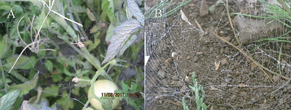
The spider was observed to wait very close to the center of the web, with its ventral side facing up. The webs have a diameter of 10-15 cm, with a single ray passing through the center and successive rays forming angles in reverse to each other, resembling a wheel. U. walkenaerius spiders have been observed to weave webs consisting of very fine fibrils in nature. Various methods have been used in order to capture images of these webs, which are very fine and almost invisible. Some of these methods include placing a dark background behind the web, painting it with spray paint, and spraying it with water. In the present study, the best method for photographing the webs of U. walkenaerius spiders was found to be spraying water onto the web. As the water is absorbed, the silk fibrils of the web thicken and clearer images of the web can be taken (Fig. 1).
The U. walkenaerius specimens that were identified were observed to adapt to the prepared terrariums and start weaving webs on the third day. It was observed that these webs were horizontal, wheel-shaped, and close to the ground, similar to those found in nature.
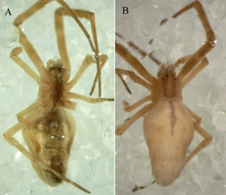
In the U. walkenaerius species, females have an average size of 6.5 mm. They have eight eyes arranged in two rows (Fig. 2). The prosoma is dark gray and the opisthosoma is covered with white hairs, including some dark stripes running longitudinally.
Fig. 1.
a) The image of U. walkenaerius web, which is woven between the branches of annual plants in nature, is displayed with its natural structure near the center of the web. b) The image of the spider’s web sprayed with water to make it more visible reveals the fine silk threads more clearly.
The species U. walkenaerius, like other cribellate spiders, have a calamistrum on their metatarsi. In U. walkenaerius, the calamistrum consists of 30-34 bristles that are arranged regularly (Fig. 3). There are long hairs on the left and right of the Bristle row. When these long hairs were examined at higher magnification, many small hairs lined up parallel to the long axis were noted (Fig. 3f). Their surfaces are also not flat, and short thick structures in the form of seta are seen on the surface (Fig. 3e)
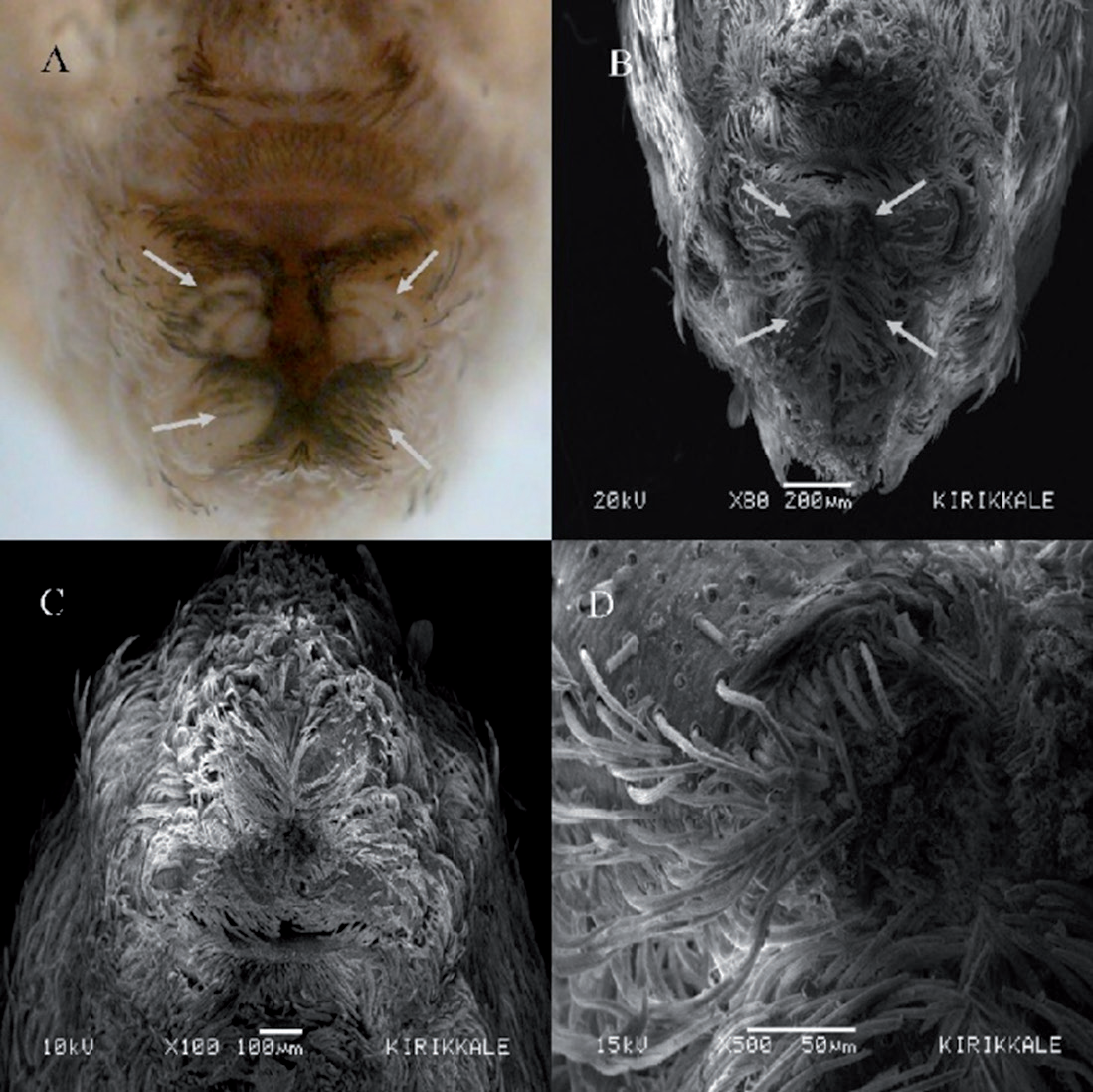
The spinnerets are on the ventral posterior end of the abdomen. A pair of anterior and posterior spinnerets forming the spinning apparatus can be easily seen, whereas a pair of median spinnerets couldn’t be observed. The posterior spinnerets are in one piece, whereas the anterior spinnerets consist of three intertwined segments. The anterior spinnerets have a whole oval cribellum and produce delicate silk. When examined under SEM imaging, the posterior end of the abdomen was found to be covered with regular and dense hairs, as is throughout the abdomen (Fig. 4).
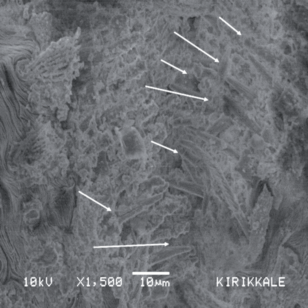
Observing spigot morphology by using light microscopes is quite difficult. Even the best light microscope does not allow for the observation of some distinguishing details on the spinnerets. The structure of the spigot as imaged by using SEM imaging is shown in Fig. 5.
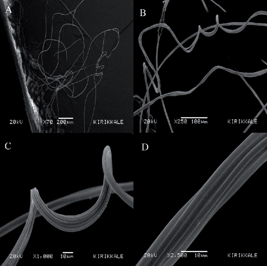
When examined with the naked eye, the web of U. walkenaerius appears to have a thin and porous structure in the form of a wheel. When a piece of the web was examined under SEM, it was noticed that different silk fibrils were used in its construction (Fig. 6 (a-b)). Examining different parts of the web at 2,500x magnification, thick fibers consisting of 6-8 fibrils were observed to be sometimes straight and sometimes spirally curved (Fig. 6 (c-d)). Some silk fibrils were also seen to have an oval shape. Droplet-shaped secretions that provide adhesiveness to the web on the silk fibers of U. walkenaerius were not observed. Instead, the web threads stick to each other via their hairy structures. In addition, it is noteworthy that there are crimped silk fibrils in the web structure of U. walkenaerius (Fig. 6 (b-c)). Actually, examining these crimped fibrils, it was seen that much thinner silk fibers (at least 8-9 thin silk fibers) were arranged side by side to form these silk fibrils.
Fig. 3.
a) Light microscope image of the calamistrum (arrows) belonging to the 4th walking leg of U. walkenaerius. b) SEM image of the calamistrum belonging to U. walkenaerius. c) Image of the calamistrum consisting of 30-34 bristles belonging to the 4th walking leg of U. walkenaerius. (d-e) Images at higher magnification.
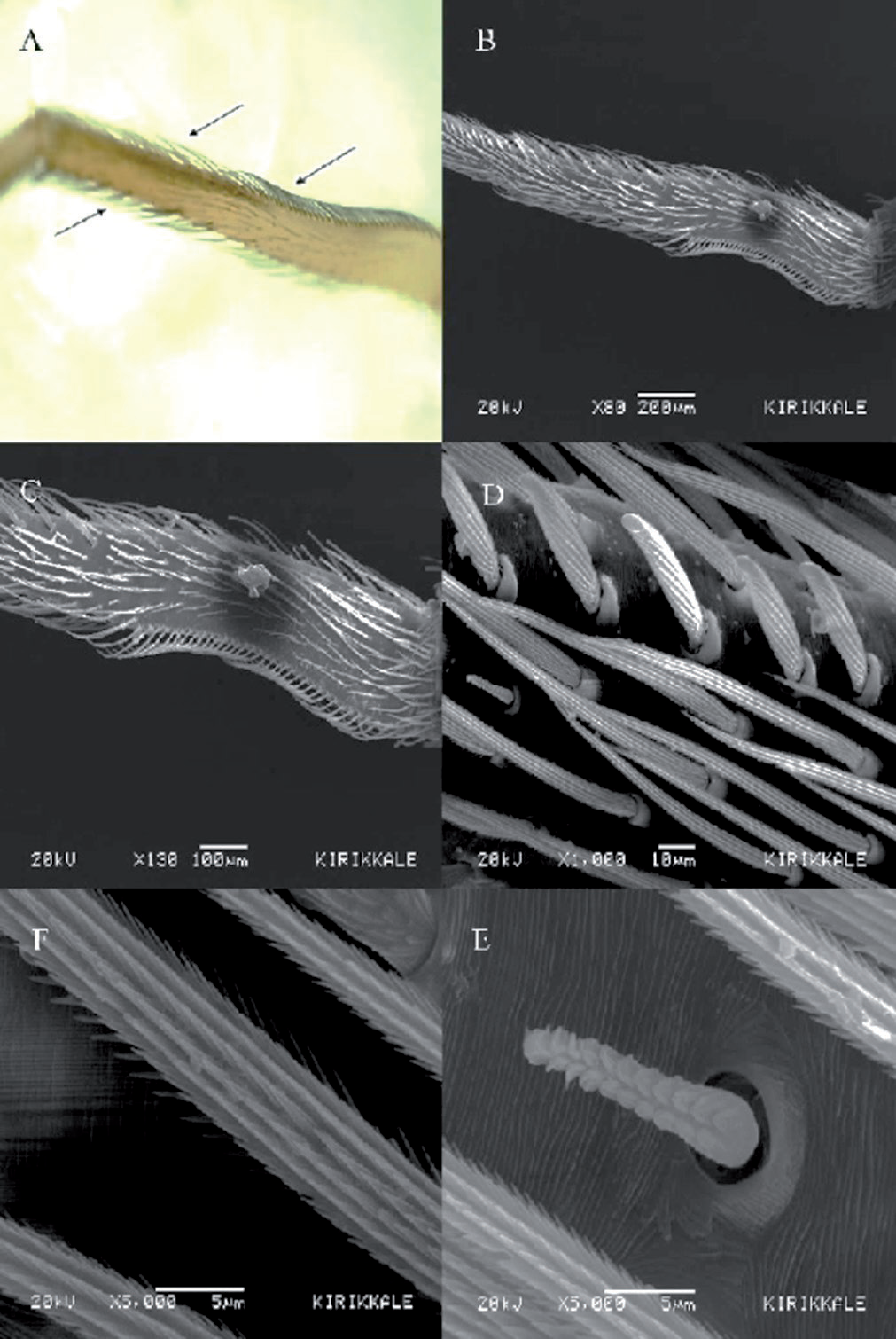
Fig. 4.
a) Image of the spinnerets of U. walkenaerius taken using a light microscope. (b-d). Detailed image of spinnerets of U. walkenaerius.
The success of spiders in the evolutionary process is mainly associated with their venom and silk-spinning capabilities. Uloboridae spiders, which do not have venom, have developed unique strategies to catch, hold, and digest their prey during the evolutionary process. These strategies include behavioral changes such as resting and feeding positions. Some Uloboridae species stand in a hidden position on their webs to hide their front legs to protect themselves from predators. In the field and laboratory parts of the present study, all U. walckenaerius spiders were seen to be in a cryptic posture position. Uloborus species typically exhibit a more cryptic posture characterized by sharply elongated and curved first legs. Dense setal tufts that are proximal to the rapidly flexing leg segment provide external camouflage and they also hide the second legs, which are shorter [21].
U. walckenaerius spiders immobilize the trapped prey by wrapping using their web, which can reach 14 meters in length, because they cannot initially paralyze their prey [22]. For this reason, the web and silk-spinning apparatus of the spider can be considered as a good example of the evolutionary process.
U. walckenaerius has three pairs of spinnerets. However, differing from the other spiders, these spiders have two different structures, named cribellum and calamistrum. The number and arrangement of the spigots, which are the exit points for the produced silk, determine the shape of the web.
Previous studies revealed that the chemical and mechanical properties of spider silk can vary. Both biotic and abiotic factors affect the silk production and, consequently, the mechanical properties of the fibrils [23]. U. walckenaerius webs seen in the field were found to be healthier and larger when compared to those observed in laboratory conditions. Spiders started spinning webs on the third day in the laboratory. It shows that stress factors also affect the quality of the web. Furthermore, the webs that they weave in nature are constructed more durable because of abiotic factors such as wind, temperature, and soil.
U. walckenaerius spiders spend the entire day in their webs in nature. Similarly, U. walckenaerius spiders observed in the laboratory were also found to spend almost the entire day in their webs. It was determined that U. walckenaerius used these webs as a shelter, trap for prey, and cocoon. This finding is consistent with the literature [24]. Moreover, U. walckenaerius spiders were also observed to weave horizontal webs in the laboratory, as in nature.
No method developed for taking photos of the webs could be found in the literature. Three different methods to be used in taking photos of spider webs in nature were specified in the literature. The first method is to place a dark plate behind the web and take the photo. However, in our fieldwork, it was determined that this method was not reliable because the web could be deformed while placing the plate. The second method is to spray paint on the web. However, this method did not sufficiently improve the visibility of the webs. The third and final method was to spray water on the web. Water sprayed on the web is absorbed by the silk fibers. This method had fibers expand and it resulted in a better image. In addition, the water-sprayed web became brighter and more visible. This photographic method used here, as the most reliable method, is consistent with the literature data [25].
The species U. walckenaerius differs from other spiders; their webs contain trap wool or cribellate fibers instead of adhesive droplets [26, 27]. These spiders catch their prey by trapping them in these fibers. The hairy fibrils of U. walckenaerius are less elastic than the spider webs containing droplet silks [28]. Spiders with capture fibrils weave webs with finer fibrils that are harder to see. This may be an adaptation to prevent prey from noticing the web. Prey, which cannot see the web, would be more easily entangled in it.
Fig. 6.
SEM images of the web of U. walkenaerius. a) General view of the web. b) Image of straight and crimped silk fibrils. c) Crimped fibrils. d) It can be seen at a higher magnification levels that a single silk fiber consists of 8-9 finer fibrils.
The morphology of the calamistrum varies between different spider species. In U. walckenaerius species, the calamistrum has evolved to form a single fibril by combining numerous fibers. These fibrils, which seem to have a smooth surface, are actually covered with fine hairs. These hairs entangle insect hairs and make them ineffective [29].
Cribellate spiders secrete nanofibers to catch their prey. During this process, the fibers are extruded from the spigots and processed using a comb-like set of bristles called the calamistrum, which plays an important role in nanofiber processing on the metatarsus. Previous researchers reported that the silk secreted by spiders for silk-spinning purposes is one of the strongest fibers [30]. However, when compared to natural fibers, artificial/human-made fibers exhibit poorer mechanical properties. This result indicates that the web-weaving process itself has a significant impact on the mechanical properties of the fibers.
Conclusions
In this study, the web structure and spinning apparatus were photographed using various microscopy techniques. The web structure and silk-spinning apparatus of U. walckenaerius, a species that has no venom gland, have evolved to empower them in nature. Understanding the spiders’ silk processing or silk-spinning process and implementing biomimicry in spinning technology might pave the way for new materials and practices [31]. The assumed role of calamistrum in nanofiber processing is still debated. However, the calamistrum plays an important role in curling the fibers. Without a calamistrum, the fibril assembly mechanism would be disrupted, and it would cause the loss of its characteristic structure [32]. The calamistrum actually consists of hairs that are bent and curved, facing toward the tarsus, on the metatarsus of the 4th walking leg, distant from the opisthosoma [33]. Moreover, they are covered with underlying structural components. Some spider species even have tooth-like extensions that can support the processing of nanofibers independently of the threads [34]. These species-specific differences in calamistrum morphology have an unpredictable effect on the nanofiber processing.

