

Introduction
In contemporary material science, nanotechnology is one of the most active research fields. Based on certain traits, including size, distribution, and shape, nanoparticles display entirely unique and superior properties. Nanoparticles and nanomaterials are rapidly finding new uses. Applications for nanocrystalline silver particles include biomolecular detection, diagnostics, antibacterial use and treatments, and catalysis [1]. However, the issue of environmental contamination is a significant one, considering the chemical processes used to create nanomaterials produce a number of dangerous byproducts. Thus, there is a need for green synthesis, which encompasses ways of synthesizing nanoparticles that are safe, nontoxic, and environmentally benign, and * Corresponding author: Dr. Huma Ali, Assistant Professor, Department of Chemistry, MANIT, Bhopal. have long-term commercial viability [2]. For the synthesis of silver nanoparticles that offer several advantages of being environmentally friendly and compatible for pharmaceutical and other biomedical applications, green synthesis uses eco-friendly, nontoxic, and secure resources such as plant leaf extracts, bacteria, fungi, and enzymes.
Nanoparticles (NPs) are smaller than proteins, lipids and nucleic acids, ranging from 1/100th to 1/10,000th of the size of human cells [3]. Because of their small size, NPs can undergo implausible and amazing interactions with a number of biomolecules within and on the surfaces of the human cells [4]. These physical and chemical properties of NPs, including their biological, catalytic, electronic, magnetic, mechanical and optical properties, could be beneficial in various medical applications such as drug targeting, drug delivery, and drug formulations. Thus, there has recently been increased attention on NPs by various industries around the world [5]. The shape, size and the surface morphology of these particles have been found to be vital in tuning the properties of nano-sized metal particles and their potential applications in different fields [6]. Several methodologies including physical, electrochemical, sonochemical, photochemical, and biochemical processes have been investigated for development of a suitable method of metallic NPs synthesis; however, most of these methods have utilized toxic and hazardous chemicals, had a high cost of production, or were subject to difficulty in purification of the end product [7,8]. Recently, the synthesis of NPs using various biological materials such as bacteria, fungi, yeast, and plant extracts employing green chemistry procedures has received a great deal of attention because of the wide range of natural resources used, with significant bioactive compounds and the availability of simple, inexpensive, eco-friendly, dependable and size-controlling approaches [9–11]. NPs produced using plants and their extracts, especially those with medicinal potential, have been found to have a more rapid synthesis process, display more stability, possess a better controlled shape and size, and to contain higher levels of important reducing agents in comparison to those produced from other organisms [12,13].
Two or more drugs that individually produce overtly similar effects will sometimes display greatly enhanced effects when given in combination. When the combined effect is greater than that predicted by their individual potencies, the combination is said to be synergistic. A synergistic interaction allows the use of lower doses of the combination constituents; a situation that may reduce adverse reactions [14]. Drug combinations are quite common in the treatment of cancers, infections, pain, and many other diseases and situations. The determination of synergism is a quantitative pursuit that involves a rigorous demonstration that the combination effect is greater than that which is expected from the individual drug’s potencies. The basis of that demonstration is the concept of dose equivalence, which is discussed here and applied to an experimental design and data analysis known as isobolographic analysis. That method, and a related method of analysis that also uses dose equivalence, are presented in this brief review, which provides the mathematical basis for assessing synergy and an optimization strategy for determining the dose combination [15].
In recent years, scientists have focused their attention on nanotechnology and nanobiotechnology applications in biomedical research, such as cell detection and monitoring systems within the body, and drug and therapy delivery systems. The term nano can be thought of as a separate state of matter aggregation in all of its states, including solid, liquid, gas, and plasma. Cytotoxicity research is an important part of the testing process for new nanomaterials in biological and medical applications [16,17]. MTT (2-(4,5-dimethyl-2-thiazolyl)-3,5-diphenyl-2H-tetrazolium bromide), nitroblue tetrazolium (NBT), and second-generation tetrazolium salts, such as XXT (2,3-bis- (2-methoxy-4-nitro-5-sulfophenyl)-2H-tetrazolium-5- carboxanilide, MTS (5-[3-(carboxymethoxy)phenyl]- 2H-tetrazolium inner salt), -2H-tetrazolium inner salt, WST-1 (sodium 5-(2,4-disulfophenyl)-2-(4-iodophenyl)- 3-(4-nitrophenyl)-2H-tetrazolium inner salt) and -3-(4,5-dimethyl-2-thiazolyl)-2-(4-sulfophenyl)-2H-tetrazolium inner salt) are basic cytotoxicity assays.
The size of nanoparticles is one of the factors that impact their toxicity. Smaller NPs are more likely to slip past physiological barriers. Phagocytosis and other mechanisms can allow small nanoparticles to enter cells [18]. Adhesive contacts are determined by the ability of NP to enter cells. Furthermore, unlike bigger nanoparticles, NPs less than 100 nm are not phagocytized, but instead enter through receptor-mediated endocytosis pathways. In the lack of particular cell surface receptors, NP absorption can occur. Some nanoparticles with a specific crystalline structure do not display toxicity because of the crystalline structure effect on NP toxicity, but other allotropes can have a significant impact on cell viability and have an influence on human physiology [19].
Materials and methods
Preparation of aqueous extract of onion and garlic outer peel
The bulbs of the Allium cepa L. (Onion) and Allium sativum (Garlic) were purchased from the Big Bazaar, Bhopal, and Madhya Pradesh, India during the winter of 2022. The outer dried peels of both were collected, cleaned, washed twice and were cut into small pieces separately. Both dried peel materials were ground into a soft powder by using an electrical grinder. The extract was produced by mixing 10 gm of each peel soft powder in 500 ml distilled water. The solution was heated at 70 °C for 2 hours with magnetic stirring. The resulting solution was cooled at room temperature and filtered. The filtrate was stored in a refrigerator.
Preparation of Silver onion and garlic nanoparticles (OGNPs)
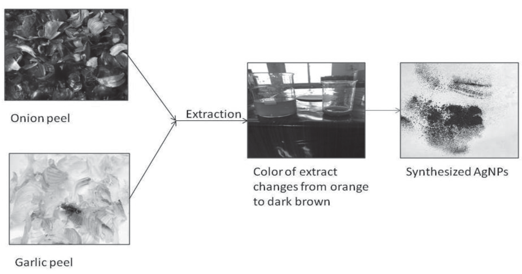
Silver OGNPs were prepared by mixing 80 ml of extract to 20 ml of 1 mM aqueous solution of silver nitrate. The mixture was stirred in water bath at 70 °C for 48 hours. Gradually, the colour of the reaction solution was changed from orange to brown, indicating the formation of silver OGNPs. After complete reaction, the solution eventually became dark brown, which may be due to the increased concentration of nanoparticles as well as particle size. The solution was centrifuged at 15000 rpm for 20 minutes then washed with distilled water and dried in a Petri dish. The Silver OGNPs powder was stored in a sealed serum bottle for further characterization. Fig. 3 shows the Silver OGNPs preparation phases using the above prepared extract from onion and garlic peel.
Cell culture
The American Type Culture Collection sold the A375 cells. Cells were subcultured in DMEM (Gibco) with 10% FBS, 100 IU/ml penicillin, and 100 g/ml streptomycin, and then incubated at 37 °C in a 5% CO2 incubator. Ag OGNPs stock suspension (1 mg/ml) in DMEM (added with 10% FBS) was diluted to concentrations of (5–40 g/ml) in order to evaluate the cytotoxicity, comet assays, and apoptosis. For each experiment, the Ag OGNP suspension was freshly prepared, diluted to the proper doses, and then immediately applied to the cells. In each experiment, a culture medium devoid of Ag OGNPs served as the control.
MTT Assay
The MTT test was used to assess the impact of Ag OGNPs (0, 5, 10, 20, and 40 g/ml) on cell viability [21]. In a nutshell, aliquots of A375 cells (5 103 cells/100 l/ well) were placed in each well of 96 multiplates after being suspended in DMEM at a concentration of 5 104 cells per ml. The media were modified the following day with various Ag OGNP concentrations, and they were cultured in a CO2 incubator for 24 hours. A 100 ml MTT solution (5 mg/ml in medium) was applied to each well after exposure, and it was then incubated for 4 hours in a CO2 incubator. The supernatant was then collected. Following incubation, the MTT solution was discarded, and the crystal that had formed was dissolved in 100 ml of DMSO. At 540 nm, the optical densities of each well were measured.
Results and Discussion
AgNPs cytotoxicity varies with temperature, time, and dose. Cytotoxicity is also closely related to other elements like cell type, size, and AgNP surface coatings [22,23]. AgNP size mediates a variety of cellular reactions, including absorption, cytotoxicity, the capacity to cross biological barriers, and immune reactions. As previously mentioned, size affects both ROS production and the breakdown of AgNPs into ions [24-26]. According to a recent study, the size of coated AgNPs is negatively connected with the levels of ROS, the ratio of apoptosis to necrosis, and the reduction in cell viability [27]. Because of their larger specific surface areas, which control oxidative stress and the rate at which AgNPs dissolve into ions in response to interfacial interaction, the smaller AgNPs exhibit increased activity. Additionally, the size of the nanoparticle affects the way cells absorb them [28,29].
Freshly prepared above mixed extract of Onion and Garlic peel was orange in colour, However, after adding AgNO3 solution and stirring at room temperature, the solution progressively turned dark brown (Fig. 1). In other words, the creation of Ag NPs and the reduction of Ag ions were both validated by an increase in colour intensity as the incubation period progressed [30]. The primary and illustrative evidence for the creation of AgNPs [31] is the colour change in the solution caused by silver nanoparticle surface plasmon excitation [32].
Characterization of Silver OGNPs
A typical method used to characterize materials at the nanoscale is X-ray diffraction (XRD). In addition to several microscopic and spectroscopic techniques, powder XRD analysis of a sample can provide useful information about it, such as phase identification, sample purity, crystallite size, and, in some situations, morphology. In order to determine whether microscopic measurements on a limited number of particles are accurate, a bulk approach known as UV-visible spectroscopy must be used. This method analyses the extinction (scatter + absorption) of light flowing through a sample. The size, shape, concentration, aggregation state, and refractive index close to the nanoparticle surface all affect the peculiar optical properties of nanoparticles. An important tool for detecting, describing, and researching nanomaterials is UV-visible spectroscopy. On a Systronic, UV-Visible spectroscopy analysis was performed. Within an hour of the reaction, silver ions were reduced, and silver nanoparticles were created. AgNO3 was used to maintain control. The UV-Vis spectra were taken at various times following the start of the reaction. Due to the surface plasmon resonance of AgNPs, the absorption spectra of AgNPs generated in the reaction medium had absorption maxima in the range of 422 to 472 nm.
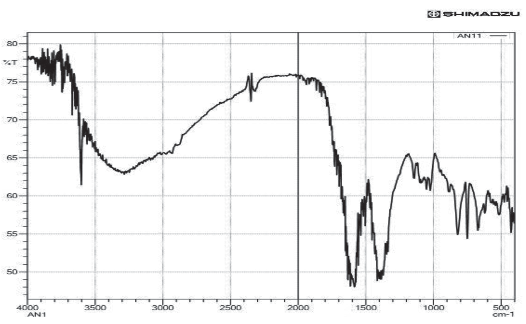
The produced dried Ag OGNPs underwent FTIR examination using the potassium bromide (KBr) pellet technique, and a Fourier transform infrared spectrometer was used to capture the spectra. The produced silver OGNPs solution was centrifuged at 15000 rpm for 20 minutes in preparation for FTIR measurements. To remove any loose proteins or enzymes that weren’t encapsulating the silver nanoparticles, the pellet was washed three times with 5 mL of deionized water. The pellet was vacuum-dried and then examined with an infrared spectrometer for Fourier analysis. Fig. 2 depicts the results of an FTIR analysis used to describe the AgNPs that were found in the extract that was previously prepared. Significant absorption bands were seen in AgNP solutions at approximately 1,025, 1,074, 1,320, 1,381, 1,610, and 2,263 cm-1. The detected peaks, which are from aromatic rings and alkyne bonds, respectively, signify -C-O-C, ether linkages, -C-O, germinal methyls, and -C=C- groups. These bands stand for stretched vibrational bands that effectively cap and stabilise the AgNPs that have been produced.
As a bulk approach, powder XRD analysis results can be compared with microscopy data to determine whether microscopic findings on a small sample of particles are typical of the sample as a whole. XRD was used to identify the silver nanoparticle’s size and makeup. It was performed using the Shimadzu XRD-6000/6100 model with 30 kv, 30 mA, and Cu kv radians at a 2 angle. A quick analytical method that can reveal the dimensions of unit cells is X-ray powder diffraction, which is primarily used to determine the phase of crystalline materials. The average bulk composition of the studied material is found after it has been finely processed. Using Debye Sherrer’s equation [20], it was possible to determine the particle or grain size of the particles on the silver nanoparticles. SEMs are a particular kind of electron microscope that produce images of samples by raster-scanning them with high-energy electron beams. In this experiment, lyophilization was carried out after the manufacture of silver nanoparticles utilising the aforementioned extract from plants. The pellet was analysed using a SEM. A very tiny amount of the sample was used to create thin films on a copper grid that had been coated with carbon; any excess solution was blotted away before the film on the SEM grid was allowed to dry for SEM and EDX analysis.
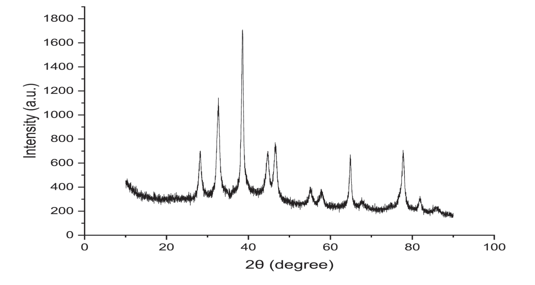
The presence of silver colloids in the sample was confirmed by the XRD, as shown in Fig. 3. The XRD pattern showed Braggs reflections at 2 values of 28.0, 32.0, 49.0 and 65. These Braggs reflections amply demonstrated the existence of the (111), (311), (200), and (220) sets of lattice planes and further supported the identification of these structures as the face-centered-cubic (FCC) structure of silver. The silver nanoparticles produced in this current synthesis are therefore crystalline, as demonstrated by the XRD pattern. Additional as-yet-unassigned peaks were seen in addition to the Bragg peaks indicative of FCC silver nanoparticles, indicating that the bioorganic phase crystallized on the surface of the nanoparticles.
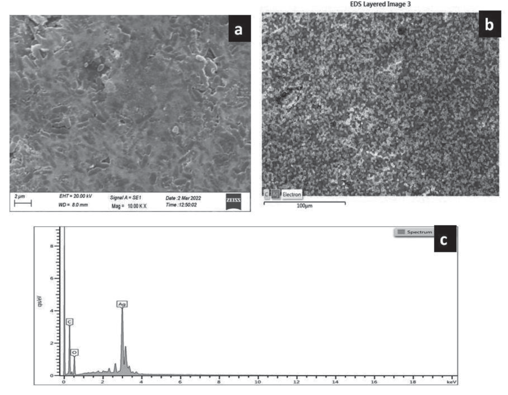
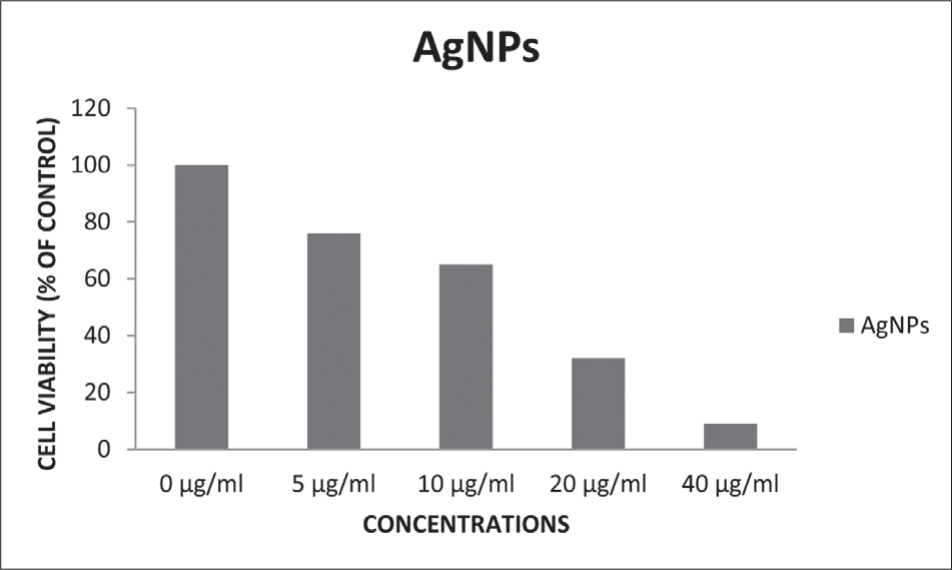
Fig. 4a depicts an Ag nanoparticle SEM image. The image clearly demonstrates that the synthetic material contains tiny grains of substance. These components are combined to create Motichoorboondi-like shapes that are spherical in shape [33]. Its energy-dispersive X-ray (EDAX) image and spectrum, which depicts the material’s elemental analysis, is shown in Fig. 4b and 4c. Strong oxygen and silver peaks may be seen in the resulting spectra.
Cells that have been exposed to nanoparticles can be tested for viability using the MTT assay. Cellular enzymes convert the yellow dye 3-(4,5-dimethylthiazol-2-yl)-2,5- diphenyl tetrazolium bromide (MTT) to the blue product formazan. The amount of blue formazan is proportional to the number of live cells because the transformation can only occur in these cells. The cell viability of A375 cells was reduced significantly in dose dependent manner due to green synthesized AgNP exposure, Fig. 5. The viability of A375 cells was decreased (to 76%, 65%, 32%, and 9%) for 24 hours at all concentrations (5 µg/ml, 10 µg/ml, 20 µg/ml, 40 µg/ml) respectively through MTT test.
Conclusion
The current work demonstrated a quick and environmentally friendly way to make silver nanoparticles by combining an Onion and Garlic peel extract. Our findings imply that the AgNPs can elicit cytotoxic effects, reducing tumour formation and thereby efficiently controlling advancement without damage to normal cells, as a result of the synergistic impact of combination extract of Onion and Garlic peel.

