

Introduction
Cancer is recognized as one of the leading diseases that cause cell death globally in the last few years [1, 3]. World Health Organization (WHO) 2020 statistics reveal that 1 out of every 5 people in the world suffer from cancer during their lifetime, 1 out of every 8 men and 1 out of every 11 women die from cancer [3]. Nowadays, it has been reported that one in every 10 women is at risk of developing breast cancer. One third of those who contract this disease are at risk of losing their lives [4, 5]. Clinical science faces challenges in developing new, targeted and effective cancer treatment modalities with low side effects, high selectivity [6, 8].
Antimicrobial resistance (AMR) is a very important problem with a rapidly increasing prevalence worldwide due to poor clinical outcomes, increased risk of recurrent infections and death. Despite the increasing problem of resistance development due to excessive and irrational use of antibacterial in recent years, a new antibacterial drug could not be used despite the developing technology. This causes the AMR problem to grow rapidly [9]. Antibiotic resistance, especially MDR, is a public health concern is the source. MDR infections have more severe clinical consequences and higher treatment costs than other infections, and it is feared that the spread of pan-resistant strains (pathogens resistant to all currently available drugs) will render some diseases incurable in the near future [10]. At this point, P. aeruginosa is an opportunistic bacterium capable of causing serious systemic infections in critically ill individuals. The increasing incidence of infections caused by P. aeruginosa and the development of multidrug-resistant (MDR) strains, which are resistant to almost all currently available drugs in hospitalized patients, is a concern due to its widespread distribution, its capacity to persist in moist environments and its inherent resistance to a wide range of antibacterial and antiseptic agents [11]. In this regards, chemotherapy-induced Febrile Neutropenia (FN) is common in cancer patients and predisposes to serious, potentially life-threatening infections. The common feature that increases the risk of infection in these patients is impaired immunity, especially the presence of neutropenia. In addition to the changing causative spectrum of infections in neutropenic and cancer patients, there have also been significant changes in the resistance to antibiotics. Vancomycin-resistant enterococci; increased resistance to penicillin in viridans streptococci; Multiple resistance due to different mechanisms in gram-negative fermentative and non-fermentative rods creates serious problems in treatment [12]. As a result of all these, it has become necessary to design safe, effective and environmentally friendly antimicrobial and anticancer agents. The most important area that has been studied recently has started to take its place in modern medical applications as nano-biotechnology.
Nano-biotechnology is a new field in medicine that utilizes nano-sized materials for targeted cell- or tissue-specific medical interventions. Nanotechnology aims to develop and apply methodologies for the production of nanosystems that can interact with high specificity at the molecular (subcellular) level to achieve maximum therapeutic efficacy with minimal adverse effects [13]. Compared to other nanoparticles, inorganic, mesoporous Zinc Oxide nanoparticles (ZnO-NPs) have proven to be outstanding [14, 15]. They are excellent therapeutic nanocarriers because of their large drug loading capacity, adaptability for simple functionalization, adjustable particle size and shape, and biocompatibility. For example, they are powerful antibacterial agents because they can effectively suppress the growth of both gram-positive and gram-negative bacteria. The antibacterial effect of zinc oxide is due to the high sensitivity of the lipid bilayer of bacteria to the reactive oxygen species produced by these nanoparticles [16, 19]. Due to their small size (100 nm) and high monodispersity, ZnO-NPs are important in many controlled-release drug delivery applications as they simplify endocytosis and cause less cytotoxicity [20, 21]. Because of the limited solubility, instability, and side effects of current antibiotic therapy, such resistant bacterial strains must be tackled with innovative and unique methods. That’s why the world needs new antibiotic delivery technologies now more than ever. Nanotechnology has received great attention due to its beneficial physicochemical properties, drug targeting efficiency, improved absorption and biodistribution capacities [22, 26]. “Nanotechnology-based” combination therapy for cancer treatment is becoming more popular in this context because it has been shown to have synergistic anticancer benefits, reduce drug-induced toxicity, and prevent multidrug resistance due to its different modes of action [27, 28]. In this context, different investigators have demonstrated significant toxic effects of ZnONPs in various types of human alveolar, kidney, hepatocyte, breast, and colon cancer cell lines, depending on the size and concentration of ZnONPs [29, 33].
The current work aimed to examine ZnO-NPs’ antibacterial efficiency and any potential interactions with the potent antibiotic (Piperacillin-Tazobactam) when employed against MDR bacteria. Moreover, the effects of Piperacillin-Tazobactam and ZnO-NPs alone or combined on MCF-7 and HepG2 cancer cell lines were investigated, and a new perspective was tried to be gained with the new findings for treatment.
Material and Methods
Zinc Oxide Nanoparticles
Zinc oxide nanoparticles of variable size [ZnO-NPs-A (35+2.5 nm), ZnO-NPs-B (50+2 nm), and ZnO-NPs-C (65+3 nm)] were purchased from CHANGSHA EASCHEM, Co., Ltd.
Antibacterial Activity against MDR Bacteria
MDR strains of P. aeruginosa were obtained from the Laboratory of Microbiology and Public Health at COMSATS University Islamabad Pakistan. Antimicrobial activity was performed according to the method of Pang et al. [34].
Antibiotics Sensitivity Profiling
The antibiotic susceptibility patterns of the ESBLs producing P. aeruginosa were determined by the disc diffusion method on Muller Hinton Agar (MHA) plates according to the CLSI guidelines 2013. The isolates were cultured on MHA plates by using a sterilized loop. Sterile discs loaded with different concentrations of ZnO-NPs-B (200 μg/mL, 400 μg/mL, 600 μg/mL, 800 μg/mL, and 1000 μg/mL) were placed on the surface of these plates that have been inoculated by ESBLs producing P. aeruginosa. The plates were incubated overnight at 37ºC and the zone of inhibitions was calculated [34, 35].
Mammalian Trial
The COMSAT Lab was employed to get a total of 50 pregnant Wistar rats, which were kept separately in lab settings (housed in cages with proper ventilation). The entire accepted experimental protocol was followed. After receiving the necessary consent from the Institutional Animal Ethics Committee, the clinical experiment was conducted on lactating rats.
Experimental Design for Mammalian Trial
As shown in Table 1, these lactating animals were split into five groups (groups I-V) of ten rats each after giving parturition, all of which received the same husbandry practices. At around 10 days old and an hour before infection, pups from nursing mothers were taken away. Sterile deionized water was administered intramuscularly (IM) to Group I, which served as the healthy control. In groups II, III, IV, and V, P. aeruginosa colony-forming units (CFU/mL) were used to infect rats.
Comparison of Nano-Treatment with Commercially Available Drug
Group II functioned as the mastitis control group, receiving no treatment. Pregnant females in group III received treatment with ZnO-NPs-B (50+2nm). Piperacillin-Tazobactam was administered intramuscularly (IM) to animals in group IV while combined therapy was given to group V. All other groups received the same medication, which was administered once daily for five days (Table 1).
Induction of Mastitis
The intraperitoneal administration of ketamine at 30 mg/kg body weight rendered the rats unconscious before they were positioned on the dorsal surface. A magnifying glass was used to examine the exposed ventral area. After using 70% ethyl alcohol to disinfect the mammary gland region, 10 L (CFU/mL) of P. aeruginosa culture was injected into the left fourth and fifth (L4, L5) and right fourth and fifth (R4, R5) teats. To serve as the standard control group, all 10 rats in group I were infected with 10 L of sterile deionized water in the left fourth and fifth (L4, L5) and right fourth and fifth (R4, R5) teats.
Table 1.
Group information and intervention procedure for the experimental trial (n = 10).
Histopathology
For histological research, a total of 5 mammary glands were selected at random. According to the procedure previously mentioned [36], tissue was obtained for histological analyses. After the blood had been taken out of the tissue framework, these samples were given a formalin treatment. Following a 24-h 10% formalin treatment, the tissue samples were moved to a fresh 10% formalin solution and left there for 21 days. Following this technique, the samples were stained using the standard H and E method following the previously described method [36].
Cell Line Culturing
MCF-7 Breast cancer cell line was cultured in RPMI supplemented with 10% Fetal Calf Serum (FCS) and 5% GPPS antibiotics in a 75cm3 culture flask at 37ºC in the presence of 5% CO2 in a SHELLAB incubator (USA).
The HepG2 [HepG2] (HB-8065) liver cancer cells lines, were cultured on Eagle’s Minimum Essential Medium (EMEM) (ATCC 30-2003) with 10% Fetal Bovine Serum (FBS) (ATCC 30-2020) that has not been heating inactivated.
Synergistic Effect of ZnO-NPs with the Drug
For the determination of the effect of ZnO-NPs (A-C) alone and in combination with Piperacillin-Tazobactam on the viability of the MCF-7 and HepG2 cell line, the following methodology was adopted. Cells at concentration 1×10⁻⁵ were added in 96 well culture plates, and various concentrations of ZnO-NPs (A-C) alone; in combinations with Piperacillin-Tazobactam and Piperacillin-Tazobactam alone (along with negative and positive control) were incubated for 24 h at 37ºC in presence of 5% CO2 in SHELLAB incubator (USA). The test compounds ZnO-NPs (A-C) at a concentration of 100 µg/mL, 200 µg/mL, 300 µg/mL, 400 µg/mL and, 500 µg/mL were used in duplicate. The various concentrations of Piperacillin-Tazobactam (µM) in ZnO-NPs (A-C, µg/mL) were used for anticancer activity as; 0.5 µM/50 µg/mL, 1 µM/100 µg/mL, 1.5 µM/150 µg/mL, 2µ M/ 200 µg/mL, and 2.5µ M/250 µg/mL respectively.
Cell Viability Assay
The percent cell viability (anticancer activity) of ZnO-NPs and Piperacillin-Tazobactam in alone and synergism were assessed against the MCF-7 breast cancer cell line and HepG2 liver cancer cell line through MTT assay [37, 38]. Cell viability after treatment was determined through MTT assay as described by Terry L Riss Promega Corporation (Sigma USA). A total of 10µl MTT (5 mg/mL) was added to the wells of the plate which were incubated for 2-3 h at the same incubation conditions. Afterward, solubilization solution (40% v/v) Dimethylformamide (2% v/v) glacial acetic acid (with 16% w/v SDS) in an equal ratio was added to the test plate and optical density (OD) was recorded at 550 nm using PLATOS R 496 plate reader (Cyprus). Cell viability was calculated using following the formula.
Results
In vitro Study on Mammalian Model
The findings of the present study demonstrated that all concentrations of ZnO-NPs-B (50+3 nm) employed in this study (200 μg/mL, 400 μg/mL, 600 μg/mL, 800 μg/mL, and 1000 μg/mL) exhibited antimicrobial activity against P. aeruginosa with a dose-dependent increase in inhibition zones on agar plates. The average zones of inhibition shown by ZnO-NPs against P. aeruginosa at 200 μg/mL, 400 μg/mL, 600 μg/mL, 800 μg/mL, and 1000 μg/mL were 10.32±0.51 mm, 18.21±0.46 mm, 24.45±0.49 mm, 32.63±0.55 mm, and 39.81±0.52 mm, respectively. The mean values were statistically significant (P<0.05) from each other (Table 2) with higher zones of inhibition found at higher concentrations of ZnO-NPs.
Clinical Signs
Following IM injection of P. aeruginosa culture in rats for 6, 12, 24, and 48 h, the clinical symptoms of mastitis were seen. In the current investigation, clinical findings included reddening of the mammary gland surface and apparent swelling and pus flowing out. Within 24 to 48 h of infection following inoculation, groups II, III, IV, and V had overt indications of infection/mastitis. In groups III and IV, the clinical signs started to diminish within two days after the commencement of the ZnO-NPs (50+3 nm) therapy and disappeared after three days, whereas in group V, the clinical indications faded three to four days after the start of the IM Piperacillin-Tazobactam + ZnO-NPs treatment.
Mammary Gland Weight
Additionally, after treatment the mammary gland weights in groups III, IV, and V were significantly different from those in group II and from each other mammary gland weights in groups III-V did not substantially differ from those in group I, indicating that the weight of mammary glands following treatment with was close to that of group I apart from group V which was almost identical proving the efficiency of combination therapy of nano+antibiotics against strain.
Bacterial Load of Mammary Glands
After homogenizing the samples, L5 mammary glands from all the rats were obtained to estimate the bacterial loads, and bacterial loads (log10 CFU/g) are shown in Table 3. Table 4 displays the bacterial load (log10 CFU/g) for groups I to V. In comparison to groups III, IV, and V, group II had a much greater bacterial load. The difference in the mean bacterial load between groups III, IV, and V was statistically significant, and the lower values in group V than in group III and IV suggested that ZnO-NPs (50+3 nm) + Piperacillin-Tazobactam treatment was more effective in reducing the bacterial load showing the efficiency of combination therapy.
Cell Viability/Proliferation Assay
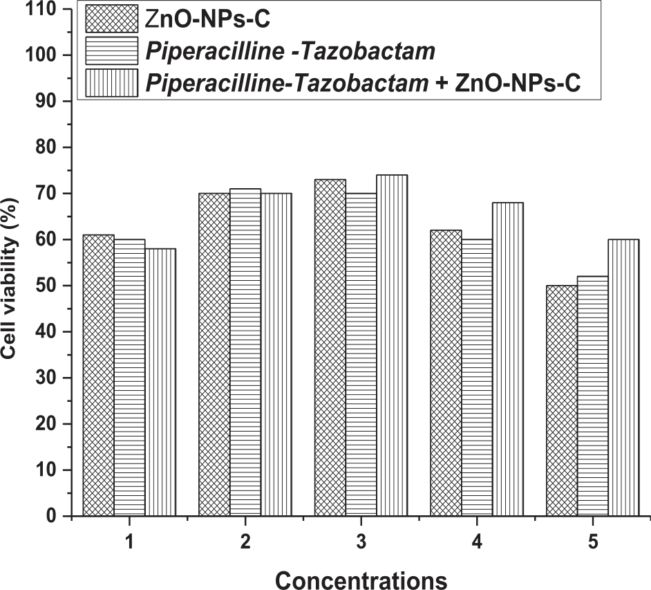
The cell viability for “ZnO-NPs-A and D” increased from treatment 1 (100 ppm, 0.5 µM/50 ppm, 2 µM) to treatment 3 (300 ppm, 1.5 µM/ 150 ppm, 6 µM) as shown in Fig. 1 and then decreased afterward. Furthermore, the commercial drug showed the highest results at treatment 3 i.e. 72% cell viability followed by ZnO-NPs-A+D (69%), while Zn1+D showed the best results at treatment 4 i.e. (70%) followed by the commercial drug (64%). The cell viability for “ZnO-NPs-B and D” increased from treatment 1 (100 ppm, 0.5 µM/50 ppm, 2 µM) to treatment 3 (300ppm, 1.5 µM/ 150 ppm, 6 µM) as shown in Fig. 2 and then decreased afterward. Furthermore, the commercial drug showed the highest results at treatment 2 i.e. 75% cell viability while the treatment combination ZnO-NPs-B+D showed the best results after treatment 2 till treatment 5 i.e. 75%,85%, 80%, and 64% respectively, though after treatment 3 the combination performed better but the cell viability declined significantly. The cell viability for “ZnO-NPs-C and D” increased from treatment 1 (100 ppm, 0.5 µM/50 ppm, 2 µM) to treatment 3 (300ppm, 1.5 µM/ 150 ppm, 6µM) as shown in Fig. 3 and then decreased afterward. The commercial drug showed the highest results at treatment 2 i.e. 71% cell viability followed by Zn-1 and D (70% each) while the treatment combination ZnO-NPs-C+D as compared to others, showed the best results for treatment 2-5 i.e. 70%,74%, 68%, and 60% respectively.
Fig. 1.
% cell viability with different concentrations ZnO-NPs-A and Piperacillin-Tazobactam in alone and synergism. *1. (100 µg/mL, 0.5 µM/50 µg/mL, 2 µM) 2. (200 µg/mL, 1 µM/ 100 µg/mL, 4 µM) 3. (300 µg/mL, 1.5 µM/ 150 µg/mL, 6 µM) 4. (400 ppm 2 µM/ 200 µg/mL, 8 µM) 5. (500 µg/mL, 2.5 µM/ 250 µg/mL, 10 µM).
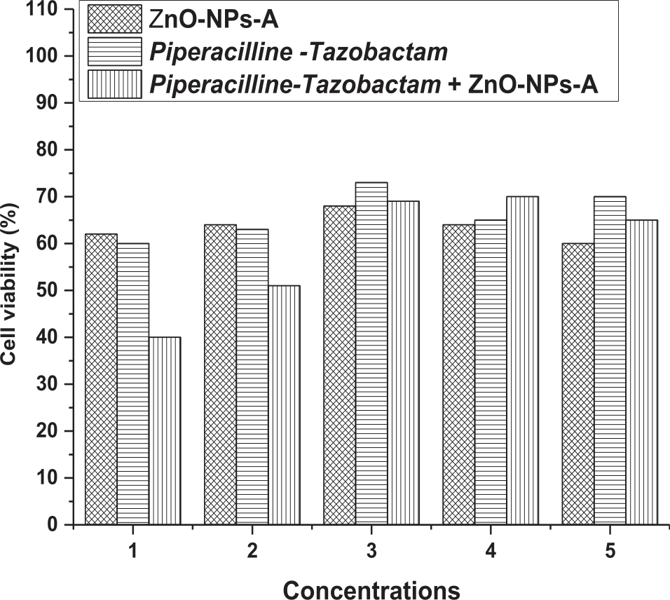
Fig. 2.
% cell viability with different concentrations ZnO-NPs-B and Piperacillin-Tazobactam in alone and synergism. *1. (100 µg/mL, 0.5 µM/50 µg/mL, 2 µM) 2. (200 µg/mL, 1µM/ 100 µg/mL, 4 µM) 3. (300 µg/mL, 1.5 µM/ 150 µg/mL, 6 µM) 4. (400 ppm 2 µM/ 200 µg/mL, 8 µM) 5. (500 µg/mL, 2.5 µM/ 250 µg/mL, 10 µM).
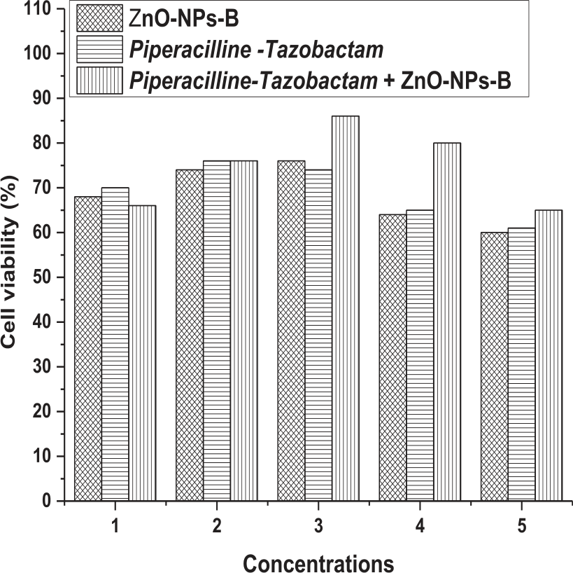
Comparing all the treatments, it is quite significant that ZnO-NPs-B+D performed better and is recommended. The synergic impact of ZnO-NPs-B and Piperacillin-Tazobactam on cell viability was 85% which is the highest for the therapy as compared to ZnO-NPs-A (70%) and ZnO-NPs-B (74%), while the conventional medication i.e. commercially available drugs performed well but not better than the combinations. It is thus recommended the use of Nanotechnology with commercially available drugs.
Fig. 3.
% cell viability with different concentrations ZnO-NPs-C and Piperacillin-Tazobactam in alone and synergism. *1. (100 µg/mL, 0.5 µM/50 µg/mL, 2 µM) 2. (200 µg/mL, 1 µM/ 100 µg/mL, 4 µM) 3. (300 µg/mL, 1.5 µM/ 150 µg/mL, 6 µM) 4. (400 ppm 2 µM/ 200 µg/mL, 8 µM) 5. (500 µg/mL, 2.5 µM/ 250 µg/mL, 10 µM).
Cell Cytotoxicity
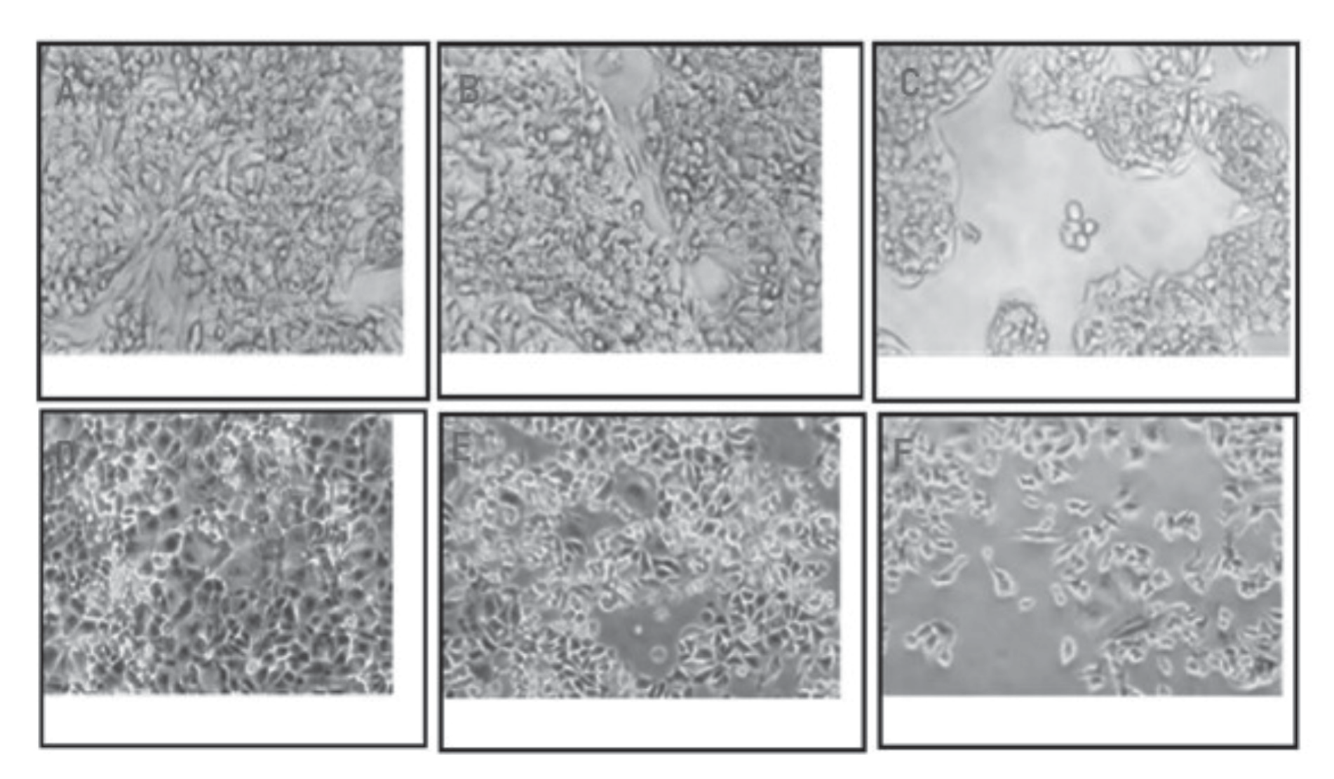
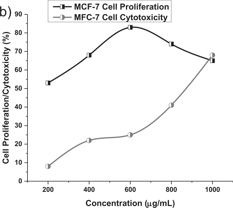
The findings of the current investigation demonstrate that all concentrations of ZnO-NPs-B (50+3 nm) employed in this study (200 μg/mL, 400 μg/mL, 600 μg/mL, 800 μg/mL, and 1000 μg/mL) exhibited antimicrobial activity against P. aeruginosa, with a dose-dependent increase in inhibition zones on agar plates. The average zones of inhibition displayed by ZnO-NPs against P. aeruginosa at 200 μg/mL, 400 μg/mL, 600 μg/mL, 800 μg/mL, and 1000 μg/mL were 10.32±0.51 mm, 18.21±0.46 mm, 24.45±0.49 mm, 32.63±0.55 mm, and 39.81±0.52 mm, respectively. The mean values were statistically significant (P<0.05) from each other (as shown in Table 2), with higher zones of inhibition observed at greater concentrations of ZnO-NPs. To assess the safety of utilizing Zinc oxide nanoparticles (ZnO-NPs) in the medical industry, their cytotoxicity was evaluated on cell lines. The results of the study revealed that at lower concentrations of ZnO-NPs, cell viability remained unaffected and cytotoxicity did not increase. However, when higher concentrations of ZnO-NPs were used, cell viability decreased and cytotoxicity increased. The concentration of 600 μg/mL was identified as the half-maximal inhibitory concentration (IC50) (Fig. 4). The data depicted in Fig. 4 showed that up to the third concentration, or 600 μg/mL, cell proliferation and cytotoxicity were positively correlated, but after this concentration, they were negatively correlated. Concentrations below 200 μg/mL and above 1000 μg/mL were excluded from the analysis as they demonstrated insignificant fluctuations and poor cell proliferation. The intersection of the graph lines for cell proliferation and cytotoxicity is known as the IC50, which was found to be 600 μg/ mL in this study, indicating a 50% suppression of cell growth. Furthermore, the application of nanoparticles in combination with drugs was found to inhibit the growth of cancer cells (Fig. 5). Fig. 5 demonstrated that although the commercially available drug was capable of controlling cancer cell proliferation (Control column), the effect was not significant when compared to the application of nano + drug (Inhibitor column). In contrast, the untreated cell lines had a high cell proliferation rate.
Discussion
In the current study, P. aeruginosa, one of the major mastitis-causing pathogens identified and isolated from clinical cases of bovine mastitis in the Gilgit, Chitral, and Kashmir valley areas [36], which is one of the major mastitis-causing pathogens, was used to evaluate the antimicrobial activity of ZnO-NPs in vitro. The goal of the current investigation was to identify an alternate non-antibiotic therapy regimen against P. aeruginosa based on the resistance profile of that organism. Nanoformulations are efficient against mastitis in laboratory mammalian models, according to a comparison of ZnO-NPs’ efficiency against P. aeruginosa. Even while prior research [36] claimed that other nanoparticles were ineffective against P. aeruginosa and other strains, none of these studies used a mammalian model, particularly concerning ZnO-NPs and cancer cell lines. Given the considerable decrease in mammary gland weights, it can be assumed from the current study that ZnO-NPs are effective in stopping inflammatory processes. Zinc oxide nanoparticles (ZnO-NPs) are a promising antibacterial agent due to their high surface area-to-volume ratio and unique physical and chemical properties. It is believed that ZnONPs generate reactive oxygen species (ROS) that damage bacterial cell membranes and disrupt cellular functions. Earlier studies showed that ZnO-NPs generated ROS that caused oxidative stress in bacterial cells, leading to cell death. It was also found that the antibacterial activity of ZnO-NPs was enhanced by exposure to ultraviolet radiation, which increased the generation of ROS [39]. ZnO-NPs disrupt the bacterial DNA replication, and repair mechanisms by inhibiting the activity of DNA gyrase, an enzyme involved in DNA replication and repair. ZnO-NPs may also interact with bacterial cell walls, disrupting their structure and function [40]. ZnO-NPs have been found to interact with the peptidoglycan layer of bacterial cell walls, leading to structural changes that made the bacteria more susceptible to other antimicrobial agents. In addition, ZnO-NPs may have immunomodulatory properties that contribute to their antibacterial effects. ZnO-NPs have been shown to enhance the activity of immune cells, such as macrophages and neutrophils, which are important for fighting bacterial infections [41]. Earlier studies claimed that Zinc can enhance the phagocytic activity of macrophages, which increases their ability to clear bacterial infections [42]. In conclusion, the mechanism of action of ZnO-NPs as an antibacterial agent is multifaceted and may involve the generation of ROS, disruption of DNA replication and repair mechanisms, interaction with bacterial cell walls, and immunomodulatory effects. The present study also demonstrated the use of ZnO-NPs as a drug carrier to check the viability of MCF-7 and HepG2 cell line with MTT assay by co-delivering Piperacillin-Tazobactam with ZnO-NPs. The ZnO-NPs-A, ZnO-NPs-B, and ZnO-NPs-C were designed for protected delivery of Piperacillin-Tazobactam to the tumor site. Compared to only delivering ZnO-NPs, the dual delivery achieved the synergistic inhibition of tumor growth in vitro. Piperacillin-Tazobactam (PT) inhibits cancer cell growth by targeting the cell membrane integrity of cancer cells. Previous studies found that it inhibited the growth of colorectal cancer cells by disrupting the integrity of the cancer cell membrane, leading to cell death [43]. PT has also induced apoptosis in non-small cell lung cancer cells by activating caspase-3 and caspase-9, two enzymes involved in the apoptotic pathway [44]. Similarly, ZnONPs are considered to induce apoptosis in breast cancer cells by generating ROS that causes DNA damage, and activation of the apoptotic pathway also inhibits the growth of prostate cancer cells by inducing cell cycle arrest at the G1 phase [45]. Mousa et al. [8] studied the antibacterial activity of Zinc Oxide nanoparticles and ovarian cancer. Notably, they reported that synthesized ZnONPs induced 50% inhibitory concentration (IC50) at a concentration of 27.45 μg/ml. On the other hand, they found that the inhibition diameter ranged from 20.16±0.16 to 27±0.57 mm for Staphylococcus aureus and between 25.66±0.33 and 31±0.33 mm for Escheria coli. They determined that the antagonistic effect of ZnONPs differed statistically with neomycin, cefaclor and cefadroxil. Mousa et al. [8] noted that the use of 27.45 μg/mL ZnONPs was sufficient to achieve a significant effect on SKVO3 ovarian cells and both Gram-positive and Gram-negative bacteria. Krishnamoorthy et al. [46] have nominated ZnONPs for use as an alternative antibacterial agent against β-lactam-resistant Gram-negative and pathogenic bacteria. In addition, ZnONPs have been shown to enhance the activity of immune cells, such as natural killer cells, which are important for fighting cancer [47]. This study determines that it is a good strategy to provide safe and effective antitumor immunity. These findings are aligned with the previously reported findings of former researchers [48]. These results also follow the same pattern of previous researchers who worked on encapsulated antibiotics bioinspired in calcium carbonate nanoparticles [49]. Furthermore, previous studies have also reported that the antimicrobial activity of Piperacillin/Tazobactam indicates that the combination may constitute an alternative to third-generation cephalosporins in the treatment of complicated infections [50]. Our findings, which base our assessment of clinical recovery on a considerable reduction in mammary gland weights following treatment with ZnO-NPs, are consistent with earlier findings with Cu-NPs [51]. In comparison to groups III and IV, group II had a much greater bacterial burden. The difference between the group III, IV, and group V mean values for the bacterial load was statistically significant, and the lower values in group V than in group III and IV suggest that ZnO-NPs were more effective in reducing the bacterial load in group V than Piperacillin-Tazobactam was in group IV. On nutrient agar plates, group II also had a considerably larger number of colonies that were counted. Colonies decreased considerably in groups III and IV compared to group V, demonstrating ZnO-NPs’ superior antibacterial activity to Piperacillin-Tazobactam in preventing infection. These results concur with those of Brasil et al. [52], who showed that nanosilver particles had a bactericidal effect in a murine mastitis model. The ZnO-NPs’ inherent ability to disrupt the machinery that makes proteins and to cause tiny gaps in bacterial cell walls may be the cause of their bactericidal effect [53, 55].
Conclusions
This study also concluded that nanomaterials like ZnO-NPs can be designed to meet the aims of modern therapy and diagnosis. The ZnO-NPs also provide a targeted combination treatment method that allows for the safe transport and release of cytotoxic agents to the core of the tumor microenvironment [56]. This study concludes that the dosage and biological action can be minimized and efficiency can be enhanced respectively by using ZnO nanoparticles as potent drug vehicles in the treatment of life-threatening human diseases (infections) as depicted by cancer cell lines (MCF-7 and HepG2). Considering the extremely promising properties of ZnO NPs and their inherent nature, such as selectivity and toxicity towards cancer cells, this is indeed a desired outcome, making them an unquestionably important tool for next-generation cancer therapy. This study with zinc oxide will be informative about the studies to be done with other antibiotics such as Ceftazidime/Ceftazidime-Tazobactam, and Ticarcillin/Ticarcillin - Clavulanate.

