

Introduction
Neonicotinoids are systemic insecticides with broad-spectrum efficacy and relatively low toxicity to mammals, fish, and birds. However, they have been linked to harmful effects on pollinating insects, including honey bees. Dinotefuran, a third-generation insecticide, is widely acceptable due to its efficacy and low toxicity to non-target species. Neonicotinoids mostly target the nervous system of insects by binding to specific receptors known as nicotinic acetylcholine receptors (nAChRs). These receptors are blocked upon binding with Dinotefuran, leading to paralysis and ultimately death in insects. It is reported that neonicotinoids may induce oxidative stress in cells, resulting in immunomodulatory effects and affecting the central nervous system of the target species [1, 2]. It’s important to note that the impact of neonicotinoids is not limited to insects. Some studies have also suggested potential effects on non-target organisms, including birds, fish, and mammals [3]. However, the exact cellular mechanisms and long-term effects on these organisms are still being studied. Regulatory authorities and researchers continue to evaluate the risks and benefits of neonicotinoids to determine appropriate usage and minimize potential harm to pollinators and other organisms in the environment. Despite their widespread use, there is limited experimental data on the impact and mechanism of action of neonicotinoids on animals. This study aims to evaluate the effect of acute exposure to dinotefuran on different mammalian tissues.
Materials and Methods
Chemical Reagents
Dinotefuran [1-methyl-2-nitro-3-(tetrahydro-3-furyl methyl) guanidine] was provided by “Four Brothers” pesticide suppliers L. Ascorbic Acid (Merck), Formalin Buffer (Reidel-de Haen), Ethanol (VWR), distilled water, and Diethyl Ether (Merck Germany).
Methodology
Thirty-five Sprague Dawley rats, of male or female sex, weighing approximately 80±20 g, and aged around 4±0.5 weeks, were acclimatized in accordance with European Union guidelines for animal research at the Laboratory animal housing facility at UVAS, Lahore. The rats were kept in steel cages, and the temperature was maintained at 21±2ºC with 50% humidity. They were provided with a basic diet and water ad libitum for a 12-hour adjustment period. Subsequently, the rats were distributed randomly into three groups, designated as the control, exposed, and vitamin supplemented groups. There were fifteen rats in each group and five in each subgroup (E1, E2, E3 & V1, V2, V3). Control (Group C) also had five rats. The first subgroup of group E, E1, received LD10 (400 mg/kg), the second subgroup received LD25 (1000 mg/kg), and the third, E3, received LD50 (2000 mg/kg). All the subgroups of “V” were exposed in the same manner as those of “E” and received a vitamin C supplement (100 mg/ 350 mL of water) additionally.
Twelve hours after the exposure, 5 mL of blood was collected directly from the cardiac puncture of randomly selected rats from each group. The blood sample was divided into two halves for further analysis of serum and whole blood.
Zero mortality was observed after 48 hours, but four rats belonging to different groups showed symptoms of anorexia and lethargy. All the rats were anesthetized and euthanized as per ethical guidelines for organ extraction. The bones and vital organs, except the brain, were preserved in formalin for subsequent analysis [4]. All procedures were conducted in accordance with the institution’s code of animal research, ensuring humane practices.
Histopathological analysis of the tissues from the liver, kidney, and heart was performed using the method devised by [3] Cui et al. in 2009. The left tibia was removed, cleansed, and kept in boiling water for 10 minutes [4]. The weight/length index and robusticity index of the removed bone were determined using the following formulas:
Length Index =
Robusticity Index =
Results and Discussion
Zero mortality or weight loss symptoms were recorded after 48 hours of exposure. Similar results were also reported after the chronic exposure of Imidacloprid (10 and 20 mg/kg/60 days) [6] alone and also in combination with Fipronil [7].
Blood Toxicity
CBC was conducted on fifteen sampled rats after a duration of 12 hours. The results showed a significant decrease (p<0.05) in the mean values of white blood cells (WBCs), MID cells, and granulocytes (GRA), while lymphocytosis (LYM) significantly increased (p<0.05) in the experimental group, whereas the control group exhibited normal values of blood parameters. Vitamin C supplementation also demonstrated a significantly positive effect, as shown in Table 1.
Leukocytes and their types decreased in number, which can be attributed to the cellular environment under oxidative stress, leading to individual cell lysis and depressed hematopoiesis [8]. Moreover, the transient rise in lymphocyte count may be the result of the body’s defense mechanism [9, 10]. It should be noted that these findings are based on short-term data from an acute trial [11]. Another study on the sub-lethal toxicity of orally applied cypermethrin reported a significant reduction in leukocyte count, except for lymphocytes, and a promotion in lymphocyte count (lymphocytosis) in adult Sprague Dawley rats [12].
A significant decrease in the mean values of the following parameters, red blood cells (RBCs), hemoglobin (HGB), hematocrit (HCT), and red cell distribution width (RDWc) in the experimental group in comparison to the control group has been observed (Table 2). Conversely, values of the following parameters, mean corpuscular volume (MCV), mean corpuscular hemoglobin (MCH), and mean corpuscular hemoglobin concentration (MCHC) were also mounted up in the intoxicated rat group. Vitamin C supplementation demeaned the toxicity effect of dinotefuran on RBCs and related parameters. Factors such as internal hemorrhage, depletion in hemoglobin synthesis, and elevation in hemoglobin destruction have been reported to contribute to the reduction in the erythrocyte count and hemoglobin concentration [13- 16] Toxic substances like insecticides can produce effects in blood cells and lead to hemolysis [17]. Similar results were observed with cypermethrin and imidacloprid [18].
A slight downward trend was also shown by the mean values of platelets (PLT) and platelet percentage (PCT%) in the experimental groups as compared to the control group. However, mean values of platelet volume (MPV), platelet distribution width (PDWc), and platelet large cell ratio percentage (P-LCR%) were not significantly increased in the experimental groups (Table 3). Vitamin C exhibited a non-significant impact. Toxic substances such as insecticides can have a negative impact on bone marrow cells, leading to impaired cell function, aplasia, and dysplasia [19]. The demeaning platelets may be attributed to the effect of the insecticide on bone marrow cells, affecting their formation or development and subsequently slowing down or halting hematopoiesis [8]. An elevated mean platelet volume (MPV) indicates an enhanced tendency for blood clotting, which may increase the risk of thrombosis, stroke, or other cardiovascular anomalies [11]. Previous studies have reported a decreased platelet number due to the toxicity of diazinon, imidacloprid, and other insecticides [17, 20].
Table 1.
Mean (±SD) values of WBCs and its types in different rat groups. E1 = Rat group fed with LD10, E2 = Rat group fed with LD25, E3 = Rat group fed with LD50, V1 = Rat group fed with LD10 + Vit C, V2 = Rat group fed with LD25 + Vit C, V3 = Rat group fed with LD50 + Vit C (Mean values with different subscripts differ significantly (p≤0.05).
Table 2.
Mean (±SD) values of RBCs and related parameters in different rat groups. E1 = Rat group fed with LD10, E2 = Rat group fed with LD25, E3 = Rat group fed with LD50, V1 = Rat group fed with LD10 + Vit C, V2 = Rat group fed with LD25 + Vit C, V3 = Rat group fed with LD50 + Vit C.
Table 3.
Mean (±SD) values of Platelets and related parameters in different rat groups. E1 = Rat group fed with LD10, E2 = Rat group fed with LD25, E3 = Rat group fed with LD50, V1 = Rat group fed with LD10 + Vit C, V2 = Rat group fed with LD25 + Vit C, V3 = Rat group fed with LD50 + Vit C.
Table 4.
Mean (±SD) values of Renal Function Test (RFT) in different rat groups. E1 = Rat group fed with LD10, E2 = Rat group fed with LD25, E3 = Rat group fed with LD50, V1 = Rat group fed with LD10 + Vit C, V2 = Rat group fed with LD25 + Vit C, V3 = Rat group fed with LD50 + Vit C.
| Parameter | Control Group | E1 | E2 | E3 | V1 | V2 | V3 |
|---|---|---|---|---|---|---|---|
| Serum Creatinine (mg/dL) | 0.34±0.03 | 0.47±0.05 | 0.84±0.04 | 1.11±0.03 | 0.41±0.03 | 0.77±0.04 | 0.98±0.04 |
| BUN (mg/dL) | 15.4±1.14 | 20.0±1.86 | 22.6±2.70 | 28±2.74 | 17.2±1.48 | 21±2.25 | 26±3.74 |
Table 5.
Weight / Length Index (mg/mm) of left tibia of different rat groups. E1 = Rat group fed with LD10, E2 = Rat group fed with LD25, E3 = Rat group fed with LD50, V1 = Rat group fed with LD10 + Vit C, V2 = Rat group fed with LD25 + Vit C, V3 = Rat group fed with LD50 + Vit C.
| Parameter | Control Group | E1 | E2 | E3 | V1 | V2 | V3 |
|---|---|---|---|---|---|---|---|
| W/L Index (mg/mm) | 20.63 | 18.75 | 17.36 | 16.83 | 19.14 | 17.93 | 17.13 |
Table 6.
Robusticity Index (mm/mg1/3) of left tibia of different rat groups. E1 = Rat group fed with LD10, E2 = Rat group fed with LD25, E3 = Rat group fed with LD50, V1 = Rat group fed with LD10 + Vit C, V2 = Rat group fed with LD25 + Vit C, V3 = Rat group fed with LD50 + Vit C.
| Parameter | Control Group | E1 | E2 | E3 | V1 | V2 | V3 |
|---|---|---|---|---|---|---|---|
| Robusticity Index (mm/mg1/3) | 2.229 | 2.272 | 2.279 | 2.295 | 2.263 | 2.283 | 2.285 |
Liver Toxicity
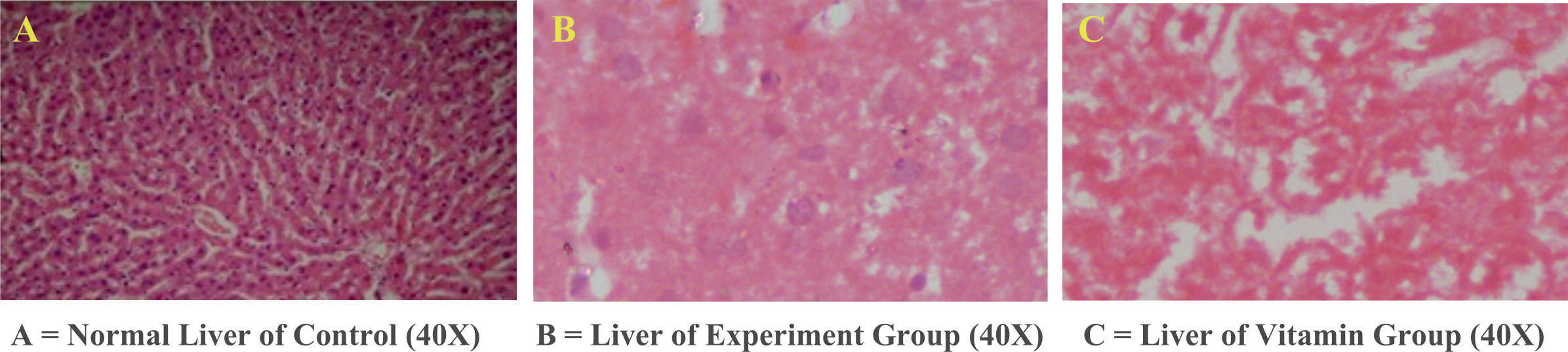
Liver tissue histopathology, initially preserved, was carried out. The serum biomarkers for the Liver Function Test (LFT) were also performed. These two procedures helped us determine the liver’s health after acute exposure of varying degrees. It was found that total bilirubin, alkaline phosphatase (ALP), alanine transaminase (ALT), and aspartate transaminase (AST) increased significantly in the experimental group in comparison to the control group. Vitamin C exhibited a significant ameliorating effect on the mentioned liver biomarkers (Table 4).
Fig. 1.
Comparative analysis of WBCs and its types in different rat groups. E1 = Rat group fed with LD10, E2 = Rat group fed with LD25, E3 = Rat group fed with LD50, V1 = Rat group fed with LD10 + Vit C, V2 = Rat group fed with LD25 + Vit C, V3 = Rat group fed with LD50 + Vit C.
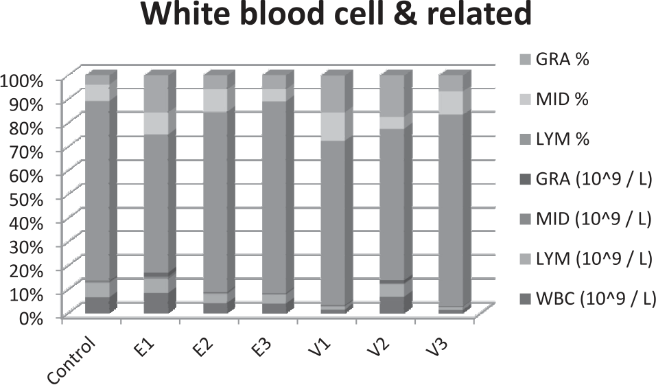
Fig. 2.
Comparative analysis of RBCs and related parameters in different rat groups. E1 = Rat group fed with LD10, E2 = Rat group fed with LD25, E3 = Rat group fed with LD50, V1 = Rat group fed with LD10 + Vit C, V2 = Rat group fed with LD25 + Vit C, V3 = Rat group fed with LD50 + Vit C.
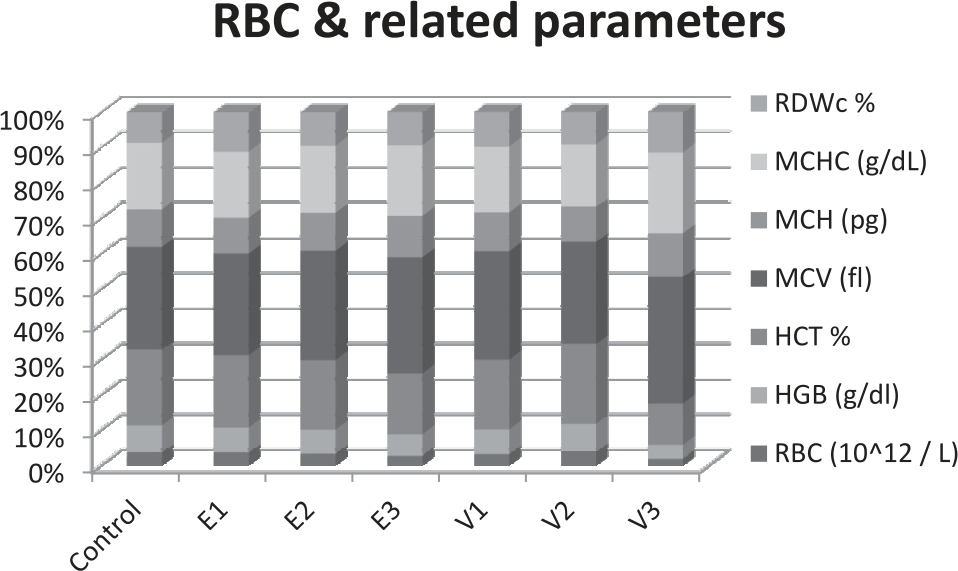
Fig. 3.
Comparative analysis of Platelets and related parameters in different rat groups. E1 = Rat group fed with LD10, E2 = Rat group fed with LD25, E3 = Rat group fed with LD50, V1 = Rat group fed with LD10 + Vit C, V2 = Rat group fed with LD25 + Vit C, V3 = Rat group fed with LD50 + Vit C.
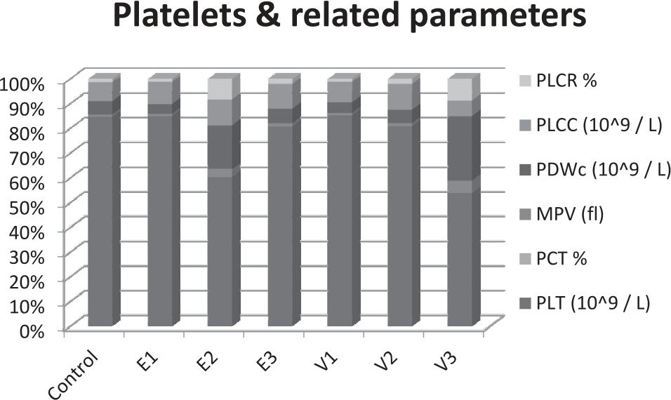
Fig. 4.
Comparative analysis of Liver Function Test (LFT) and Renal Function Test (RFT) in different rat groups. E1 = Rat group fed with LD10, E2 = Rat group fed with LD25, E3 = Rat group fed with LD50, V1 = Rat group fed with LD10 + Vit C, V2 = Rat group fed with LD25 + Vit C, V3 = Rat group fed with LD50 + Vit C.
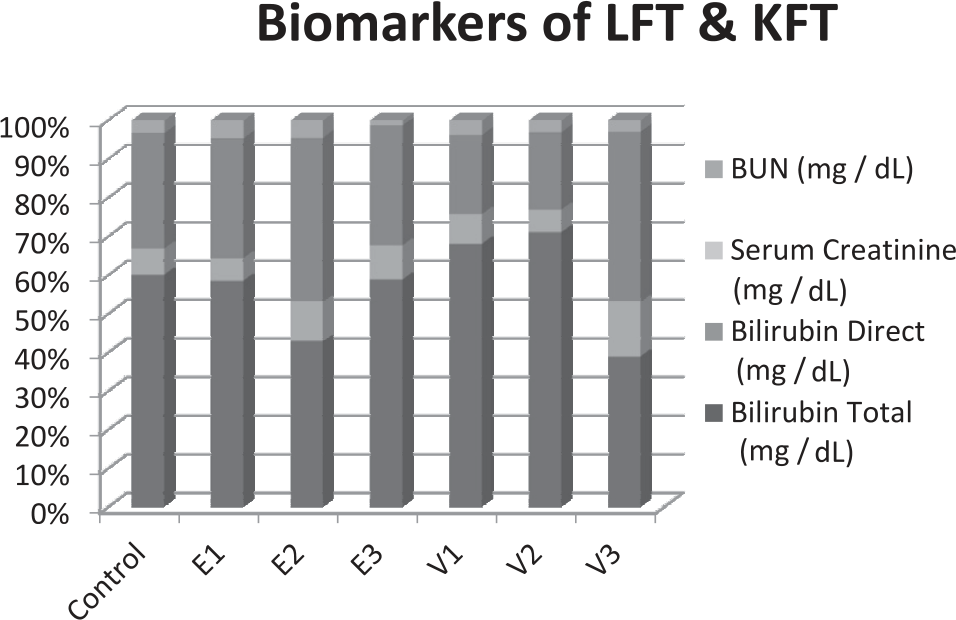
There is a fair argument that these mounted up levels of liver enzymes could be the result of the insecticide’s interference in the biosynthesis pathways of these enzymes,or any alterations in membrane permeability could be the potential cause of hepatic tissue degeneration. These abnormal findings demonstrate tissue damage and probable narcosis of hepatic tissues. These findings were supported by various [21, 22] studies of hepatic toxicity with different molecules like chlorpyrifos, imidacloprid, and fipronil. It is worth mentioning that most of the studies were carried out with chronic exposure rather than acute exposure, hence further studies are required to examine the effects of acute exposure [6, 23-25].
Histopathological examination of liver tissues revealed severe hydropic degeneration and coagulative necrosis in Experiment Group E3 (LD50), as well as mild congestion and coagulative necrosis in Group V (Fig. 5).
Renal and Cardiac Toxicity
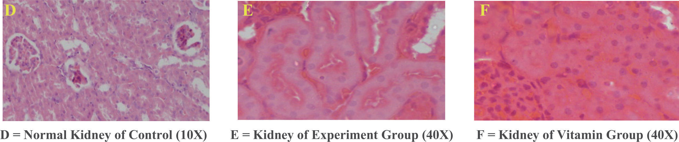
The Renal Function Test (RFT) and kidney histopathology test were performed to assess the extent of renal damage. Table 5 shows that serum creatinine and Blood Urea Nitrogen (BUN) values were significantly increased in intoxicated rat groups compared to the control group. Vitamin C also demonstrated antioxidant and protective effects on these kidney-related serum parameters. Histopathological examination of renal tissues revealed congestion peri-tubular in nature, pyknosis, karyolysis, and coagulative necrosis in exposed groups, while mild congestion and coagulative necrosis have also been observed in the ascorbic acid supplemented groups (Fig. 6).
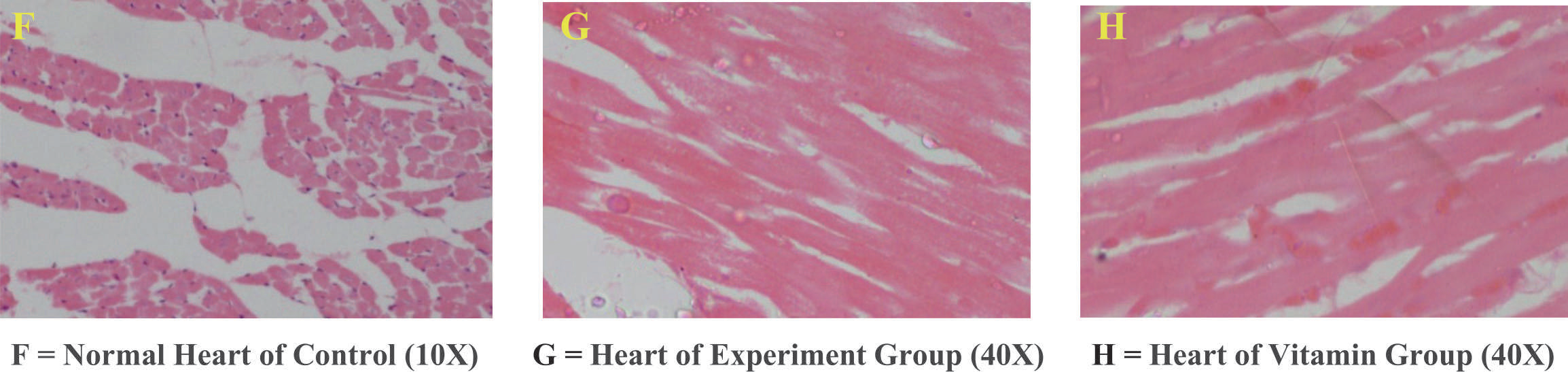
Histopathology of cardiac tissues showed loss of striated muscles and the fragmentation of cardiac cells in the exposed groups in a dose-dependent manner, whereas ascorbic acid supplementation exhibited mild congestion and cellular degeneration (Fig. 7). These results indicate the nephrotoxic potential of Dinotefuran. Hence, exposure-related alterations in different biomarkers associated with histopathology can be attributed to oxidative damage and the subsequent demeaning of the glomerular filtration rate, as reported in other studies by [26].
The tibia of the rat was used to assess the damage. The mean weight and length decreased in varyingly exposed groups as compared to the control group. Consequently, it led to a low Weight/Length Index and Robusticity Index, as indicated in Table No. 6. These findings highlight the inhibitory and protective effects of Vitamin C against the toxic effects of Dinotefuran. Overall, these results elucidate the reduced bone density [27]. It is important to note that there is currently no available data in regard to the weight/length index and robusticity index of any rat bone in relation to insecticide or pesticide toxicity.
Based on the aforementioned discussion, it is to be concluded that acute exposure to dinotefuran has an impact on the structure and functioning of vital organs in mammals, including the kidney, liver, heart, and bone [28]. It leads to alterations in hematological, biochemical, and only histopathological and bone-related parameters. Even acute exposure to Dinotefuran can be hazardous in a dose-dependent manner. Vitamin C supplementation shows potential in mitigating the toxicity of the insecticide, although the effects are dose-dependent.
Conclusions
The above mentioned study concluded that one-time exposure to Dinotefuran can damage mammalian cells in a dose-dependent manner. A strict regulatory policy must be in place when dispensing these insecticides. A regular intake of vitamin C, both by humans and livestock, can minimize damage. There is a need to explore acute exposure studies further.

