


Introduction
Bacterial infection is one of the major stressors that threaten the fish industry. Among them, Aeromonas sp. is a Gram-negative bacterium that is normally established in the aquatic environments [1]. It is one of the most common bacterial pathogens in freshwater causing infections in fish which are described by sepsis with systemic hemorrhage and could induce human and fish infections [2–5]. Aeromonas hydrophila is a pathogenic bacterium that causes epizootic ulcerative syndrome, hepatosplenomegaly in fish, hepatic energetic metabolism, and furunculosis [6]. The liver and kidney are the main internal organs affected by Aeromonas sp. [7]. This bacterium is a zoonotic organism capable of infecting animals and humans through the consumption of infected fish and inducing oxidative damage that is involved in the pathogenesis of diseases [3]. Such bacterium targets the liver and causes much damage to it and oxidative stress has different antioxidant mechanisms that make them more resistant to the hosts’ immune systems [8]. A. hydrophila infections may increase the intracellular generation of reactive oxygen species (ROS) in fish, as well as increase free radicals with changes in antioxidant defense mechanisms and infliction of oxidative stress possibly lead-ing to the oxidative damage of cellular macromolecules [9, 10]. Many bacterial pathogens, such as A. hydrophila, A. caviae, A. salmonicida, and A. veronii, are bacterial zoonosis of fish that have received increasing focus due to the identification using molecular diagnostic techniques [11, 12]. The presence of virulence factors (aerolysin, hemolysin, gelatinase, enterotoxin cytosine, and antimicrobial peptides) in A. hydrophila is responsible for infecting fishes and inducing septicemia, gastroenteritis, plus causing a Motile Aeromonas Septicemia (MAS) disease [13].
Oxidative stress and oxidant status are directly related to the initiation and progression of bacterial infectious disease [14]. Catalase (CAT) and superoxide dismutase (SOD) are antioxidant enzymes that play an important role in protecting the host cell from oxidative damage induced by bacterial infection and environmental pollution [15, 16]. Exposure of fish to stress causes the release of reactive oxygen species (ROS), which includes hydrogen peroxide and other oxidant molecules, including superoxide anion, peroxyl radical, and peroxynitrite anion [17–19]. The GPx antioxidant enzyme contains selenium in its active site and eliminates inorganic and organic hydroperoxides, reducing them to water or alcohol coupled with reduced glutathione (GSH) oxidation [20]. It is clear that each fish species exhibits a different pattern of antioxidant enzy-matic activity according to the type of injury and the type of bacteria [8].
The common carp (Cyprinus carpio) is an important fish and has a long history of cultivation, which may be followed back to 8,000 years ago [21]. C. carpio and O. niloticus are the most commonly cultivated species in Basrah, Iraq because they are resistant to aquatic stressors. Bacterial infection is a significant threat to the cultivable of C. carpio and O. niloticus causing mortality and economic loss [22, 23]. The present study is a comparative study to evaluate the response of C. carpio and O. niloticus to A. hydrophila infection on redux status, and histopathological alterations in different vital organs.
Materials and Methods
Preparation of Bacterial Isolation and Challenge for A. hydrophila.
A. hydrophila was isolated and identification of 16S rRNA using a DNA sequencer for bacterial fish diseases. The A. hydrophila strain 16S rRNA (OR398683.1) (BLAST) was used for the homological search. In addition, the Bioedit program was used for estimating the similarity matrix. (MEGA7) sequence analysis software with 1000 bootstrap values was utilized for constructing the phylo-genetic tree. Bacteria were cultured in (Tryptic soy agar) for 24 h at 30℃, and a single colony was chosen to incu-bate in (Tryptic soy broth) for 24 h at 30℃, A. hydrophila cultured broth was centrifuged for 15 min at 3000 × g at 4℃ and after that, the pellet was removed and suspended in sterile normal saline. Determination of the semi-lethal dose (LD50) was identified as 6.9 × 10⁷ CFU/ mL, which was used for injecting the fish intraperitoneally (IP) according to [24].
Experiment Design
Fingerlings of C. carpio and O. niloticus (n=60 for each species of fish) (average body weight of 20–25 g) were randomly caught from Karma-Ali River north of Basrah and each species of fish species was divided randomly into the control group (A) and infected groups (B) in triplicate. The experiment was carried out separately for each species of fish. Fish were injected with a semi-lethal dose (LD50) of the suspension containing (0.5 ml; 6.9 × 10⁶ CFU/ml) as an intraperitoneal injection. The control group was injected with 0.5 ml of a sterile saline solution (0.86%), and clinical signs and mortality were recorded daily according to [25].
Fish Samples
Healthy fingerlings of two species of C. carpio and O. niloticus with a body weight of 30-35g were collected live from the Al-Mas`hab River in the Basra governo-rate, Iraq. The healthy fish were selected for studies, which were confirmed through physical appearance, including skin luster, eyes, color, and behavior [16]. Fish were trans-ported in a plastic container with oxygen as soon as possible to the laboratory (Department of Biological Development in Shatt Al-Arab and NW Arabian Gulf, Marine Science Center, University of Basra, Basra, Iraq), and randomly distributed in an Aquarium with a capacity of (60 × 40 × 50 cm). Fish were kept in glass tubs (10 fish/aquarium; 50 L) (All species of fish were experimentally placed separately). The fish were fed during the days of acclimatization for 7 days, and before the injection, the food was cut.
Samples Collection
Three fish were randomly selected after injection from each species of fish. The fish were killed by an overdose of anesthesia (250 mg of clove oil/liter), dissected, and their internal organs (liver, kidney, and spleen) were collected at intervals for 7 days (1st, 3rd, 5th, and 7th days) after injection and part of the internal organs was kept at a tempera-ture of -80℃ (kept in liquid nitrogen) until performing the biochemical analysis. The second part of the organs was preserved in (10%) neutral buffered formalin for histological analysis.
Protein Extraction from the Liver, Kidney, and Spleen
The protein was extracted using the Protein Extraction Buffer prepared by (the Iraqi Company for Biotechnol-ogy). The extraction buffer consists of 50 mM 4- (2 hy-droxyethyle) -1-piperazineethanesulfonic acid (HEPES, pH 7.5), 150 mM NaCl, 10 mM Dithiothreitol (DTT), 1 mM polymethylsulfinyl fluoride (PMSF), 1 mM Ethylen-ediaminetetraacetic acid (EDTA) and 1% sodium dodecyl sulfate (SDS).
The protein was extracted with the following steps: First, 0.5 g of weight was taken from each tissue (liver, kidney, and spleen), each individually for each C. carpio and O. niloticus (three fish/replicas) and mashed well with the addition of 1 ml of Protein Extraction Buffer. Secondly, the mixture was transferred to liquid nitrogen and immersed deep 3 times for 5min. At last, it was kept at room tempera-ture for 20 minutes, then inserted into a cooler centrifuge at 18,000 × g for 20 min.
Calculation of Protein Concentration
To calculate protein concentration, a special kit (BCA assay) from (Thermo Fisher Scientific) was used according to the following protocol of Abd-alsahb et al. (2021).
ELISA for Antioxidant Concentration in Tissues
The antioxidant concentration in C. carpio and O. niloticus in tissues (liver, kidney, and spleen), (three fish/replicas) was determined by ELISA, Elabscience (USA) following Yin et al. [26]. The plates contain the 96-well plates, the glutathione peroxidase (GPX) kit, and the absorbance of the superoxide dismutase (SOD) kit at 505 nm and using (U/mg protein). Catalase (CAT) concentration was measured as a colorimetric method, according to the following protocol kit, expressed as units (U/mg protein) and absorbance at 405 nm [27]. The protein concentration of the supernatant for the samples was homogenized using a buffer (pH 7.5) and then centrifuged for (15 min) at 5°C at 15,000 × g, to get the supernatant. The supernatant was centrifuged for 1 h at the same status. The final supernatant was obtained, and the pellet was collected, cleaned, and then stored in the buffer (HEPES, pH 7.5), according to [24]. Each plate contained the appropriate positive and negative controls, and the results of the test samples were calculated from standard curves.
Histopathology
Tissues from the liver, kidneys, and spleen collected from the infected fish were fixed in (10%) neutral buffered formalin and prepared for further processing, sectioning with microtome, and stained with Haematoxylin and Eosin (H & E) according to Roberts [28].
Statistical Analysis
The results were compiled by one-way ANOVA followed by All Pairwise Comparison according to the Tukey test to distinguish the difference among means. The statistical analysis was conducted using SPSS V. 21 and graphs were prepared by GraphPad Prism V7. The data were presented as mean ± standard deviation.
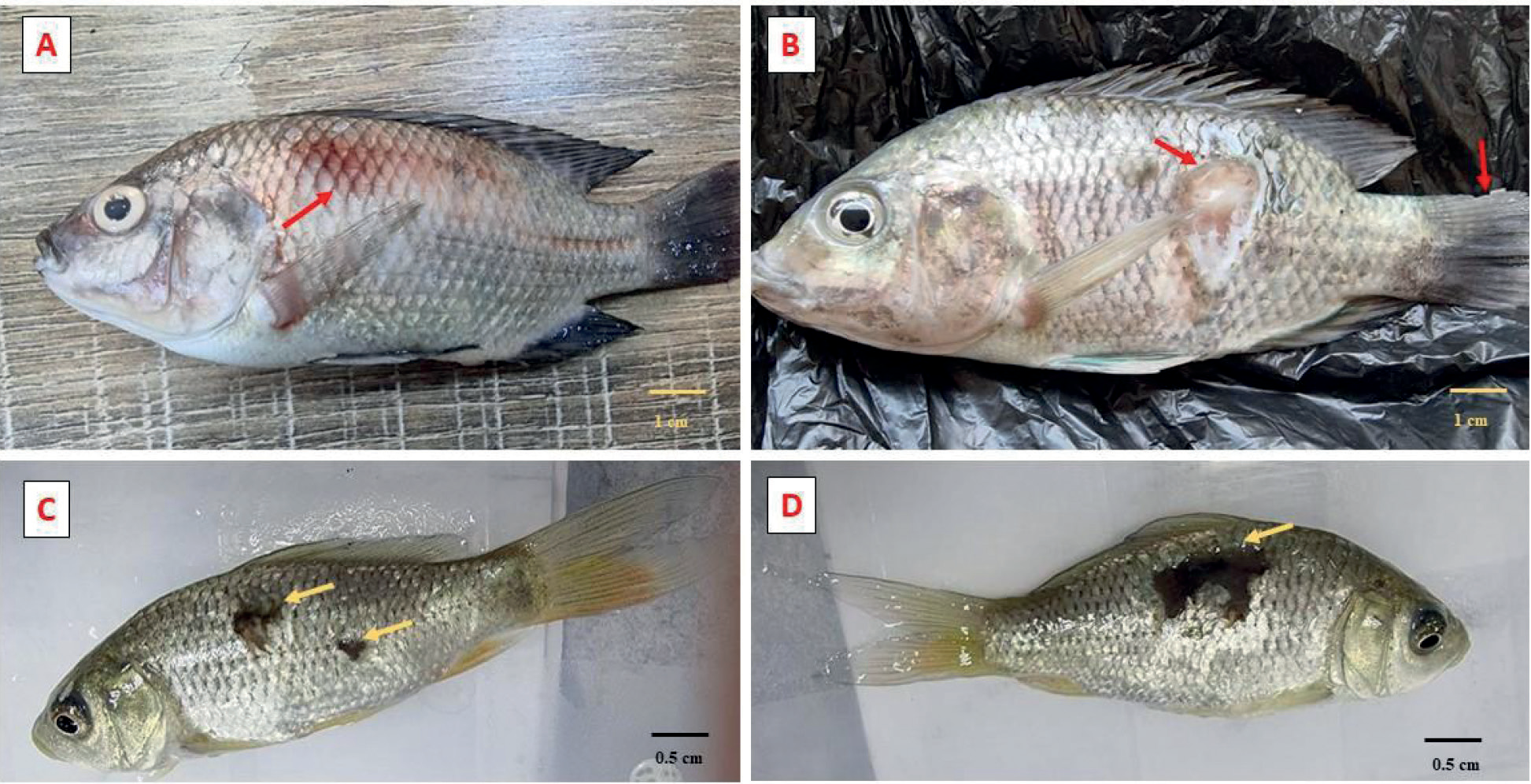
Fig. 1.
Experimentally infected Oreochromis niloticus with Aeromonas hydrophila showing severe erythema in the skin (A) and ulceration in the skin (B). Experimentally infected Cyprinus carpio with Aeromonas hydrophila showing ulcerative spots in the skin (C) and extended areas of severe ulcerations (D).
Results
Pathological Clinical Symptoms
The experimentally infected C. carpio and O. niloticus with A. hydrophila showed clearly characteristic pathological symptoms of A. hydrophila. Fig. 1. A and B show severe hemorrhagic patches and congestion on the body of O. niloticus. Fig. 1. C and D show ulcers, inflammation, and falling scales on the skin of C. carpio.
Antioxidant Activity
The antioxidant response was determined in different tissue samples (liver, kidney, and spleen) from fish C. carpio and O. niloticus infected with A. hydrophila after 0, 1, 3, 5, and 7 days of infection.
Tissue Levels of GPX Activity in Fish
The activity of GPX in the liver, kidneys, and spleen of C. carpio, significantly increased at different sampling days post-infection and peaked on the 3rd day in all tissues (Fig. 2 A, B, & C). The levels of this enzyme decreased remarkably on the 7th day with significant differences in the control. While, the level of GPX activity was significantly (p < 0.05) increased in all evaluated tissues of O. niloticus (Fig. 3 A, B, & C). There was also an increase in the activity of the GPX on the 3rd day post-infection in all tissues, and a decrease in the kidney on the 5th and 7th.
Tissue Levels of SOD Activity for Fish
The activity of SOD in tissues of C. carpio was significantly increased after the 1st day of infection and continued as over the control in the kidney and spleen (Fig. 4). Meanwhile, this increase was restored on the 7th day in C. carpio liver (Fig. 4 A).
In O. niloticus, SOD was significantly increased at the 3rd and 5th days in the liver, and 1st, 3rd in the kidney, and only at the 3rd in the spleen, and the other sampling times were maintained as in the control group (Fig. 5).
Tissue Levels of CAT Activity for Fish
The effect of A. hydrophila infection on the activity of the CAT activity in the liver, kidney, and spleen of C. carpio is presented in Fig. 6 (A, B & C). In the liver and spleen, the expression of the CAT activity was found to increase significantly (p < 0.01) after one day post-infection, meanwhile in the kidney, increased significantly starting from day three post-infection (Fig. 6 B). O. niloticus experienced a significant increase in CAT activity post-infection, which peaked on day three and then declined during the sampling time.
Histopathological Changes in C. carpio and O. niloticus Post-Infection with A. hydrophila
Histopathological changes were revealed in the liver, kidney, and spleen of C. carpio and O. niloticus, with the semilethal dose (LD50) of a pathogenic strain of A. hydrophila isolated and identified bacterial by Al-Shammari et al. [29]. The pathological state of the internal organs of C. carpio after the experimental challenge is shown in Fig. 8. A- The liver showing necrotic and denatured cells and is congested. B-Hepatic cells showing congestion of blood vessels. C- hepatocytes showing infiltration and necrosis. D- hepatocytes showing degeneration. E- Spleen hyperplasia of white pulp and appears as round or oval nodules with few masses of hemosiderin and melanomacrophages, degeneration of red pulp, an example of a fish defense mechanism. Bacteria cause effects on the cytoplasm of hepatocytes containing fats and glycogen associated with normal liver metabolism. Any defect caused by bacteria between the level of synthesis of the substance in paren-chymal cells and the rate of release of the systemic circu-lation can generally cause metabolic damage according to Dougklas et al. [30]. F - Spleen white pulp as a nodule with few masses of melanomacrophages, the red pulp, and sinusoid. G- Kidney revealing degeneration of renal tubules and has eosinophilic lining renal tubules and few melanomacrophages with globular cellular masses in hematopoietic tissues [31].
Fig. 2.
The activity of glutathione peroxidase (GPx) in the liver (A), kidney (B), and spleen (C) of Cyprinus carpio after 1st, 3rd, 5th, and 7th days of Aeromonas hydrophila infection. The experiment was performed in triplicate, and the data are shown as the mean ± SD. Values with a different letter superscript are significantly different (p < 0.05).
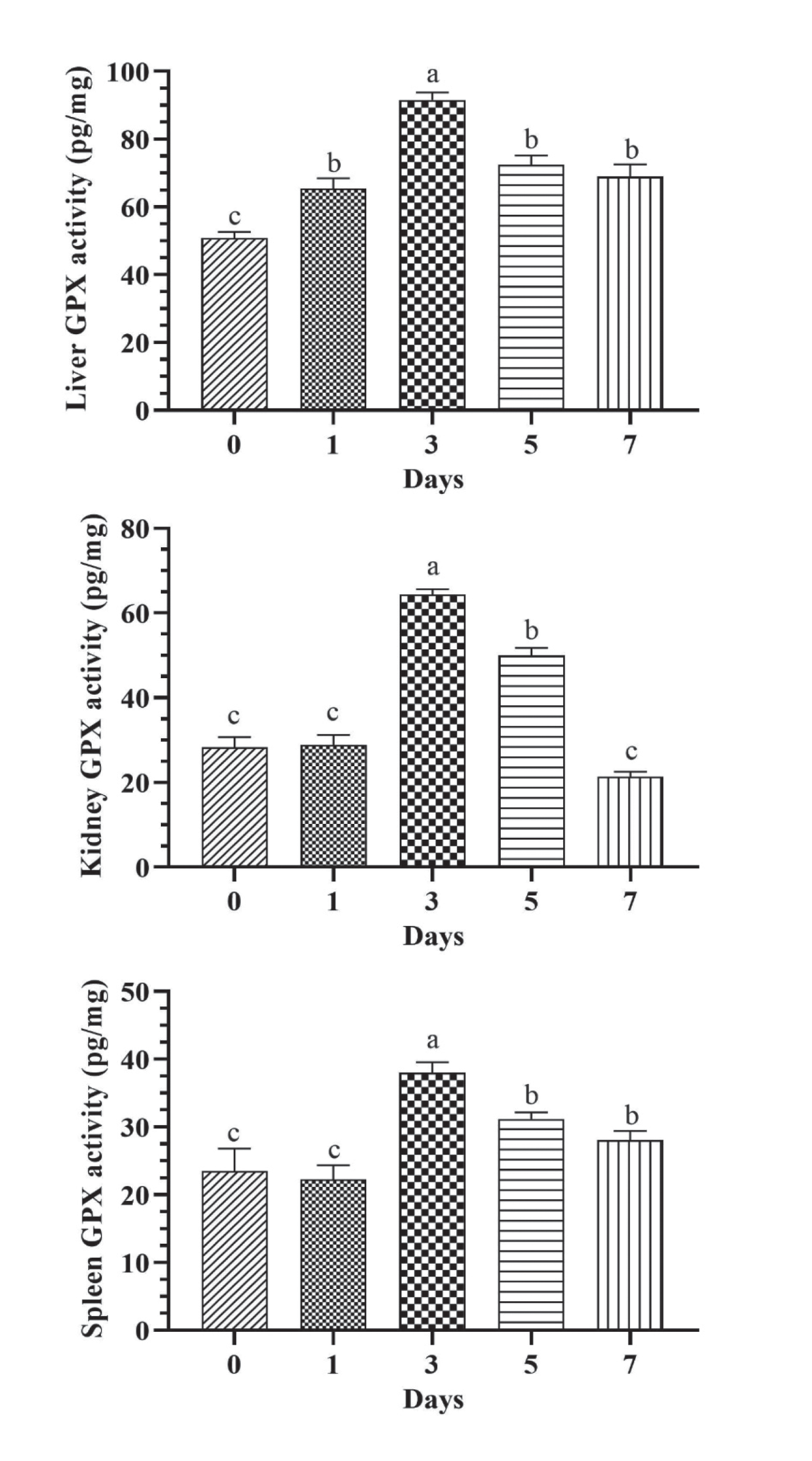
Fig. 3.
The activity of glutathione peroxidase (GPx) in the liver (A), kidney (B), and spleen (C) of Oreochromis niloticus after 1st, 3rd, 5th, and 7th days of Aeromonas hydrophila infection. The experiment was performed in triplicate, and the data are shown as the mean ± SD. Values with a different letter superscript are significantly different (p < 0.05).
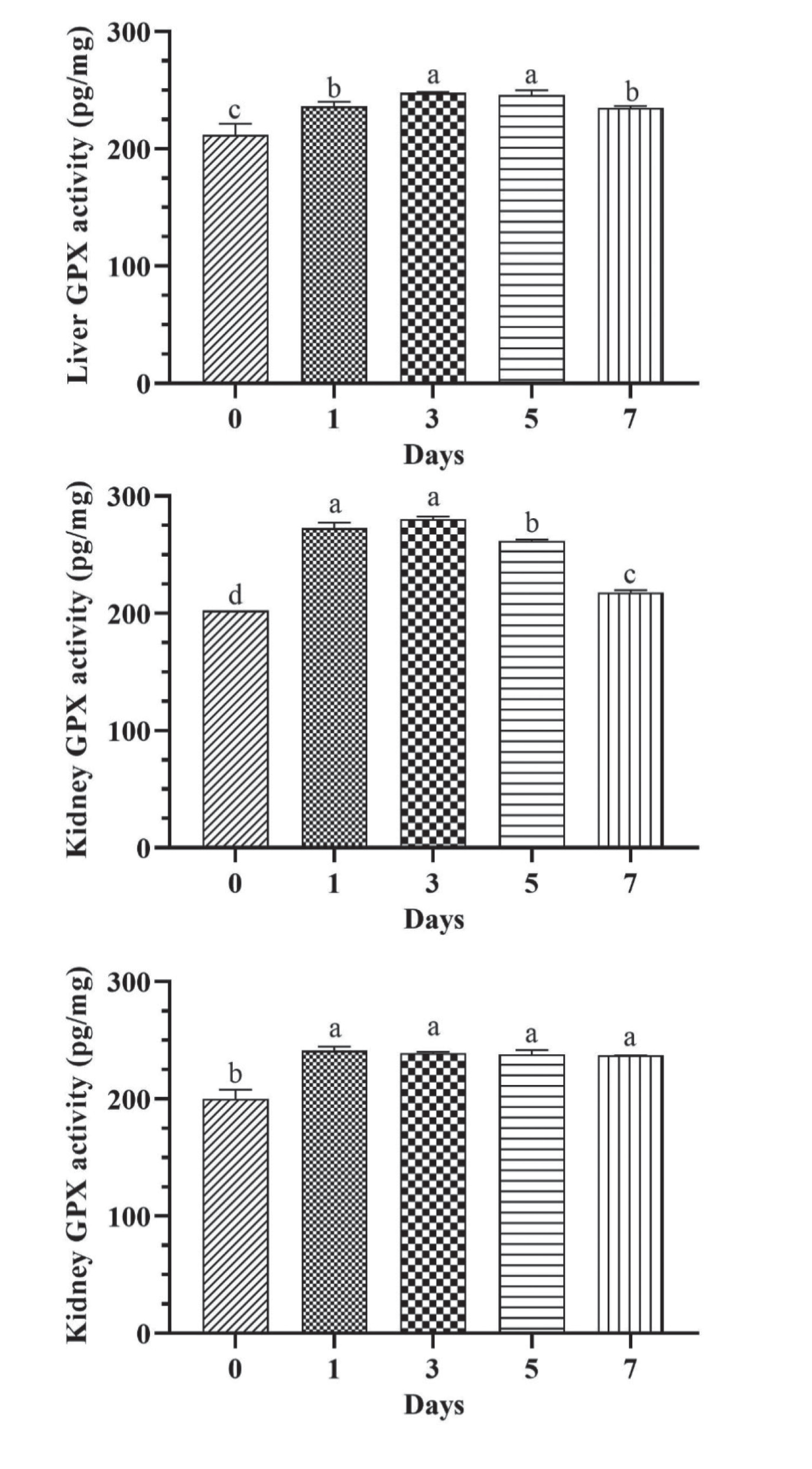
Fig. 4.
The activity of superoxide dismutase (SOD) in the liver (A), kidney (B), and spleen (C) of Cyprinus carpio after 1st, 3rd, 5th, and 7th days of Aeromonas hydrophila infection. The experiment was performed in triplicate, and the data are shown as the mean ± SD. Values with a different letter superscript are significantly different (p < 0.05).
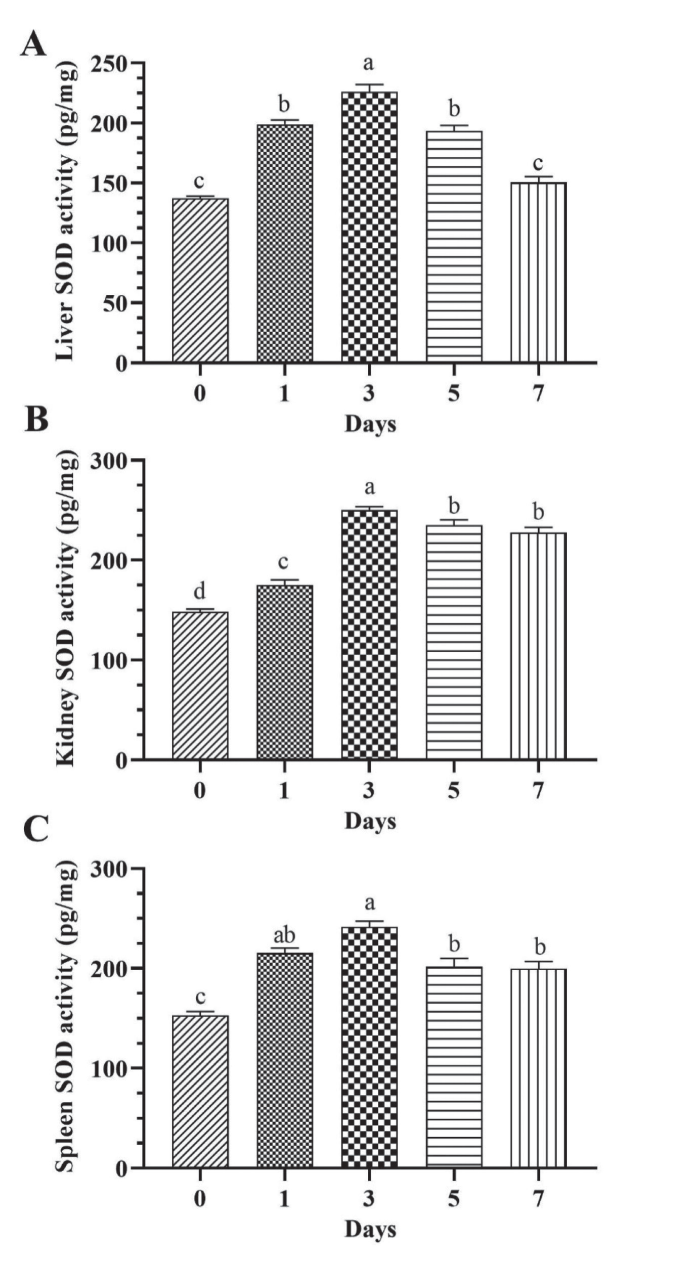
Fig. 5.
The activity of superoxide dismutase (SOD) in the liver (A), kidney (B), and spleen (C) of Oreochromis niloticus after 1st, 3rd, 5th, and 7th days of Aeromonas hydrophila infection. The experiment was performed in triplicate, and the data are shown as the mean ± SD. Values with a different letter superscript are significantly different (p < 0.05).
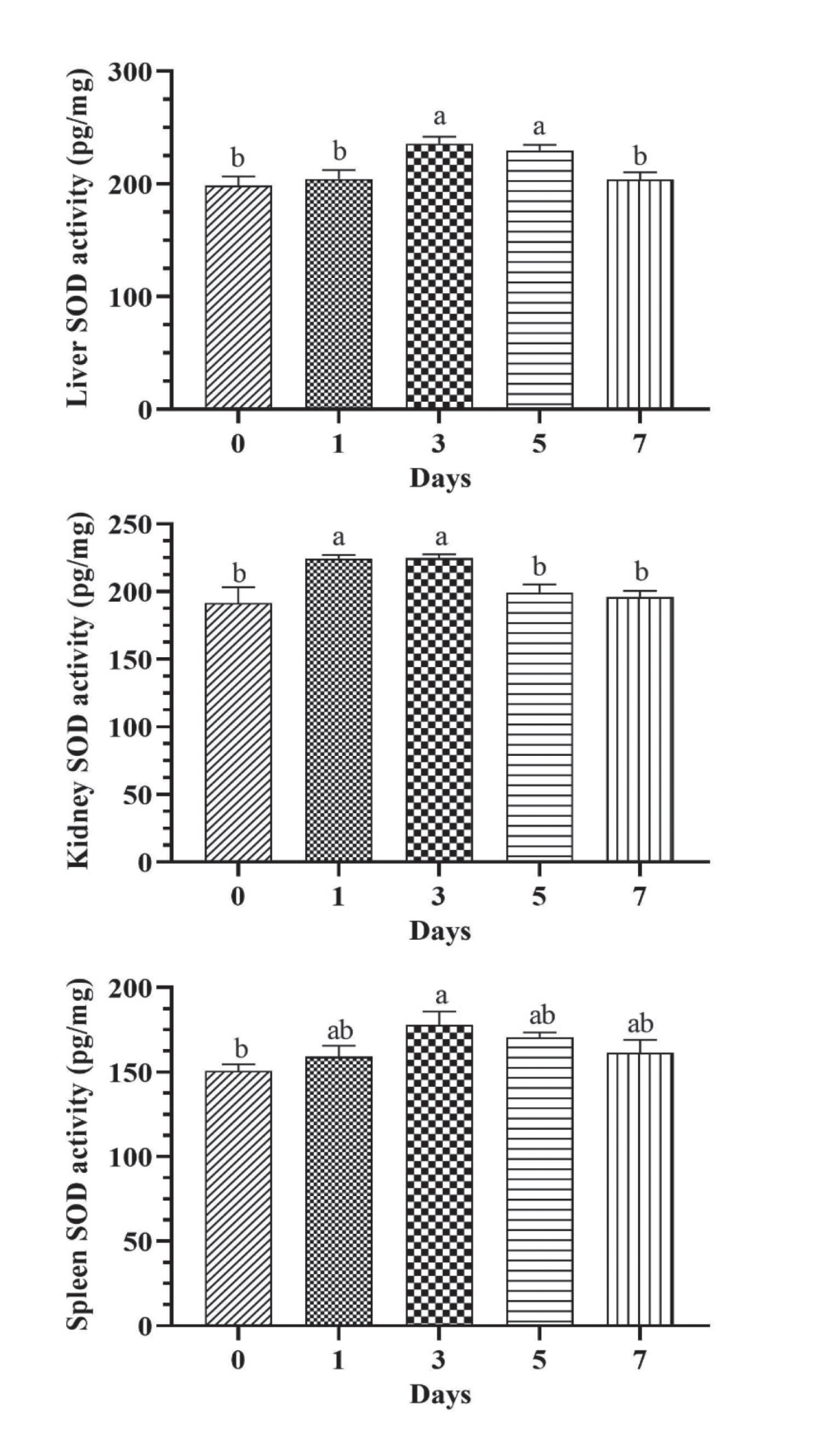
Fig. 6.
The activity of catalase (CAT) in the liver (A), kidney (B), and spleen (C) of Cyprinus carpio after 1st, 3rd, 5th, and 7th days of Aeromonas hydrophila infection. The experiment was performed in triplicate, and the data are shown as the mean ± SD. Values with a different letter superscript are significantly different (p < 0.05).
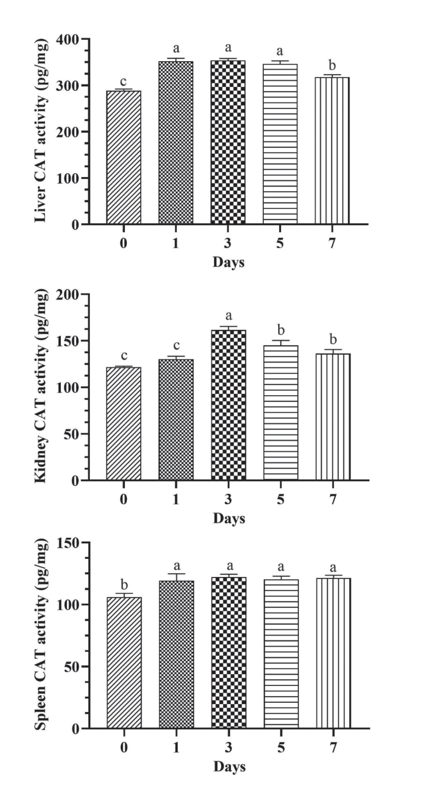
Fig. 7.
The activity of catalase (CAT) in the liver (A), kidney (B), and spleen (C) of Oreochromis niloticus after 1st, 3rd, 5th, and 7th days of Aeromonas hydrophila infection. The experiment was performed in triplicate, and the data are shown as the mean ± SD. Values with a different letter superscript are significantly different (p < 0.05).
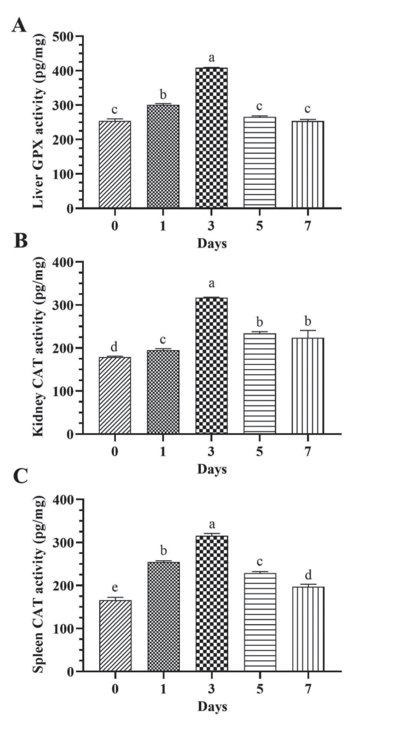
Fig. 8.
The histopathological effects of Aeromonas hydrophila infection on the liver, kidney, and spleen of Cyprinus carpio after seven days of infection. A: Hepatic cells showing denaturation and necrosis; B: Hepatic cells showing congestion of blood vessels; C: Eosinophilic hepatocytes showing infiltration and necrosis; D: Eosinophilic hepatocytes showing degeneration; E: Spleen hyperplasia of white pulp (wp) and appear as round or oval nodules (thick arrows) with few masses of hemosiderin and melanomacrophages (thin arrows), degeneration of red pulp (RP); F: Spleen white pulp (wp) as a nodule with a few masses of melanomacrophages (thin arrows), the red pulp (RP), and thick arrows pointed to a sinusoid; G: Kidney degeneration (thin arrows) of renal tubules and eosinophilic (thick arrows) lining renal tubule and few melanomacrophages (head arrows) with globular cellular masses (GC) in hematopoietic tissues; H: Kidney necrotic tissue (arrows) with large globular cellular mass in hematopoietic tissue (asterisk) X40.
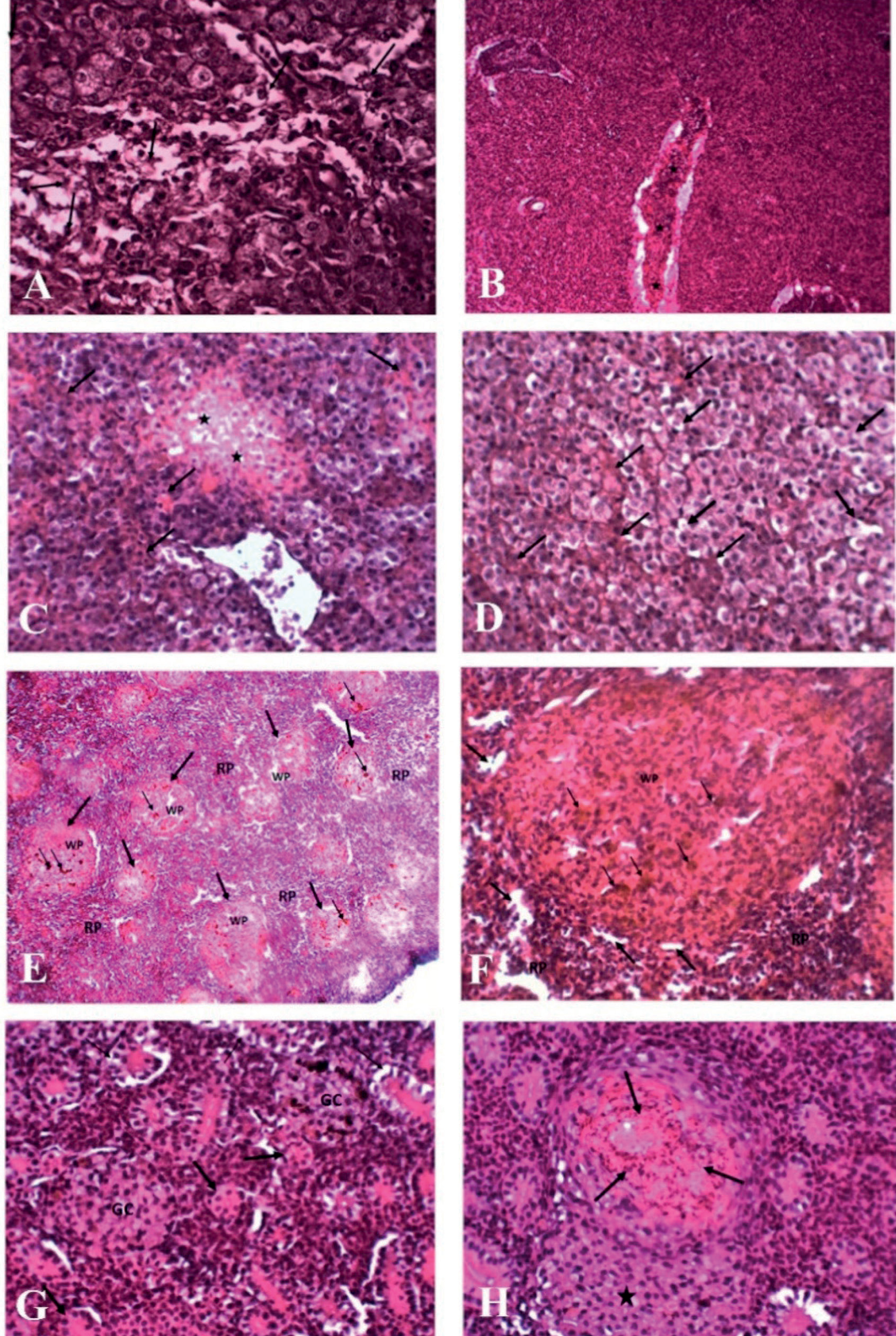
Fig. 9.
The histopathological effects of Aeromonas hydrophila infection on the liver, kidney, and spleen of Oreochromis niloticus after seven days of infection. A: Hepatic cells showing dilation of the sinusoid (thin arrows) and atrophy of hepatocytes (thick arrows); B: Hepatic cells showing massive necrosis of hepatocytes; C: Spleen more masses of melanomacrophages concentrated in white pulp (arrow) X40; D: Spleen hyaline necrosis splenic tissue (asterisks) with more masses of melanomacrophages (arrows) distributed in white and red pulp; E: Kidney necrosis (thin arrows) of renal lining epithelial tubule and degeneration (thick arrows) with eosinophilic other lining epithelial cell, collecting duct (cd); F: Kidney necrosis (thin arrows) of renal lining epithelial tubule and degeneration (thick arrows) X40.
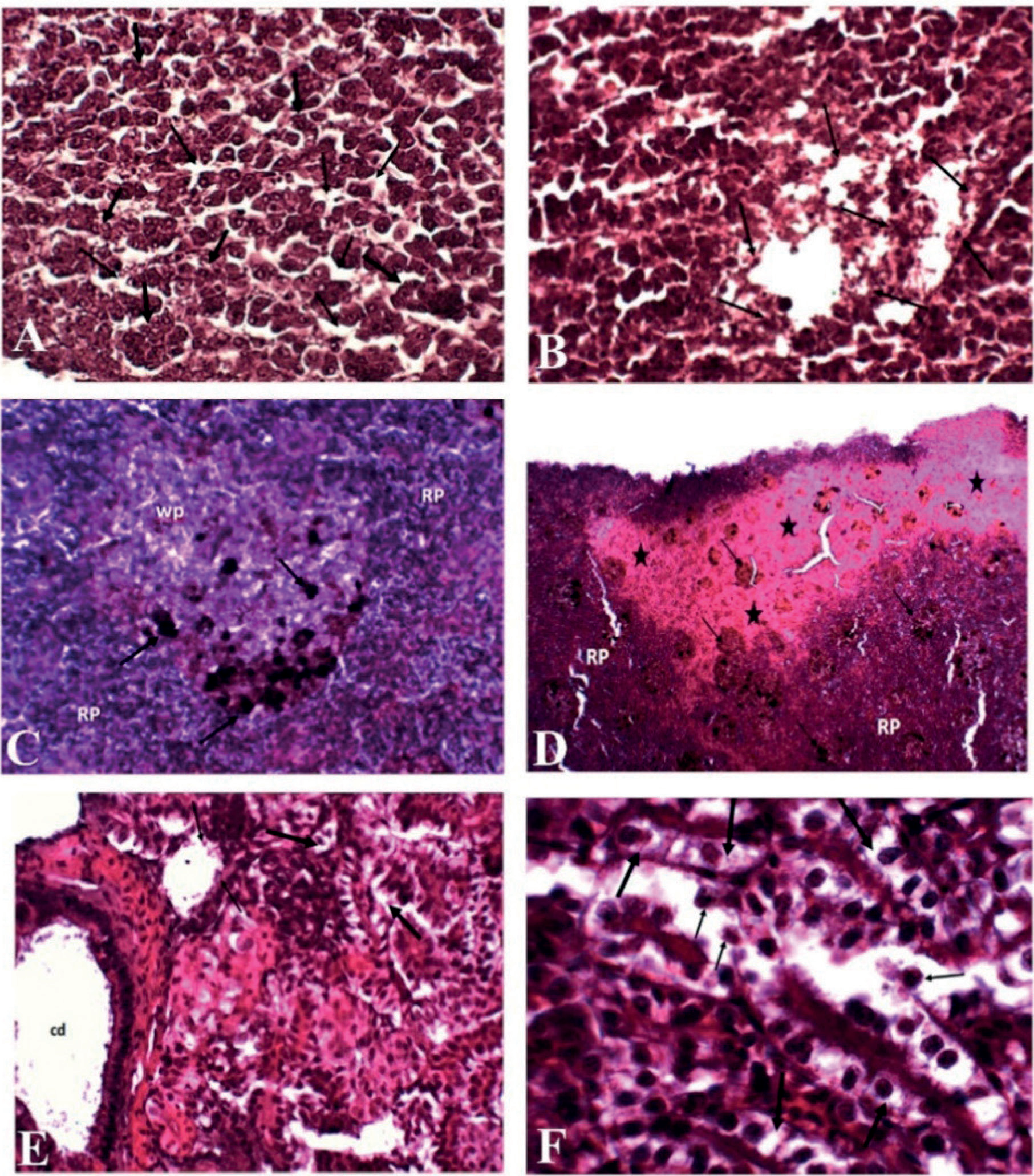
The pathological states in the internal organs of Oreochromis niloticus are shown in Fig. 9 A. The liver cells are dilated of the sinusoid and have atrophy of the hepatocytes [32]. B-Hepatic cells show massive necrosis of hepatocytes. Spleen shows: C- masses of melanomacrophages concentrated in white pulp and D- hyaline necrosis splenic tissue with more masses of melanomacrophages distributed in white and red pulp. E- kidney tissue in organs shows necrosis of the epithelial tubule of the renal lining and degeneration with other eosinophilic lining epithelial cells, collecting duct [31]. F- kidney reveals necrosis of the renal lining and degeneration.
Discussion
Fisheries are an essential part of food production that assures the nation’s nutritional security. In addition, it provides a major source of income for a significant portion of the population, especially the fishermen’s community [33, 34]. The fish has antibacterial molecules that contribute to eliminating the bacterial infection, and post the resistance of the animal. The immune system has innate and adapted responses against pathogens, which induce hyperactivity of these components and increase the high production of reactive oxygen species (ROS) even as a weapon or waste [35, 36]. In normal conditions, there is a balance between ROS and antioxidant systems, but after infection, this balance is disrupted and induces extra cell damage [37]. Therefore, the current study aims to compare the response of the most common two fish species, C. carpio and O. niloticus, which are widespread in culture in freshwater fish. The stress caused by the A. hydrophila post-experimental infection negatively influenced the antioxidant defense mechanism and resulted in histopathological changes.
Motile Aeromonas is widespread in the aquatic envi-ronment, which induces serious pathological symptoms in fish and causes numerous deaths. This could be returned to the bacterial toxins products of Aeromonas, such as dermo-necrotizing factors, extracellular hemolysin, and gelatinase, which work on hemolysis and hemolytic changes in the form of intravascular blood [38].
Antioxidant enzymes, including SOD, GPx, and CAT, are the most widely used sensitive indicators for assessing oxidative stress [39], especially after Aeromonas spp. infections [24, 40]. The SOD is the primary enzyme that transforms superoxide radicals into hydrogen peroxide [41]. Antioxidant enzymes are excreted from the cells of fish, which actively work to lessen reactive oxygen species (ROS), while the accumulation of ROS and the elevation of lipid peroxidation usually occur in response to bacterial stress [42, 43]. In this paper, we report the appearance of oxidative stress in the liver, kidney, and spleen following bacterial infection.
Furthermore, the activity of antioxidant enzymes in general, increased in the early stages after injury, especially on the 3rd day after the experimental injury, and generally decreased on the seventh day after injury to reach a significant value (P < 0.05). It is assumed that the pathogenic effect post-infection by A. hydrophila resulted in a production of ROS in the liver, kidney, and spleen in both fish. As depicted in Fig. 3. A, B, & C, there was an increase in GPx on the 3rd day in all tissues because of the rapid stress response induced by the bacterium, and then gradu-ally declined on the 7th day. It seems that the fish showed a transient stress response to stress, as reported by Yu et al. [44]. These outcomes were concurrent to those reported by Parida and Sahoo [10] who clarified that after 48 h of A. hydrophila infection, there was an elevation of liver GPx levels in Labeo rohita. In C. Carpio, it is opined that there was a formation of lipid peroxidation which was considered the first step following A. hydrophila infection, then; the cell membrane was damaged resulting in stimulating antioxidants in the liver, kidneys, and spleen. Meanwhile, O. niloticus showed a clear increase in the efficacy of GPx activity in the liver, kidneys, and spleen compared to C. carpio. This result could be attributed to increased GPx secretion in the early stage to compensate for cell damage as a result of exposure to infection. Furthermore, Almarri et al. [45] dominated the increased levels of antioxidants in O. niloticus in response to infection to displacing free radicals through their ability to activate transcription signals for antioxidants by activating the nuclear factor erythroid pathway of the protein complex. Additionally, Mahmoud et al. [46] supported our findings and found that O. niloticus is characterized by more tolerance to maintain its antioxidant levels despite stress and shows resistance and higher antioxidant activity when exposed to stress, compared to C. carpio.
Considering another antioxidant indicator, SOD activity is considered the first defense antioxidant agent and is necessary for the body to defend against reactive oxygen species by reducing the superoxide radical of hydrogen peroxide [47]. In C. Carpio, the value of SOD showed a smaller increase in the kidney and liver on the 3rd day following bacterial challenge compared to O. niloticus. The variance between the two species in the resistance against bacterium depends on the release of antioxidants at a considerable concentration, as reported by Trenzado et al. [41]. A recent study documented that C. carpio is more vulnerable to infection and symptoms compared to O. niloticus [48].
Catalase (CAT) is one of the essential enzymes for the detoxification of ROS in all organisms and it can split the level of H2O2 to harmless H2O and O2 and liver tissue may be included in energy expenditure provoked by oxidative stress [44, 49]. Tilapia has a large number of host defense peptides that work to break down bacterial cell walls [50]. In this paper, we reveal an enhanced antioxidant response in O. niloticus compared to C. carpio indicated by increased elevation in CAT level. Furthermore, the level of antioxidant capacity increased significantly on the 3rd day for all fish and clearly decreased on the day in the infected group. Wang [51] obtained similar findings after a bacterial challenge of Aeromonas veronii in sea bass. This could be rediscovered by the ability of O. niloticus to reduce cellular oxidative stress. In addition, scavengers exhibit reactive oxygen species within cells, lowering the level of cellular stress responses as clarified by Zhang et al. [52] and Sies and Jones [53]. Furthermore, a previous report by Tattiyapong et al. [54] demonstrated that O. niloticus has increased antibody responses against bacterial infection after one-time exposure.
In the present perspective, C. carpio and O. niloticus were used as in vivo models to assess the evolution of A. hydrophila. It is known that this bacterium causes oxidative stress, which has a role in autophagy in the presence of stress [55]. The occurrence of the autophagy process is associated with physiological changes in the cell as a result of the presence of reactive oxygen species [56]. Interestingly, innate immunity and autophagy are two processes with a common course and common pathological signs [57, 58].
The challenge test is a verifying tool for assessing the clinical manifestations and histopathological changes [51]. Fish challenged with A. hydrophila showed pronounced, signs of illness including, excess mucous secretion, and hemorrhages, A Similar study by Chen, et al. [3] was conducted on C. carpio and O. niloticus and revealed signs of infection by A. hydrophila including enteritis, congested liver, enlarged kidney and spleen, and visceral hemorrhage. The pathogenicity of A. hydrophila could refer to the production of cytotoxic, spore-forming enterotoxin, which is a dangerous virulence factor [59]. Likely, our results were similar to those of Mahboub, et al. [40] who showed a pronounced enhancement in C. carpio resistance against A. hydrophila. In the present results, we report major histopathological changes in the liver and kidneys of C. carpio and O. niloticus following infection by A. hydrophila. This could be attributed to the occurrence of oxidative damage in the hepato-renal organs, which, in turn, resulted in alterations in tissue architectures. Concurrent with another study, Salam et al. [60] revealed that A. hydrophila infection resulted in histopathological changes that were prominent in the liver, spleen, and kidney of both O. niloticus and C. garipinus including perivascular melanophore aggregation and vacuolar degeneration. Numerous studies of fish species showed that the liver and kidney are the only organs in which oxidative stress occurs [61]. The decrease in the effectiveness of antioxidants in some organs is a result of various stresses, such as the liver being damaged as a result of resistance and its role in osmosis, the rest of the organs are injured, and this is called organ suffering, the rate of glomerular filtration decreases and loses its func-tion, the permeability of blood vessels increases [62]. Oruç and Usta [63] proved that C. carpio is more sensitive than O. niloticus to the effectiveness of antioxidant enzymes.
Based on the results of the study, O. niloticus is dis-tinguished by high antioxidants and high resistance to A. hydrophila infection, indicated by less oxidative stress and inflammation in the tissues compared to C. carpio.
Conclusion
The current study reports on the hazardous impact of the zoonotic pathogenic A. hydrophila on altering antioxidant status and inducing histopathological changes in the liver, kidneys, and spleen of C. carpio and O. niloticus. The results showed that O. niloticus has a higher tolerance than C. carpio in response to A. hydrophila. The activities of GPX, SOD, and CAT showed high alterations in response to the bacterial infection. The histopathological findings were prominent in the liver, kidneys, and spleen of both C. carpio and O. niloticus, particularly, hepatic tissues showed vacuolar degeneration and hemorrhagic septicemia and mainly targets the liver and tissue damage in response to A. hydrophila. Fu-ture researches are recommended for evaluating the adverse impact of A. hydrophila on other fish species.

