

Introduction
Aluminum (Al) is the third most common element in the earth’s crust, which is considered toxic to plants in acidic soil and harms human health through the food chain. Al is solubilized into the soil as Al3+ under strongly acidic soil conditions having a pH below 5.0 [1]. Excess Al3+ enters the plant primarily through the roots, which interferes with morphological, physiological and biochemical processes, such as the inhibition of root elongation, the reduction of stomatal opening, the generation of reactive oxygen species (ROS), and the deficiency of some essential elements [2, 3]. Al3+ inhibits root growth at micromolar concentrations, which primarily injures the root cytoskeleton, the cell wall, the plasma membrane, DNA/nuclei, and signal-transduction pathways [4, 5]. The plant reduces aluminum toxicity by adopting several mechanisms during the evolutionary process, such as sequestering Al into vacuoles or other organelles, secreting some organic acids (OAs) to chelate Al cations, and transcriptional regulation of Al tolerance genes [6, 7]. Al toxicity leads to the decrease in crop production worldwide on acid soils, which comprise over 50% of the world’s arable lands [8]. Moreover, aluminum accumulated slowly in the human body may cause dialysis encephalopathy syndrome, Alzheimer’s disease, and other degenerative neurological diseases. Therefore, it is very necessary to develop practical ways to reduce Al toxicity in edible crops grown in acidic soils. The selection and breeding of Al tolerant crop varieties is one of the important ways for the improvement of crop production in acid soil [9]. Exogenous application of various substances has been well documented to increase the Al resistance of plants in acid soils, such as lime [10], silicon [11], boron [12], magnesium [13], sulfur [14], putrescine [15], etc. Recently, some researchers reported that colonization with arbuscular mycorrhizal (AM) fungi could help to protect host plants from Al toxicity in acid conditions [16]. Plant growth-promoting bacteria (PGPB) were also used to increase the plant tolerance against Al toxicity [17]. Zaurov et al. reported that endophyte infection increased Al tolerance in fine fescues in certain endophyte-plant combinations [18]. However, no literature has investigated the interactions about endophytes-rice combinations under Al stress.
Endophytes and their host plants have established a friendly relationship during the long period of co-evolution. Endophytes have been shown to help host plants grow well and protect host plants from the adverse effects of heavy metal pollution, salinity, and drought [19-21]. It is known that endophytes adopt different mechanisms that may be involved independently or simultaneously to support host plant growth and to mitigate environmental stress [22, 23]. For example, endophytes produce various kinds of bioactive metabolites, such as extracellular enzymes, OAs, and plant hormones [24]. Moreover, the endophytic association also elevated water-use efficiency, increased the activity of antioxidant enzymes, improved nutrient acquisition, and expressed stress-related genes [24]. Recently, the exploitation of endophytes in counteracting stressful environmental conditions on plants has become an attractive subject [25].
In previous work, we isolated and identified the endophytic fungus Glomerella sp. JP4 from the leaves of Suaeda salsa L., which supported Oryza sativa cv. Liaoxing No.1 growth under Cd stress [20]. Interestingly, at pH below 5.0, we found that Glomerella sp. JP4 tolerated the presence of Al3+ on potato dextrose agar (PDA) medium, and also improved the growth of rice seedlings under Al stress. Rice is an important staple food for people worldwide, and some varieties are sensitive to Al toxicity. Accordingly, in this study, we aimed to determine how endophytic infection detoxified the rice seedlings exposed to Al stress.
Material and Methods
Glomerella sp. JP4 and Plant Culture
Fungal endophyte Glomerella sp. JP4, which was isolated and identified according to Yu’s description [26], was stored on PDA medium at 4ºC for further use. The isolate was grown in 50 mL potato dextrose broth (PDB) medium (20% potato, 2% glucose, 1000 mL distilled water) at 25ºC for 9 days in a shaking incubator at 125 rpm, and 5% fermentation broth was utilized for the rice seedling treatments. Oryza sativa cv. Liaoxing No.1 seeds were surface sterilized using 2% sodium hypochlorite for 10 min and washed with distilled water three times. The sterilized seeds, placed in petri dishes with two pieces of filter paper moistened with sterile deionized water, were incubated in the dark for germination. Germinated seeds were transplanted in a series of pots (100 seedlings per pot) containing Hoagland’s solution in a growth chamber. The growth chamber conditions were adjusted to a photoperiod of 16 h, 800 μmol m-2 s-1 photosynthetic photon flux density (PPFD), day/night temperature of 27/20ºC, and 80% relative humidity. Fungal Endophyte Infected and AlCl3 Treatments
Five-day-old rice seedlings were selected to carry out fungal endophytic infection and AlCl3 treatments according to our previous methods [20]. Rice seedlings, raised on Hoagland’s solution with a pH of 4.5, were randomized into 2 groups. One group was used for endophyte infection by 5% fermentation broth (EI, endophyte-infected seedlings), but not the other group (EF, endophyte-uninfected seedlings). At the same time, two groups were supplemented with different concentrations (0, 40, 80, and 120 μM) of AlCl3, respectively. Thus, the experiment was performed with a 2×4 factorial plan in a completely randomized design. All measurements were conducted in three replicates. The infection rate of roots was determined according to the method of Liu and Chen [27], which reached over 90% in rice seedlings infected with fungi JP4.
Growth Determination
Shoot and root lengths from each treatment were immediately measured on sets of 10 plantlets after harvest. The dry weight of shoots and roots was then recorded after drying at 80ºC until achieving a constant weight.
Photosynthetic Pigments
A fresh leaf sample (0.1 g) was collected to extract photosynthetic pigments with acetone. Chlorophyll a+b and carotenoid (Car) content were measured spectrophotometrically according to the method of Agrawal and Rathore [28].
Measurement of Antioxidant Enzyme Activities and MDA Content
Fresh leaf or root samples were homogenized in an extraction buffer (0.1 M phosphate buffer, pH 7.8) on the ice. The homogenate was then centrifuged at 10,000×g for 15 min, and the supernatant was used as the crude extract for antioxidant enzyme analysis. The superoxide dismutase (SOD), guaiacol peroxidase (POD) and catalase (CAT) enzyme were measured using the method of Costa et al. [29], Kochba et al. [30], and Cakmak and Horst [31]. The estimation of protein content was made according to the method by Lowry et al. [32]. The MDA content was determined following the method of Heath and Packer [33].
OAs Extraction from Seedlings Roots and Leaves
Fresh 0.5 g of leaves or roots were used for OAs extraction. Samples were homogenized and extracted in deionized water. The homogenate was incubated for 15 min at 70ºC and centrifuged at 10,000×g for 15 min at 4ºC; the supernatants were then collected. The supernatant was filtered through 0.20-μm nylon filters and stored at -20ºC. The extracts were analyzed by high-performance liquid chromatography (HPLC, Agilent1200) equipped with a reverse phase C18 column (250 × 4.6 mm). Operation conditions were the column at 35ºC and a flow rate of 0.8 mL min−1. The injection volume was 20 μL, with 0.01 mM sulfuric acid as the mobile phase. Chromatograms were obtained at 210 nm with standards for citrate, acetate, oxalate, malate, tartrate, fumarate, succinate, and lactate (purchased from Sigma Aldrich).
Measurement of Al Content
Samples of shoots and roots were oven-dried at 70ºC-80ºC for 48 h to reach a constant weight. Each treatment sample (100 mg) was digested for 30 min in a high-flux closed microwave digestion instrument (MDS-2100, CEM Corp., Matthews, NC, USA) with HNO3/HCl (3:1[v/v]) to extract Al. After digestion, the samples were supplemented with deionized water to a final volume of 50 mL. The Al content of each sample was measured using an inductively coupled plasma atomic emission spectroscopy (ICP-AES) (ICP model Liberty 200, Varian Australia Pty. Ltd., Mulgrave Victoria, Australia).
Data Analysis
All test data were obtained from three independent experiments, which were shown as the mean±standard deviation (SD). Two-way analysis of variance (ANOVA) was used to analyze the effects between endophyte infection and Al stress on all experimental data by using the SPSS 19.0 statistical software package. LSD multiple range test for multiple comparisons was used to compare and detect significant differences between different treatments (p<0.05).
Results
Growth Parameters and Biomass Production
The main effects of JP4 infection and Al treatments on growth parameters and biomass production were significant (p<0.01). However, the interaction effect of the two factors was not significant except for shoot height (Table 1). The shoot height, root length, and dry weight were significantly (p<0.05) decreased for EF plants exposed to different concentrations of Al stress, except for the shoot height and root length of EF plants at 40 μM Al treatment (Table 1), compared to no Al stress. In EI plants, endophytic infection markedly (p<0.05) increased the height and the dry weight of the shoots compared to EF plants (Table 1). Likewise, the dry weight of the root was markedly (p<0.05) increased under 40 μM and 80 μM Al stress. However, root length was decreased in EI plants compared to EF plants (Table 1).
Photosynthetic Pigments
There were significant main and interactive effects (p<0.01) on photosynthetic pigments (Table 2). The chlorophyll a+b content and carotenoid content of the EF plants were significantly (p<0.05) decreased under Al stress compared to without Al stress. However, there was no significant difference between 80 μM and 120 μM Al stress (Table 2). A significant increase was observed in the chlorophyll a+b content and carotenoid content due to infection of endophytes in EI plants relative to EF plants (Table 2).
Antioxidant Enzyme Activities and Lipid Peroxidation
Two-way ANOVA indicated that the main effects of JP4 infection and interactive effects were significant at p<0.01 (Table 2) With the increase of the Al concentration, SOD activity was initially increased and thereafter was decreased in EF and EI plants. Furthermore, SOD activity showed a significant difference between EI plants and EF plants with or without Al stress (Table 2). POD activity was obviously (p<0.05) decreased in EF plants exposed to Al stress. POD activity in EF plants was not significantly different among different concentration Al treatments. POD activity in EI plants was apparently (p<0.05) increased compared to the EF plants with or without Al stress (Table 2). CAT activity was gradually diminished when the Al concentration increased in EF plants. The endophytic infection induced CAT activity in the EI plants compared to the EF plants, especially markedly enhanced CAT activity after exposure to 120 μM Al treatment (Table 2).
Table 1.
Effects of JP4 and Al treatment on shoot and root lengths, and shoot and root dry weights in rice seedlings.
Table 2.
Effects of JP4 and Al treatment on photosynthetic pigments, antioxidant enzyme activities and MDA content in rice seedlings.
The main effects of JP4 inoculation and Al treatments on MDA contents were very significant (p<0.01), but not interactive effects (Table 2). MDA content of the EF plants was markedly (p<0.05) increased as the Al concentration was increased. An apparent (p<0.05) reduction was noted in EI plants compared to the EF plants subjected to Al stress. Without Al stress, the MDA content of the EI plants was also diminished, with no apparent difference, compared to the EF plants (Table 2).
OAs Content of Leaves
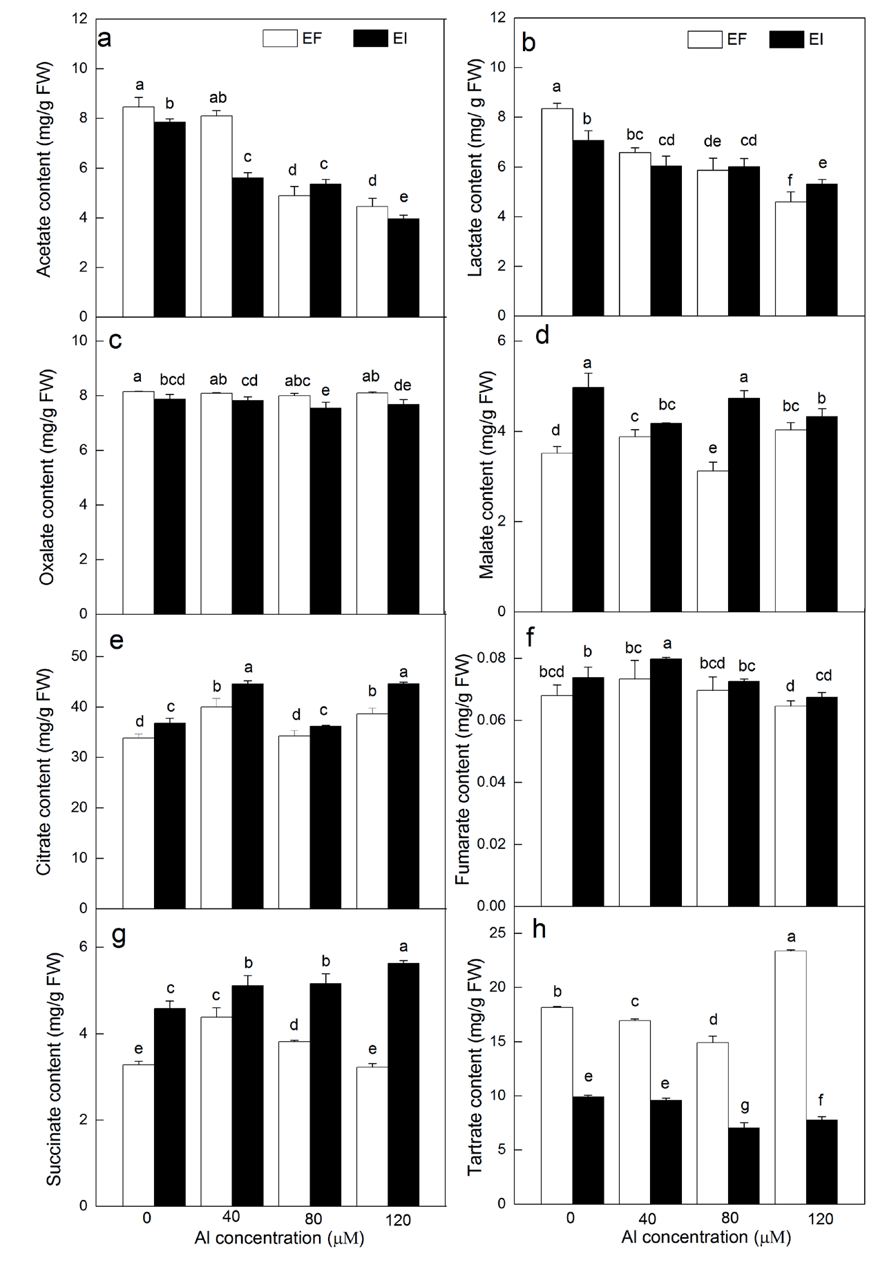
The interaction and main effects of JP4 infection and Al treatments were significant (p<0.05) on acetate, succinate, citrate and tartrate content of leaves (Table 3). When the Al concentration was increased in EF plants, acetate content (except acetate content under 40 μM Al stress) and lactate content were significantly (p<0.05) decreased, malate and citrate content (except malate and citrate content under 80 μM Al stress) was significantly (p<0.05) increased, oxalate content variation of different concentrations of aluminum was not apparent, fumarate and succinate content was first increased and then was decreased, but the changing trend of tartrate content was opposite to that of fumarate and succinate content (Fig. 1). Endophytic infection markedly decreased acetate, oxalate, lactate, and tartrate content in EI plants compared to EF plants exposed to Al stress (except lactate content under 40 μM and 80 μM Al stress). However, endophyte infection significantly (p<0.05) increased citrate and succinate content under 0-120 μM Al stress, malate content under 80 μM Al stress, and fumarate content under 40 μM Al stress.
OAs Content of Roots
The interaction and main effects of JP4 infection and Al treatments were significant (p<0.05) on lactate, oxalate, malate, citrate and tartrate content of roots (Table 3). Acetate content under 40 μM and 80 μM Al stress, lactate content under 40 μM, malate content under 120 μM Al stress, and succinate and tartrate content under 40-120 μM Al stress was significantly (p<0.05) increased in EF plants compared to plants without Al stress (Fig. 2). However, citrate content under 80 μM Al stress and fumarate content under 80 μM and 120 μM Al stress were significantly decreased. Oxalate content of roots response tendency to Al stress was consistent with that of leaves. Endophytic infection significantly (p<0.05) increased lactate, malate, fumarate, and succinate content in EI plants relative to EF plants subjected to Al stress. Oxalate content under 120 μM Al stress and citrate content 40 μM and 80 μM Al stress was also significantly increased. However, endophytic infection significantly decreased tartrate content and acetate content under 80 μM and 120 μM Al stress.
Al Content
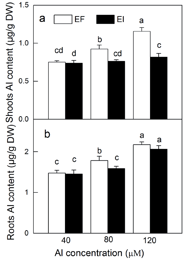
The Al content in the shoots and roots of the EI and EF plants increased gradually with increasing Al concentration (Fig. 3). The higher Al contents were measured in the roots compared to in the shoots for infection with or without endophyte. Endophytic infection decreased the Al content in the shoots and the roots in the EI plants compared to the EF plants, especially significantly decreased in the shoots and the roots in the EI plants under 80 μM Al stress, as well as in the shoots under 120 μM Al stress.
Table 3.
Results of two-way ANOVA between Al stress and JP4 inoculation on the acetate, lactate, oxalate, malate, citrate, fumarate, succinate, and tartrate content in rice seedlings.
Discussion
Evidence is increasing that Al stress inhibits plant growth and limits plant productivity in acid conditions. Our results also showed that reductions in shoot height, root length, and dry weight were observed in EF plants exposed to Al stress (Table 1). Several studies have shown that endophyte infection can help plants to grow well and to counteract stressful events [18]. In the present study, the positive effects of infection with the endophytic fungus JP4 were observed on the height of the shoots, and dry weight of the shoots and roots in the EI plants relative to the EF plants with or without Al stress. In contrast, root length was inhibited in the EI plants. Dobbelaere et al. thought that inhibition of root length was due to IAA production in wheat inoculated with Azospirillum brasilense [34].
Chlorophyll content was significantly decreased in EF plants (Table 2). Increased chlorophyll contents were observed in the EI plants relative to the EF plants with or without Al stress. Similar effects were found in endophyte-infected rice under Cd stress [20]. Li et al. also found higher levels of chlorophyll in EI Oryza sativa L. exposed to osmotic stress [35]. Our data also indicated that carotenoid content was the same trend as chlorophyll content in all treated plants (Table 2). The increased carotenoid content of EI plants may protect the lipid phase of the thylakoid membrane.
An obvious increase in leaf MDA content, which was considered to be a characteristic of lipid peroxidation, was observed in EF plants exposed to Al stress (Table 2). Our results agreed with Cakmak and Horst that Al stress enhanced the level of MDA in soybean [31]. It is well known that Al acting as a catalyst in reactive oxygen species (ROS) yields induced oxidative damage in plant cells. The enhanced level of MDA could be attributed to the poisoning ROS presence or inhibition of antioxidative defense systems. The present study also recorded that MDA content was lower in the EI plants compared to the EF plants. To eliminate the excess of ROS or reduce damage effects, plants have evolved some defense mechanisms including enzymatic antioxidants and non-enzymatic antioxidants. In the current study, SOD activity was higher, whereas POD and CAT activity lower in the EF plants subjected to Al stress (Table 2). SOD, POD and CAT activity was higher in the EI plants compared to the EF plants under Al stress, which is consistent with our previous study on the endophyte-infected rice under Cd stress [20]. Pan et al. also reported that endophytic infection increased antioxidant enzyme activity in tall fescue under salinity stress [36]. Plants combat the adverse effects of stress by increasing the activity of antioxidant enzymes [37]. Hence, our results demonstrated that the endophytic fungus JP4 infection was beneficial to triggering antioxidant systems to eliminate the ROS in rice subjected to Al stress.
Fig. 2.
Effects of endophytic infection on the acetate a), lactate b), oxalate c), malate d), citrate (e), fumarate f), succinate g) and tattrate h) content of rice roots under Al stress. Different letters indicate significant differences at p<0.05.
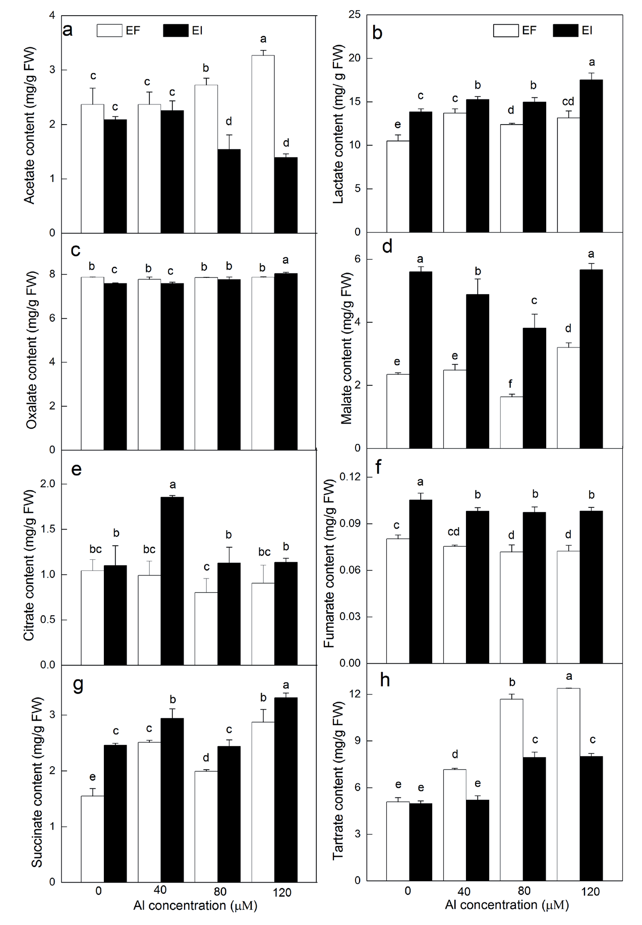
Accumulation of OAs is an important process involved in the internal and external detoxification of Al [5]. In the present study, we investigated the effects of Al stress on the contents of OAs in the leaves and roots in the EI and EF plants. Citrate, fumarate, and succinate contents of the leaves were increased, as well as lactate and succinate contents of the roots in EF plants under Al stress. The largest amount of citrate in the leaves was found among the eight OAs measured, but not in the roots. Lots of citrates were exuded from the roots of pasture and grain legumes and rice [38, 39]. Fumarate content was the lowest in the leaves and roots among the eight OAs investigated. Succinate was only accumulated in the leaves and roots in response to Al stress, which is consistent with the results in maize exposed to Cu stress [40]. Delhaize et al. found malate was secreted only from the root of wheat exposed to Al stress [41]. Ryan et al. reported that malate was not an effective organic acid to detoxify Al ions in wheat [42]. However, in this paper, the higher the malate content in the leaves and roots was observed except for 80 μM Al treatment relative to that without Al stress. Some researchers also reported that alfalfa and Hevea brasiliensis released oxalate to detoxify Al [43, 44]. However, our data indicated that oxalate content did not change in the leaves and roots. These results might be attributable to the different contributions of OAs to detoxify Al in organ type or species.
Fig. 3.
Effects of endophytic infection on theAl contents in the shoots a) and the roots b) of rice under Al stress. Different letters indicate significant differences at p<0.05.
Endophytes can regulate organic acid metabolism in quantity and quality [45, 46]. Endophytic infection accumulated some OAs in leaves and in roots of the EI rice seedlings exposed to Pb stress compared to that of the EF plants [47]. Similarly, in the present investigation, under Al stress, inoculation of Glomerella sp. JP4 significantly increased the contents of citrate and succinate in leaves. Moreover, lactate, malate, fumarate, and succinate content in roots were also enhanced due to the inoculation with Glomerella sp. JP4. These results suggested that more kinds of OAs in roots were needed to resist Al stress relative to that in leaves. In plants, some homologs, such as ScALMT1, MsALMT1, AtALMT1, BnALMT1/2, GmALMT1, ScALMT1, HlALMT1, and TaALMT1, regulates the secretion of organic acids in Al tolerance [3]. However, under endophyte infection, which homologs are involved and how to regulate the secretion of organic acids remain poorly understood in rice.
Crops can take up Al from Al–contaminated area and translocate it to edible portions, enhancing the exposure risk for humans. In the current study, the Al content was determined to clarify the detoxification effect of the Glomerella sp. JP4. Infection of endophytic fungus JP4 decreased root Al absorption in varying degrees, which corroborated previous observations recorded by Wang et al. in maize under Cd stress [48]. Endophytic fungus JP4 infection significantly restricted the Al transfer from the roots to the shoots under 80 μM and 120 μM Al stress. Some transporters, such as Nramp aluminum transporter 1 (OsNrat1 gene coded) in rice, transporter (AtOT gene coded) in Arabidopsis thaliana are involved in the uptake and subsequent sequestration and root-to-shoot translocation of Al [49, 50]. However, the mechanisms by which endophyte infection restricted the Al transfer from the roots to the shoots at molecular levels are still unclear, and further research should be needed.
Conclusions
Infection with the endophytic fungus Glomerella sp. JP4 had positive effects in improving Al stress tolerance by employing several different mechanisms, such as by triggering antioxidant systems and increasing photosynthetic pigments. Furthermore, endophytic fungus JP4 infection also increased the accumulation of some OAs in leaves and roots to detoxify Al, decreased root Al absorption, and restricted the Al transfer from the roots to the shoots. Therefore, utilizing endophytic fungus JP4 is a promising approach because of its great application value on Al-contaminated areas.

