

Introduction
Concerns over Bisphenol A (BPA) have increased in the last four decades due to its widespread use and consequent release to the environment [1]. It is obvious that the increase in plastic production every year will bring BPA accumulation to critical levels in aquatic environments. The annual production of BPA, which is the most produced bisphenol component, has exceeded 5 million tons [2]. It is widely used in industry as a raw material for various plastics such as powder paints, paper coatings, plastic bottles, and polycarbonate plastics [3]. In addition, the main usage area of BPA is industrial activities in epoxy resin production. BPA reaches the aquatic ecosystem through sewage system as a result of the degradation of synthetic plastics, with the discharge of industrial wastewater in the production processes, and with animal feces [4]. It has been reported that the presence of BPA in the river varies between 5 and 320 ng/L [5]. Not only the presence of BPA in natural waters, but also studies of BPA residue on aquatic organisms have been reported [6].
Animals and potentially humans are systematically, developmentally and reproductively adversely affected by BPA. BPA is responsible for altering the functioning of estrogenic and androgenic hormones and are known as endocrine disrupting chemicals (EDC), which are commonly found among identified pollutants in soil and water bodies [7]. Exposure to BPA adversely affects fish growth, reproduction and survival rate [8]. The effect of BPA is more in larval fish than in adults [9]. The lethal media concentration (LC50) of BPA in aquatic invertebrates was between 0.96-2.70 mg/L, while it ranged from 6.8 to 17.9 mg/L in fish [10].
BPA exposure always poses a danger up to death for organisms in aquatic ecosystems. The toxic effect of BPA can be seen in natural conditions as well as among the future problems for aquaculture. In this context, Danube sturgeon (Acipenser gueldenstaedtii) is a species worth researching with its aquaculture potential and its presence in natural waters. In addition, A. gueldenstaedtii, which is critically endangered species, has delicious meat and caviar with high commercial value, revealing its significance for aquaculture and fisheries [11]. Since it takes many years to reach sexual maturity, the physiological responses of this fish to reproductive toxic substances such as BPA should be investigated. This study reveals the LC50 concentration of A. gueldenstaedtii larvae against BPA. In addition, the morphological and histopathological alterations of the larvae were also examined in the BPA concentration at the 24th hour. The lack of previous studies on the potential effect of BPA, especially on sturgeon larvae, is an important novelty that will fill this gap in the literature.
Experimental
Experimental Design and Water Quality
Trials were established in toxicology laboratory, Recep Tayyip Erdoğan University, Rize, Türkiye. Sturgeon larvae (<1 g; older than 4 days”90 day/ºC”) were inseminated from broodstock in Aquaculture Application and Research Center. A total of six different concentrations (Control, 100, 200, 400, 800, 1200 µg/L) including control were designed in triplicate. 10 fish were monitored for 72 hours for each replicate. Larvae were fed with Artemia nauplii 6 times a day. The study was carried out with a 12 h light and 12 h dark photoperiod. Behavioral changes and deaths of the fish were regularly monitored and noted.
Stock solution was prepared by dissolving BPA with ethanol (99% purity, analytical grade, Sigma Chemicals, Inc. St. Louis, MO, USA). The study was carried out with underground water in each 1 L beaker. Aeration is provided with air stones placed in each beaker at a flow rate that will not adversely affect the larvae. Daily water changes were applied to prevent the larvae from being affected by the water quality. Water quality parameters were monitored daily. Accordingly, pH, dissolved oxygen (DO; mg/L), temperature (°C), salinity (‰) and electrical conductivity (EC; µS/cm) were measured instantaneously with a portable multi-parameter (Hach, HQ40D 58258- 00).
Histological Examinations
At the end of the 72th h, the fish exposed 800 µg/L BPA were anesthetized for histopathological examination. Gill, liver, kidney, and intestinal tissues were fixed in 10% neutral buffered formalin during one day. Subsequently, samples were placed to 50% ethanol solution for two days. Tissues were treated with alcohol (%80, %90, %95, and %100 for 15 min, respectively) and xylene series (10 min×2 times). Tissues were placed in the paraffin at +65ºC for 1 night after series. Next day, paraffin has cooled and samples with a thickness of 0.5 microns were taken to microscope slide from paraffin-blocked tissues using a microtome. Preparates were treated by xylene (1.5 h×2 times) and stained by hematoxylin (10 min) and eosin (5 min). Finally, tissues on the preparate were examined by light microscopy [12].
Statistical Analyzes
Water quality parameters were presented in mean ± standard deviation. The LC50 value of BPA on sturgeon larvae was tested by Probit analysis. Normality of variance were detected based on Kolmogorov Smirnov test. One-way ANOVA were used to determine differences between concentration groups for the water quality. A maximum p value of 0.05 was considered in order to interpret statistical differences. All data-set were analyzed by SPSS 25 software package for Windows (Version 25, IBM Corp., Armonk, New York, USA).
Results and Discussion
Water Quality Monitoring
Water quality parameters were monitored daily during 72 hours of acute exposure. Water quality parameters are among the limit values suitable for the survival standards of A. gueldenstaedtii larvae (7.29-7.25 mg/L, 23.2-23.6ºC, 2.48-2.66‰, and 4.17-4.46 mS/cm for DO, temperature, pH, salinity, and EC, respectively). Due to the use of groundwater as a water source, the EC and salinity values are slightly higher. However, these values are also below the limits that will adversely affect the larvae. No significant differences were observed between groups for all water quality parameters (p>0.05). Although the larvae continued to feed intensively during the study, daily water changes were provided to prevent possible poor water quality. Therefore, it was ensured that histological and physiological alterations were only caused by BPA. Previous studies have revealed that water quality is adversely affected by acute BPA exposure [13]. Since A. gueldenstadii, which spends most of its life in the Black Sea, is an anadromous species [14], the high salinity and EC in the study did not adversely affect the larval life.
Table 1.
LC50 values of A. gueldenstaedtii larvae in 24th, 48th, and 72nd h.
Fish Behavior and LC50
Abnormal behavior was observed in fish exposed to BPA concentration during the acute trial. Especially, abnormal behaviors such as low mobility or stationary on the bottom were observed at high concentrations. Although DO concentration remained at normal levels, only gill movement was observed depending on time. The LC50 values of A. gueldenstaedtii larvae exposed to five different BPA concentrations at 24th, 48th and 72nd hours were 803.4 µg/L, 63.1 µg/L, and 39.6 µg/L, respectively (Table 1). After 72 h, the fish were examined under a stereo microscope. Fish in the BPA group produced more mucus secretion than the control group.
Swimming behavior in fish often changes during stress. Therefore, slowing or accelerating the swimming movement in toxic studies provides information about the toxicity of the material. BPA exposure has been reported to slow down the swimming movement of fish in previous studies [15]. In the present study, larvae exposed to BPA remained stable at the bottom and only provided rapid gill movement. This low mobility and poor swimming were observed in our previous study [13]. Several studies have been reported showing that rapid operculum movement occurs under stress [16, 17]. Toxic substances disrupt nerve cells, axons and the myelin sheath, altering the synthesis of neurotransmitters involved in behavior [18]. In general, toxic substances cause neurotoxic effects in fish and thus pose behavioral differences [17]. Skin mucus is a potential matrix for minimally invasive environmental monitoring in fish [19]. Skin mucus in fish has the ability to reflect the effects of stress on various biological conditions such as respiration, reproduction, resistance to disease and nutrition [20]. In the present study, the larvae of A. gueldenstadii exposed to BPA secreted more mucus than the control [17]. An increase in mucus secretion as a behavioral response was mentioned in another acute study (96 h exposure) as a result of exposure to BPA [13]. It is an expected hypothesis that mucus secretion increases with exposure to a toxic substance. However, future studies on the content of secreted mucus will be needed. Fish mucus contains a number of compounds such as glycoproteins, immunoglobulins, proteolytic enzymes and pheromones [21]. Therefore, the composition of the mucus will be an important indicator for effect of the toxic substance on fish welfare.
In the current study, LC50 values show a high mortality in fish, especially after the 24th hour. While the alarm reaction and resistance phase of acute stress ended within the first 24 hours, high mortality rate was observed with the exhaustion phase in the following process. BPA is classified as “moderately toxic” and “toxic” to the aquatic environment with an LC50 value of 1-10 mg/L [22]. Accordingly, the current study showed that the LC50 value is less than 1 mg/L even at 24 hours. Therefore, we advocate the highly toxic effect of BPA for A. gueldenstadii larvae. LC50 variation of BPA for fish is related to age, sex, size, physiological status, water quality and genetic background of the fish [23]. In addition, the current study will shed light to researchers and commercial fish farming facilities with the dose response model in terms of BPA toxicity.
Histopathological Findings
The histopathological whole body view of A. gueldenstaedtii larvae were presented in the Fig. 1a). There are no significantly histopathological differences between gill tissues of control and BPA groups (Fig. 1(b, c)). Mild vacuolization was observed in the gill tissues of control group, while moderate symptom was detected in the BPA group. The exposure of BPA caused symptoms of vacuolization and melanomacrophage centers in the liver of larvae (Fig. 1(d,e)). Similar symptoms were observed in the kidney of larvae (Fig. 1(f,g)). In addition, the degradation/distortion of goblet cells was detected in the intestine tissues of larvae exposed to BPA (Fig. 1(h,i)). The number of goblet cell per ville was counted 11.39±2.2 and 5.61±1.7 in the control and BPA groups, respectively. Accordingly, the number of goblet cell per ville was significantly less compared to control group (p<0.01; t = -8.713).
Fig. 1.
The histopathological view of A. gueldenstaedtii larvae. a) Whole body of larvae; b) Gill tissue in control group; c) Gill tissue in BPA group; d) Liver tissue in control group; e) Liver tissue in BPA group; f) Kidney tissue in control group; g) Kidney tissue in BPA group; h) Intestine tissue in control group; i) Intestine tissue in BPA group. Arrow: vaculization, star: melanomacrophage centers, and red circle: goblet cells in the ville.
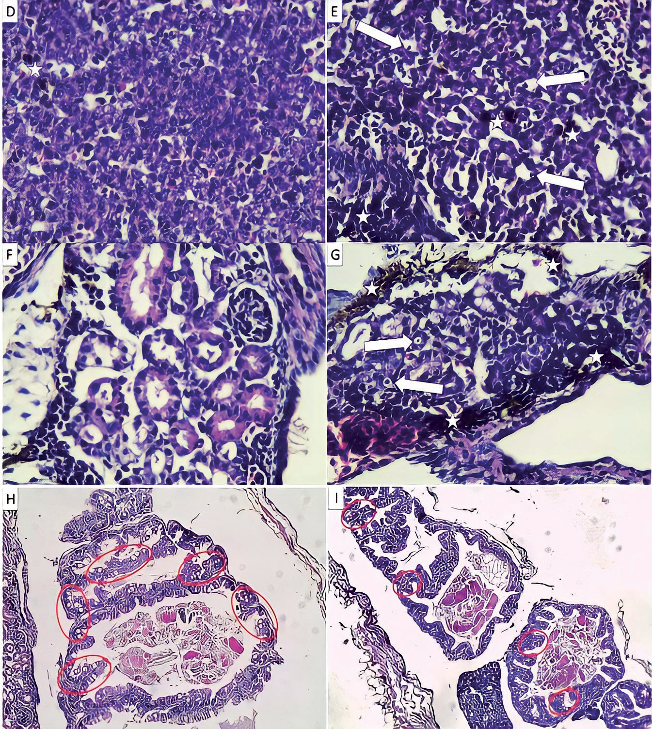
Histopathological studies are often preferred to demonstrate the health status of a population. Based on a previous study, the whole body histological view of a healthy A. gueldenstadii larvae was presented in the current study [24]. The gills are the primary organs in contact with water, so they are the most important markers for histological studies [25]. In the present study, we focused on the histopathological alterations of LC50-72 h concentration due to current study can be an inspiration to future studies. In this context, vacuolization symptoms were observed in the gill tissues of the control and BPA groups. There is a high correlation between the toxic effect of a pollutant and the severity of the lesion [26]. Although there was no significant difference between the gill tissues of the fish in the control and BPA groups, the BPA group was slightly more adversely affected than the control. It has been discussed before that BPA causes vacuolization in the gill and liver tissues of fish [8, 27]. In fish, the liver is the main organ for detoxification of xenobiotic pollutants such as BPA [26]. Therefore, the liver of fish is an important indicator for estimating the water quality of the aquatic environment. In the present study, vacuolization and severe melanomacrophage centers were observed in the liver of the BPA group. Although the presence of melanomacrophage centers in tissues is an expected situation, similar to our study, severe melanomacrophage center is an indicator of a response to toxic substance. Because macrophages are indicator cells of innate immunity in fish and other vertebrates and have the ability to phagocytosis, they have functions such as post-inflammatory repair, tissue regeneration and elimination of old cells [28]. In fish, the kidney is represented as a hematopoietic and osmoregulatory organ [26]. The kidneys are another organ that shows the effect of the toxic substance because it collects a large part of the post-branchial blood [29]. In the current study, vacuolization and melanomacrophage centers were observed in the kidney tissue, similar to the liver. However, melanomacrophage centers in the kidney tissue were more severe in the BPA group than in the liver. When fish encounter toxic substances in the aquatic environment, they react by secreting mucus by the skin goblet cells. On the other hand, mucus-secreting goblet cells in the digestive system produce a lubricant that protects the mucous membrane from physical and chemical effects during the digestion [30]. The mucus secretion of goblet cells is involved in the absorption of digestible substances in fish and all other mammals [31]. The distortion effect of potentially toxic substances on intestinal goblet cells has been discussed previously [32]. In the current study, the number of goblet cells per ville in the intestine was found to be significantly lower in the BPA group (p<0.01). This may be related to BPA degrading or distorting sensitive goblet cells.
Conclusions
In conclusions, this study presented the LC50 value of BPA for A. gueldenstadii larvae and explained its toxic effect on gill and visceral organs histopathgologically. Especially the lethal effect of BPA on the larvae was remarkable at the end of the first 24 hours. On the other hand, histopathological adverse effects of BPA on liver and kidney were observed. Negative results of BPA on goblet cells in the intestinal tissue were also highlighted. Future studies should focus on the effects on reproduction activity, rather than the lethal or acute toxic effects of larvae exposed to BPA.

