


Introduction
Microalgae constitute a highly diverse group of photosynthetic organisms, encompassing both eukaryotic and prokaryotic taxa. This diversity is reflected in their wide range of physiological, morphological, and genetic traits, which equip them with the capacity to biosynthesize a diverse array of bioactive compounds. Notably, within the extensive spectrum of algal organisms, Chlorophyta emerges as one of the largest phyla, characterized by a plethora of distinct species distributed across geographically expansive regions [1]. The significance of microalgae extends to their role as a valuable source of polyunsaturated fatty acids (PUFA). Consequently, the extraction and purification of these PUFA compounds from various algal strains has become a burgeoning research area. This field is gaining increasing recognition due to its potential to address the dietary needs of a growing global population [2].
PUFAs are a category of fatty acids characterized by the presence of two or more double bonds in their acyl chain. PUFAs can be further classified into two primary groups based on the length of their carbon backbone: short-chain polyunsaturated fatty acids (SC-PUFAs), comprising 16 or 18 carbon atoms, and long-chain polyunsaturated fatty acids (LC-PUFAs), with more than 18 carbons. Extensive research has established that PUFA consumption is associated with a multitude of health benefits for humans. These benefits encompass improvements in metabolic rates, the regulation of blood pressure and glucose levels, and protection against various diseases, including certain types of cancer. These findings underscore the significant role of PUFAs in promoting human health and well-being [3].
Palm oil milling operations produce a substantial amount of palm oil mill effluent (POME), which stands out as the most voluminous waste in this process. The unregulated release of POME into the environment poses a significant threat to both human health and the ecosystem. This is primarily due to its elevated levels of toxins as well as the presence of organic and inorganic materials. It is imperative to emphasize the essential need for treating POME before discharging it into the environment in order to safeguard human health and preserve the natural surroundings [4]. In a prior research investigation, a microalgae species, Chlorella vulgaris, isolated from the coastal areas of Malaysia, exhibited the capability to thrive and synthesize PUFAs using POME as a nutrient source [5]. Furthermore, several studies have substantiated the beneficial impact of dark stress fermentation on enhancing both the quantity and quality of lipids within microalgae [6].
The objective of this study is to isolate microalgae with the ability to produce PUFA from water utilized in betta fish cultivation. Furthermore, the research aims to assess the potential of PUFA production by these isolated microalgae when utilizing POME as a substrate under dark stress conditions.
Experimental Procedures
Sample Collection and Isolation
Samples were collected from the betta fish farm and transported to the laboratory at the Faculty of Science and Digital Innovation, Thaksin University, Thailand. The collected sample was centrifuged at 10,000 rpm for 5 min to separate the supernatant. Subsequently, it underwent 5-wash cycles with sterile distilled water to remove any contaminated sediment. The microalgae cells were inoculated into BG11 agar (Himedia, India) and cultured for 5 days under a continuous white fluorescent lamp at room temperature [7]. The microalgae were isolated until a single colony with a green color formed on the medium surface.
The pure culture was inoculated into BG11 broth and cultured for 5 days under a continuous white fluorescent lamp at room temperature. Subsequently, 10 mL of 5 day old microalgae culture (OD680 = 0.1, 0.02 g/L) was inoculated into 90 mL of BG11 broth. The growth potential was monitored every 24 h for a duration of 5 days [8]. The microalgae biomass was determined using spectrophotometry at 680 nm, and the calculated OD680 value of 1.0 corresponds to a dry mass of 0.19 g/L.
Oil Content and Profile
The Bligh and Dyer method was adapted for crude oil extraction from algae. Microalgae cells were dried at 60ºC for 48 h and 1 g of dried material was placed into a 50 mL centrifuge tube. Subsequently, 12.5 mL of n-hexane was added to the algae biomass [9], which was then soaked for 6 h under continuous shaking at 150 rpm at room temperature in a rotary shaker. The mixture was centrifuged at 5,000 rpm for 10 min to separate the microalgae cells. The lipid portion with the solvent was transferred into a separating funnel, and 10 mL of sterile distilled water was added to the mixture. The oil content in the organic solvent phase was collected from the separating funnel. The oil phase was aspirated in a laboratory chemical fume hood overnight. The content percentage was calculated using Equation (1) [10].
Gas chromatography-mass spectrometry (GC-MS) was utilized for the analysis of the fatty acid profile. A 1 µL sample of the oil was injected into a gas chromatograph equipped with a DB-Wax column (60 m x 250 µm x 0.25 µm). The injector temperature was set at 250ºC, with a purge flow rate of 50 mL/min for 2 min. The column temperature was initially held at 50ºC for 1 min, followed by an increase of 25ºC/min to 200ºC, and then a gradual increase of 3ºC/min to 230ºC, held for 18 min. The total running time was 35 min [11].
Identification
The microalgae-producing PUFAs were initially identified using morphological characteristics under a light microscope (Olympus, Japan). The selected microalgae in BG11 were centrifuged at 5,000 rpm for 10 min. The pellet was placed on the BG11 agar and incubated under a continuous white fluorescent lamp at room temperature. The single colony of microalgae was placed on a microscope slide to observe the morphological characteristics. The characteristics were confirmed at the TISTR Algal Excellent Center (ALEC), Thailand.
For molecular identification, 1 g of wet algae cells was used for genomic DNA extraction with the InvitrogenTM genomic DNA kit (Thermo Fisher Scientific, United Kingdom). The purity of the genomic DNA was assessed using a Thermo Scientific NanoDrop (Thermo Scientific, United States). The 18S rDNA gene was amplified using the universal primer pairs NS1 (GTAGTCATATGCTTGTCTC) and NS8 (GTAGTCATATGCTTGTCTC). The PCR reaction was carried out in a 0.2 mL tube, consisting of 5 µL of PCR master mix (Mg2+ plus), 4 µL of dNTPs (2.5 mM), 1 µL of each primer (20 µM), 1 µL of DNA template, 0.2 µL of Taq polymerase (5 U/µL), and 37.8 µL of DNA-free water. The PCR conditions were as follows: 94ºC for 30 sec, 50ºC for 30 sec, and 72ºC for 2 min, for a total of 30 cycles.
POME Preparation
POME was collected from Trang Palm Oil, Southern Thailand, and stored in a plastic container at -25ºC to preserve the quality of the wastewater. For preparation, the POME was centrifuged at 4,000 rpm for 10 min to remove the sediment and increase the light penetration of wastewater. The centrifuged POME was sterilized and used as an algae medium. The 10 mL of microalgae (OD680 = 0.1, 0.02 g/L) was inoculated in the 90 mL of diluted wastewater at various concentrations at ratios of 1 : 1 (50 Ml : 50 mL), 1 : 2 (33 mL : 67 mL), 1 : 3 (25 mL : 75 mL), and 1 : 4 (20 mL : 80 mL). The mixtures were incubated under a continuous white fluorescent lamp at room temperature. The algae growth was measured at 680 nm using UV-Vis spectrophotometry. The algae dried weight was calculated based on OD680, as one OD equals 0.19 g/L [8].
Effects of POME Ratio
The 10 mL of microalgae (OD680 = 0.1, 0.02 g/L) was inoculated in the 90 mL of diluted wastewater at various concentrations at ratios of 1 : 1 (50 Ml : 50 mL), 1 : 2 (33 mL : 67 mL), 1 : 3 (25 mL : 75 mL), and 1 : 4 (20 mL : 80 mL). The mixtures were incubated under a continuous white fluorescent lamp at room temperature for 5 days. The algae growth was measured at 680 nm using UV-Vis spectrophotometry.
Effect of Dark Stress
The 10 mL of microalgae (OD680 = 0.1, 0.02 g/L) was inoculated in the 90 mL of diluted wastewater at the optimal concentration. The mixtures were incubated under a continuous white fluorescent lamp and dark stress (absolute darkness) at room temperature for 5 days. The algae growth was measured at 680 nm using UV-Vis spectrophotometry. The fatty acid yield was extracted and measured.
Results and Discussion
Isolation, Oil Content, and Identification
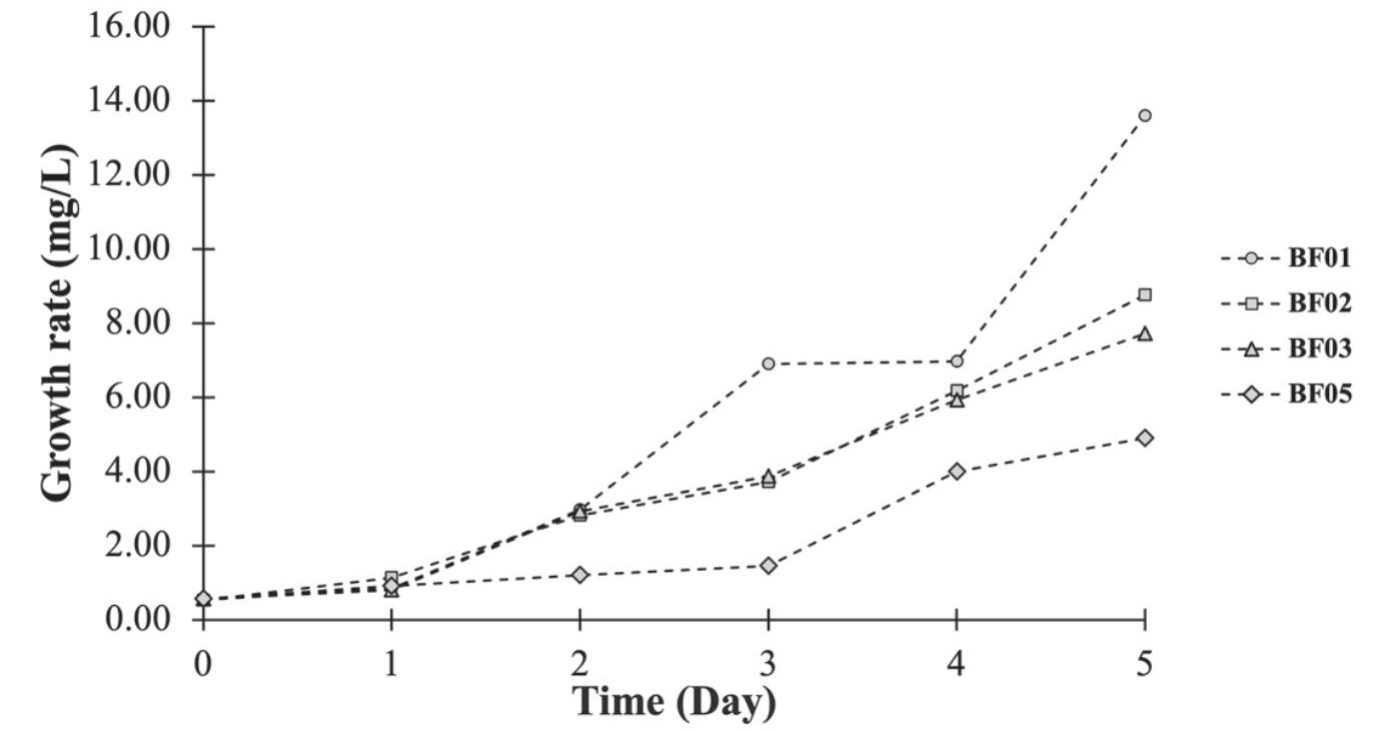
In our study conducted at the Betta Fish Farm, we isolated four distinct strains of green microalgae. We assessed their growth rates using UV-Vis spectrophotometry at 680 nm [8]. The results are presented graphically in Fig. 1. Among the isolated microalgae strains, BF01 demonstrated the most robust growth, with an average growth rate of 13.60±2.09 mg/L. Following closely was BF02, with a growth rate of 8.76±2.02 mg/L, while BF03 exhibited a growth rate of 7.71±1.35 mg/L. Finally, BF05 displayed the lowest growth rate among the strains at 4.90±0.68 mg/L. These findings highlight the varying growth rates of the microalgae strains, with BF01 showing the highest growth potential and BF05 the lowest.
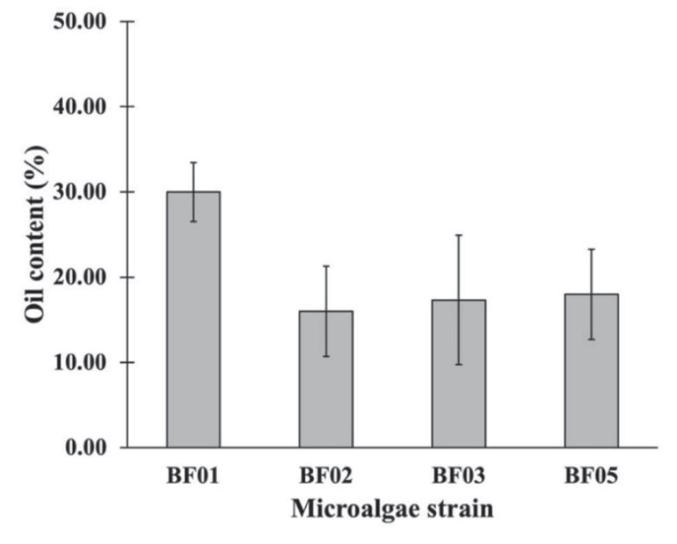
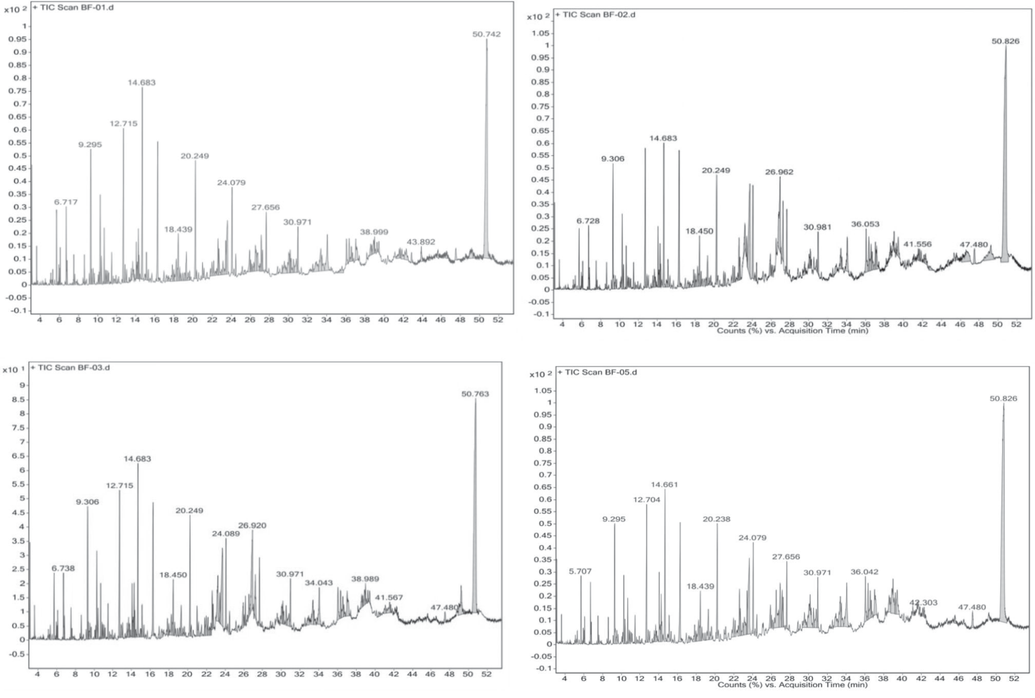
In our investigation, the analysis of oil content in the microalgae strains was conducted using dried cell samples. The outcomes of this analysis are depicted in Fig. 2. Our findings revealed that the microalgae strain BF01 exhibited the highest oil content, reaching a maximum of 30.00±3.46%. Following BF01, the order of oil content in the strains was as follows: BF05 with 18.00±5.29%, BF03 with 17.33±7.57%, and BF02 with 16.00±5.29%. This data underscores the variations in oil content among the microalgae strains, with BF01 displaying the highest oil content, while BF02 had the lowest among the strains assessed. The chromatogram illustrating the oil content is presented in Fig. 3. A comprehensive list of the fatty acid composition is provided in Table 1.
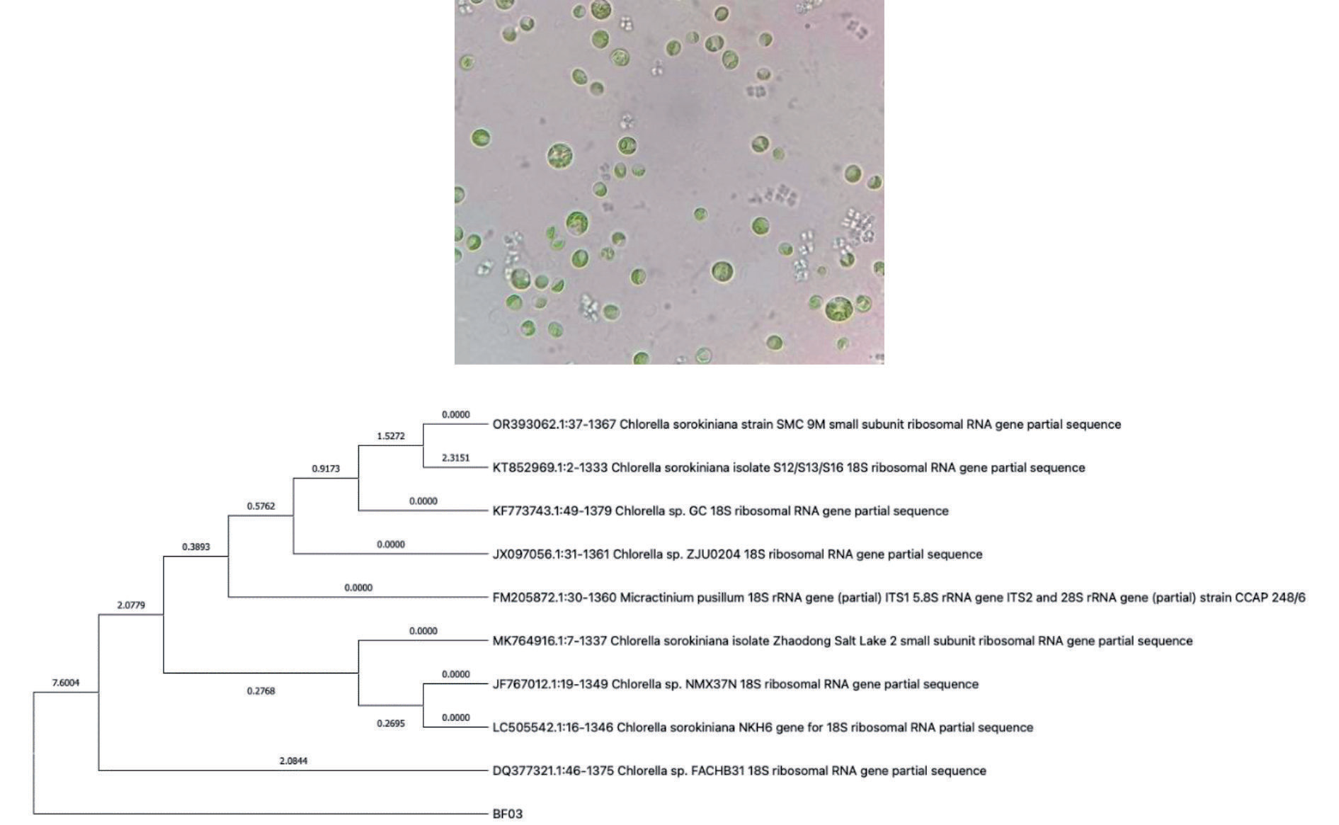
Morphological examination of microalgae strain BF03 was conducted using a light microscope. The observations revealed distinctive characteristics of BF03, notably its slightly oval-shaped cells, which measured approximately 2 - 15 µm in size. Furthermore, it was noted that BF03 lacked flagella, as illustrated in Fig. 4. These characteristics align with the typical features of Chlorella sp. To confirm the identification, molecular analysis was undertaken by sequencing the 18S rDNA. The obtained nucleotide sequence was then compared with existing sequence data available in GenBank at NCBI. The phylogenetic relationship of BF03 was elucidated and is depicted in Fig. 4. The result showed the BF03 is similar to Chlorella sp. FACHB31 (Accession number DQ 377321.1) with 95.43%. This comprehensive analysis solidifies the identification of BF03 as a member of the Chlorella species and provides valuable insights into its morphological and genetic characteristics.
Table 1.
Fatty acid profile of algae oil content.
On the other hand, the marine microalgae Nanochloropsis oculata and Chlorella sp. have been shown to exhibit high oil content, often reaching up to 50%. While also producing α-Linolenic acid, even under conditions of low nutrient levels and salinity [11]. The research conducted by Maneechote et al. demonstrated that freshwater microalgae isolated from the local lake, specifically Scenedesmus sp. SPP and Chlorella sp. PPS, exhibit significant promise as sources of PUFAs [12]. Furthermore, the microalgae Thalassiosira weissflogii TRG10-p105, obtained from seawater in Peninsular Malaysia, have demonstrated the ability to produce substantial amounts of PUFAs and carotenoids. This particular strain holds promise as a potential candidate for large-scale cultivation in environments with high ammonium content. This discovery holds significant relevance for our research discussion [13].
Effect of POME Ratio and Dark Stress
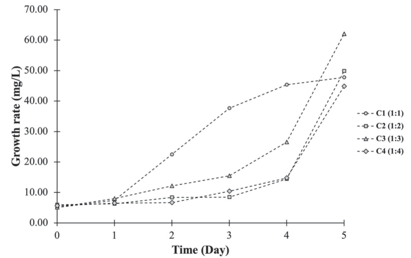
In our investigation, we assessed the impact of different POME ratios on growth rates. The results demonstrated that the 1 : 3 POME ratio yielded the highest growth rate, with a remarkable average of 62.02±5.09 mg/L. Following this, the 1 : 2 ratio achieved a growth rate of 49.86±8.18 mg/L, the 1 : 1 ratio reached 47.74±3.31 mg/L, and the 1 : 4 ratio displayed a growth rate of 44.84±2.64 mg/L (Fig. 5). These findings underscore the significance of POME ratios in influencing the growth rates of the microalgae, with the 1:3 ratio providing the most favorable conditions for growth, followed by the other tested ratios in descending order.
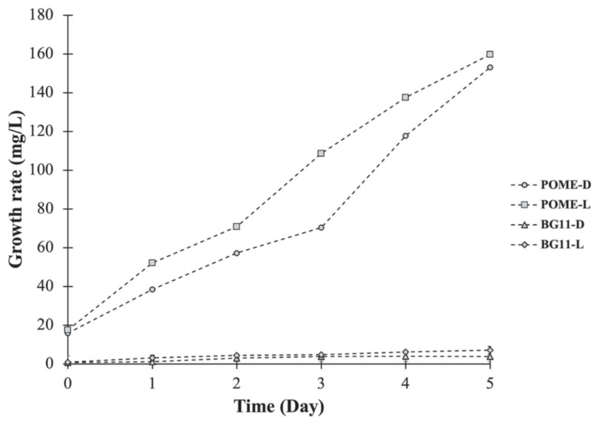
In our investigation into the effects of dark stress, microalgae strain BF03 displayed successful growth under sterile POME conditions, both in the absence of light (dark stress) and in well-illuminated conditions. The growth rates were impressive, with BF03 achieving 153.03±36.16 mg/L in dark stress and 159.75±19.04 mg/L in light conditions. Comparatively, when BF03 was grown in the fundamental BG11 medium, the growth rates differed significantly. In dark stress conditions, BF03 exhibited a growth rate of 3.85±0.61 mg/L, while under well-illuminated conditions, it reached a growth rate of 7.08±1.90 mg/L (Fig. 6). These results highlight the remarkable adaptability of BF03, which thrived in both dark stress and light conditions when grown in sterile POME, achieving notably higher growth rates compared to its performance in the standard BG11 medium. When microalgae strain BF03 was cultivated in a POME culture under dark stress conditions, it exhibited an impressive oil content of 20.97±0.10%. This surpassed the control group, which consisted of BF03 in the BG11 medium under well-illuminated conditions, by 21%. Similarly, in the POME culture under light conditions, BF03 displayed a noteworthy oil content of 19.10±0.50%, which was 10% higher than the control. These results underscore the potential of BF03 to yield elevated oil content, particularly in POME cultures under both dark stress and light conditions, surpassing the oil content achieved in the standard BG11 medium under well-lit conditions.
Similar to the study of Desjardins et al., the results showed that dark stress cultivation can improve lipid quantity and quality of produce from the microalgae Coccomyxa sp. The data indicated that dark stress mainly favored the production of PUFAs and resulted in a lower C:N ratio [6]. On the other hand, the microalgae Chlorella sorokiniana CY-1 were cultured in POME. The microalgae produced 2.12 g/L of biomass and exhibited an 11.21% lipid yield in pretreated POME [8].
Furthermore, subjecting Nannochloropsis oceanica, isolated from seawater, to dark stress treatment has the capacity to stimulate the production of omega-3 fatty acids. [14]. On the other hand, heterotrophic microalgae like Crypthecodinium cohnii have exhibited the ability to yield significant quantities of docosahexaenoic fatty acids, a type of PUFA, through dark fermentation within the anaerobic digestion process [15].
Conclusions
This study has established that cultivating Chlorella sp. BF03, which was initially isolated from a betta fish farm and grown in POME, led to significant enhancements in both biomass quantity and PUFA content. The best results were obtained when Chlorella sp. BF03 was grown in POME with a dilution ratio of 1 : 3, yielding the highest biomass of 153.03±36.16 mg/L. Despite the abundance of nutrients in POME that support the growth of PUFA-rich Chlorella sp. BF03. This study contributes new insights into the utilization of Chlorella sp. BF03, isolated from a fish farm, for PUFA production under dark stress conditions, using POME as a low-cost substrate.

