

Introduction
Plant diseases have been with humanity since agriculture began. Plant diseases cause substantial annual losses. Plant disease might be a significant cause of reducing the yearly level of food production in the world, which, depending on the source, is estimated at the level of 10-40% [1-3].
The economic importance of plant diseases in agricultural production in terms of their impact on the national economy cause a shortfall in the quantity of the crop, a decrease in its value, and damage to the appearance and characteristics of the plant product, such as distortions, and thus affect the consumer [4].
Soil microbes can make plants more resistant to epidemic diseases, opening a new door to plant disease resistance and maintaining plant health, opening new possibilities for sustainable food production. Soil fungi are the beginning of the food chain that helps other soil organisms and supports healthy soil functions. They are microscopic plant-like cells that may be unicellular or grow in the form of long, thread-like structures, and they do not depend on certain types of plants. As they are slow growing [5].
Due to the wide range of biocontrol features, including parasitism, antibiosis, the synthesis of secondary metabolites (SM), and the stimulation of plant defense systems, fungi from the genus Trichoderma are among the most employed and researched microorganisms as BCA. Well-known mycoparasites include a number of Trichoderma species. Due to the wide range of biocontrol features, including parasitism, antibiosis, the synthesis of secondary metabolites (SM), and the stimulation of plant defense systems, fungi from the genus Trichoderma are among the most employed and researched microorganisms as BCA. Well-known mycoparasites include several Trichoderma species. Due to the wide range of biocontrol features, including parasitism, antibiosis, the synthesis of secondary metabolites (SM), and the stimulation of plant defense systems, fungi from the genus Trichoderma are among the most employed and researched microorganisms as BCA. Well-known mycoparasites include several Trichoderma species [6].
Trichoderma has gained immense importance in the past several decades due to its antagonistic ability against various plant pathogens and growth promotion in crop plants. Some species of Trichoderma viz., Trichoderma harzianum, T. viride, T. virens, and T. kongsi are well-known antagonists. They are being utilized to control plant pathogens under field conditions. Extensive use in agriculture is directly related to its biocontrol and biostimulation abilities. Trichoderma is recognized as the genus with the most significant promise for biocontrol since it has the most isolated antifungal bioactive chemicals [7-9]. 50-60% of the fungal BCAs are Trichoderma species, according to Rush et al., 2021 [9]. There are currently at least 77 commercial Trichoderma-based bio fungicides on the market worldwide, including seven that the European Commission has approved for use in EU member states [9].
Insects and nematodes are just a few pests that fungi may effectively suppress biologically [10-12]. It has been demonstrated that some Trichoderma species efficiently eradicate pests like Tetranychus urticae and several insects that harm crucial crops [11].
Trichoderma viride is an active antagonist in moist soil but is inhibited under very wet conditions when pH is 5.4 or above. Furthermore, two fungi, Physalospora rhodian and Diaporthe citri affected the vessels of citrus tissues. These pathogens generally show leaf dryness and shedding, twigs, and turns, a decline in tree growth, and reduced yield [13]. Mycoparasitism by Trichoderma is a sequential process that involves detecting the fungal host, chemotrophic growth towards the target, lysis, and assimilation of the intracellular content [13].
The present study was taken up to prove T. viride antagonistic and T. harzianum as biocontrol agents and establish a methodology for inoculation of vegetables and serial plants that were infected by some pathogens to be cured by Trichoderma fungi strains in combination with some fungicides to reduce diseases and improve future industrial-scale mass production to formulate microbial pesticide packages that enabled application irrespective of seasonal /biological/ physiological constraints.
Material and Methods
Seeds
Seeds of Wheat (Triticum aestivum) and maize (Zea mays) cultivars were obtained from the local market (Khartoum).
Pathogenic Fungal Isolates
Three fungal isolates, including Phylaspora rhodina, Diaporthe citri, and Nattrassia mangiferae were recovered from plants showing typical symptoms of wilt and root- rot and wood decay at the soil surface. The fungi were revived and isolated by placing infected plant tissues (after surface disinfection with 1% sodium hypochlorite for 2 min) on potato dextrose agar (PDA) and incubating them at 35ºC for five days. Morphological identification of pathogenic fungal isolates was based on characteristics of the macroconidia, phialides, microconidia, chlamydospores, and colony growth traits. The forma specialism of these pathogens was identified using pathogenicity tests. Based on these tests, several aggressive isolates were selected and used in this study.
Soil Samples
Samples of soil were taken from the rhizosphere soil of plantation areas in Al-Qaid North of Hail region, Saudi Arabia where wheat and Zea mays were cultivated. Soil samples were taken at a depth of 3-5 cm and laid out in sterile containers and transported to the laboratory; 5 g of root and closely adhering soil was suspended in a flask containing 50 ml of sterile distilled water. Flasks were placed on a rotary shaker at 200 rpm for 10 minutes. After collection, they were air-dried and ground into powder. A stock solution of the sample was created by combining 90 mL of distilled water with 10 g of a soil sample that had been pulverized. Then, samples were made with successive dilutions of 101, 102, and 105. These serial dilutions were used as preparation to isolate Trichoderma.
Isolation of Trichoderma
Trichoderma harzianum Selective medium (THSM) is recommended for selective isolation of T. harzianum. The medium consisted of the following (g/Liter): Magnesium sulfate heptahydrate 0.2; Dipotassium hydrogen phosphate 0.9; Ammonium nitrate 1 g; Potassium chloride 0.15; Glucose 3 g; Rose Bengal 0.15 g and Agar 20 g. The fungicides used were Propamocarb and Quintozene. To the THSM, Propamocarb p (1.2 ml equivalent to 772 g of active ingredient/L autoclaved medium at 50ºC were added. 25.54 g (The equivalent weight of dehydrated medium per liter) were suspended in 960 ml distilled water. Then we heated the medium to boiling in order to dissolve it completely. The contents were then sterilized by autoclaving at 15 lbs pressure (121ºC) for 15 minutes, then cooled to 45-50ºC. The antimicrobial and fungicidal ingredients (all amounts are per liter) included: 0.25 g of chloramphenicol, 9.0 ml of streptomycin stock solution (1% wt/vol), 0.2 g of quintozene, and 1.2 ml propamocarb, all in 40 ml of sterile distilled water, and the mixture was added to the cooled basal medium. Then rehydrated contents of one vial of T. harzianum were added aseptically.
Identification of the Isolates of Trichomoniasis
Trichoderma spp. have previously been identified using conventional morphological and cultural methods. Conidiophores, phialides, and conidia were arranged this way, and hyphae’s coloration and linear growth patterns are examples of cultural characteristics.
Five mm diameter mycelial plugs were taken from the edges of CV8 agar cultures four to five days old. With the mycelia in contact with the medium, one plug was positioned in the middle of a 9-cm petri dish. For each isolate, three replicate dishes per treatment were infected. The plates were then incubated for 5 to 7 days at 37ºC in complete darkness. Colony diameters were measured for all treatments when colonies on non-amended control dishes had reached the edge in two perpendicular directions. The two readings were averaged after subtracting the mycelium plug’s diameter. By dividing the colony diameter in amended dishes by the colony diameter in non-amended control dishes, the relative growth of an isolate on amended media was computed and expressed as a percentage.
Trichoderma Soil Inoculation
109 cfu/ml conidial suspension of Trichoderma was diluted in 5 liters of water to prepare a solution strength of 2X10⁵ cfu/ml. For each pot, 100 ml of solution was used, which accounted for 2X10⁷ cfu of Trichoderma per pot. 100 ml of the solution was used to drench the soil per pot [14, 15].
Assessment of Trichoderma for Their Biocontrol Efficiency
Mycelial plugs (5 mm diameter) from each Trichoderma spp. were cut from the margin of 4- to 5-day-old CV8 agar cultures. Five plugs were placed apart onto a 9-cm petri dish with the mycelia in contact utilizing the medium. For each isolate, three replicate dishes per treatment were infected, with the suspension of fungal pathogens Phylaspora rhodina, Diapothe citri, and Nattrassia mangifera. Five Zeya maize or wheat seeds were embedded into CV8 cultures (0.8%) and left to sprout. Inoculated dishes were then incubated at 37ºC for 21 days in darkness. The efficiency of the Trichoderma strain was selected according to the plant seedling health.
Soil Infestation with Trichoderma
Mycelial plugs of three old cultures of Trichoderma grown on PDA medium were put into sterilized maize meal sand medium (maize meal 10 g, and 90 g and water 16 ml) in 250 ml conical flask and incubated at room temperature for nine days. Before application into the soil, the Trichoderma biomass was mixed with sterilized field soil. The observation was recorded.
In vivo Experiment of Trichoderma:
A. Trichoderma’s efficacy in controlling root and stem rot diseases was tested after applying and sowing Wheat and Maize seeds into artificially made Nattrasia mangifera infested with suitable modification. The earthen pots (25 cm diam.) were filled with naturally infested field soil where N. Mangifera root rot was found a year ago. The pathogen culture was isolated from the affected plant (Moringa sp.) stem, subculture, and inoculated at room temperature for 15 days. This mixture was incorporated into pot soil at 10-20 cm depth. Then 20 seeds of susceptible Wheat and Maize were sown into this soil, and after thirty days, all the infected and healthy plants were removed.
B. Trichoderma’s efficacy in controlling wood decay diseases was tested after being applied to Moringa tree by tainting the bark with 109 cfu/ml conidial suspension of Trichoderma superinduced to sterilized sand soil.
Effect of Trichoderma on Diapothe citri
Conidiomata sporulating on PDA were scattered or aggregated, black-deeply embedded in the medium, becoming erumpent at maturity. Conidiomata had an elongated black neck and were sub-globose and/or variable in shape. Conidial mass was initially hyaline to yellowish, becoming white to cream conidial droplets exuding from central ostioles after 25 days in light at 25ºC. Alpha conidia were aseptate, hyaline, smooth, ovate to ellipsoidal, mostly bi-guttate, apex bluntly rounded, base sub-truncate, × 2.6±0.5). Beta conidia were aseptate, flexuous, flexible to slightly curved or hamate, smooth, hyaline, apex acutely rounded, base truncate.
The antagonistic effect of Trichoderma viride on the growth and formation of the two isolates was tested by the culture of T.viride (blotter test method). Several Petri dishes were powered with a PDA medium. Then a 7 mm disk was cut from the edge of seven days culture of both fungi (Physalospora rhodina and Diapothe citri). The inoculums were placed with mycelium in contact with agar in Petri-dish. Then a disk of Trichoderma was placed 3 cm from the inoculums, which were already placed at a side of the petri-dish; the plates were inoculated at 28±2ºC for P. rhodina and 20±2ºC for D. citri. The effect of T.viride on the two fungi was assessed according to the method of Azam and Khan (1973).
Five mm diameter mycelial plugs were taken from the edges of CV8 agar cultures four to five days old. With the mycelia in contact with the medium, one plug was positioned in the middle of a 9-cm Petri dish. For each isolate, three replicate dishes per treatment were infected. The plates were then incubated for 5 to 7 days at 37ºC in complete darkness. Colony diameters were measured for all treatments when colonies on non-amended control dishes had reached the edge in two perpendicular directions. The two readings were averaged after subtracting the mycelium plug’s diameter. By dividing the colony diameter in amended dishes by the colony diameter in non-amended control dishes, the relative growth of an isolate on amended media was computed and expressed as a percentage.
Results and Discussion
Due to genetic, environmental, and nutritional factors, the fungus has displayed varied morphologies on various cultivation media. Trichoderma cultures recovered from Hail soil samples showed green colony pigmentation following incubation for seven days at 28ºC on potato dextrose agar (PDA) [17]. Rhizospheric isolates showed inverted colonies with a pale or yellowish color, fast growth, loosely organized conidia, and effused conidiation at 30 and 37ºC. Trichoderma spp. were ellipsoidal, oblong, and bowling pin phialides; five isolates from the groundnut rhizosphere showed various morphological and microscopic characteristics Table 1.
All isolates of T. harzianum grew considerably faster on PDA than did the pathogens in the same conditions of culture. Our isolates of T. harzianum TII were better than those of TI and TIII at stopping pathogens from growing and making spores. Its rapid growth gives Trichoderma an essential advantage in the competition for space and nutrients with plant pathogenic fungi, even before it deploys its arsenal of mycotoxins. Colonies on non-amended colony diameters were measured for all treatments when the edge of the control dishes had been reached. The two readings were averaged after subtracting the mycelium plug’s diameter. By dividing the colony diameter in modified dishes by the colony diameter in non-amended control dishes, the relative growth of an isolate on amended media was computed and expressed as a percentage in Table 2.
Table 1.
Morphological characteristics of some Trichoderma isolates.
Table 2.
Effect of volatile metabolites produced by T. harzianum strains on wood decay pathogens mycelia growth (%).
| Trichoderma harzianum strains | Plant Pathogen | ||
|---|---|---|---|
| TIII | TII | TI | |
| 79.66a | 71.67a | 75.00a | Diporthe citri |
| 76.66a | 70.00a | 81.00a | Phylospora rhodina |
| 72.00a | 63.33a | 73.33a | Nattrassia mangifera |
In vitro results obtained showed that T. hazianum had an antagonistic effect on both isolates, Physalosporarhodina and Diapothe citriso. The fungus T. hazianum inhibited the growth of the two virulent fungi Figs 1, and 2. In vivo, the affected Moringa tree is attacked by the target fungus Nattrassia mangifera by binding carbohydrates from its cell walls to the lectins of the host. Once contact has occurred, most Trichoderma species coil around the fungal target and form appressoria. This result explained the degradation of the N. mangifera fungus Figs 3(a-b).
The antagonistic agent that was used in this study: Trichoderma spp (3 isolates) previously tested against Phylaspora rhodina, Diapothe citri and Nattrassia mangifera. The isolates are also shown to be effective against several pathogens ongrown on wheat and maize plants Table 3, 4. Therefore, thirty days after planting, the results showed that N. mangifera was not significantly reduced plant height and weight of shoots and roots for all plants. This results in a variable degree of wilting of the infected plants. Control plants remained asymptomatic. N. mangifera was consistently isolated from infected plants. The plants treated with Trichoderma showed no symptoms and higher weight of shoots and roots.
The two isolates of Physalospora rhodian and Diapothe citri were used to test the effect of T. harzianum on them. The result showed that Trichoderma infects a plant and often follows severe mechanical injuries to the peel and occurs at the stem or styler end and Trichoderma may not spread into a healthy plant [18]. Recently, about thirty-four isolates have been obtained from different crop fields identified as Trichoderma species using ITS-5.8s region sequence analysis [19].
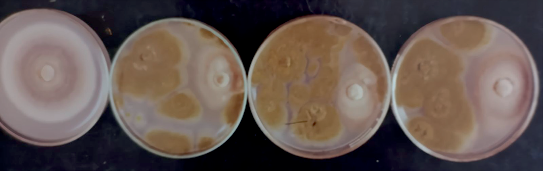
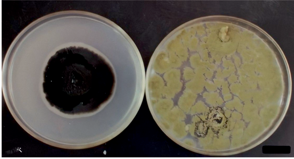
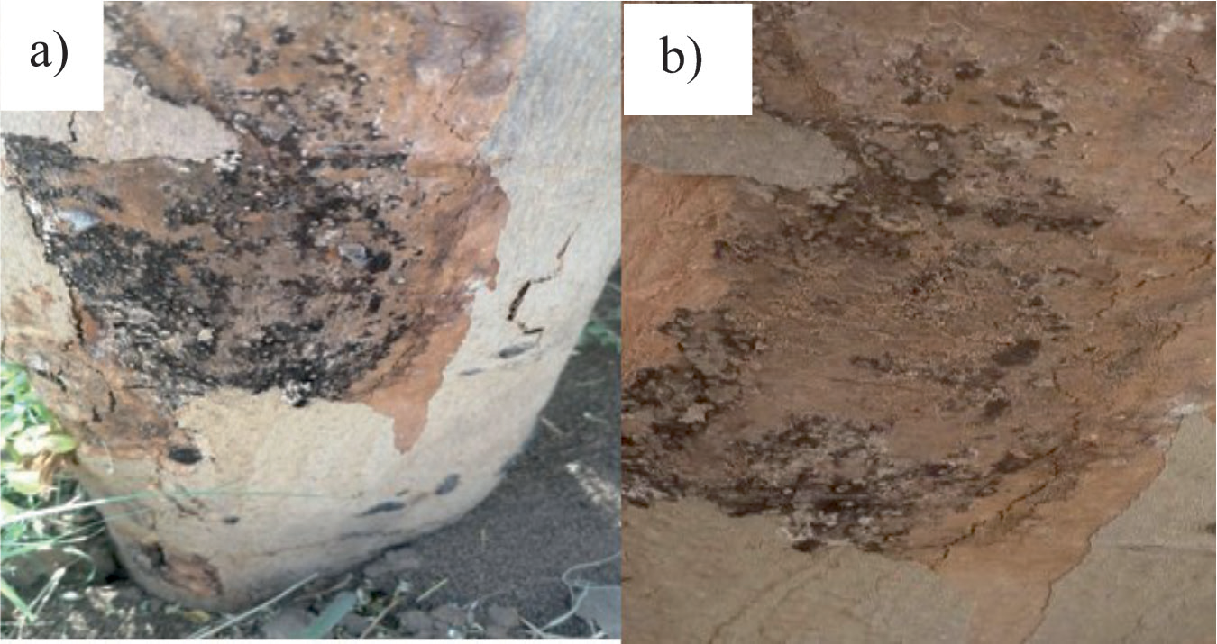
Table 3.
Effect of Trichoderma strain (TII) on the root growth of wheat.
| Treatments | Root dry weight (g) | Root Length/ Density (cm/cm3 ) | |
|---|---|---|---|
| T1 | NS | 0.6 1.7 1.38 | 7.10 14.49 9.4 |
| T2 | NS | 0.3 0.6 0.7 | 6.6 10.1 5.6 |
| T3 | NS | 0.3 0.1 0.36 | 3.4 1.8 2.2 |
| T4 | NS | 1.9 1.4 1.7 | 7.3 9.9 15.3 |
| CV% | 0.5 | ||
| SE± | 0.8 |
Table 4.
Effect of Trichoderma strain (TII) on the root growth of Zea mays.
| Treatments | Root dry weight (g) | Root Length/ Density (cm/cm3 ) | |
|---|---|---|---|
| T1 | NS | 2.6 2.2 3.3 | 7.96 6.36 6.33 |
| T2 | NS | 0.9 1.2 1.7 | 5.1 6.1 4.2 |
| T3 | NS | 2.2 1.5 0.8 | 4.5 5.5 3.4 |
| T4 | NS | 3.9 3.1 3.5 | 8.1 8.5 6.6 |
| CV% | 0.5 | ||
| SE± | 0.8 |
Several strategies have been used to obtain new and better antagonistic strains of Trichoderma with enhanced properties. The ability of Trichoderma spp. strains to make cell wall degrading enzymes (CWDEs) is part of what makes them useful as biological control agents in biotechnology. Early attempts to improve strains focused on making more of different CWDEs. In recent years efforts have been directed toward obtaining strains with altered intracellular signaling pathways. These pathways control the broader aspects of mycoparasitism, so mutants show more exciting and informative phenotypes than those that over-express CWDEs. In addition, some strains isolated by classical genetics have good antagonistic properties.
Potential sources of significant antimicrobial compounds against gram-negative and -positive bacteria, fungi, and yeast include Trichoderma species [20]. Trichoderma strains’ extracted peptides demonstrated antibacterial efficacy against S. aureus earlier in 1995 [21]. T. harzianum produced 44.06 μg/mL of the well-known antifungal drug, cyclosporine. A novel diterpene with an unusual fused 6-5-6-6 ring structure, known as Trichodermanins C-E has also been discovered from a fungus. T. harzianum “Cytotoxicity assay using three cancer Cell lines showed significant activity in Yamada et al. (2005) [22].
Several research findings have been reported that Trichoderma species control various diseases of different crops. Trichoderma species may be a source of significant antibacterial compounds effective against gram-negative and gram-positive bacteria, fungi, and yeast [20]. The well-known antifungal medication cyclosporine was generated at 44.06 g/mL by T. harzianum [16]. A complex network of multiple processes creates the triangle between plants, Trichoderma, and pathogens. Trichoderma spp. Function as powerful, inexpensive, and environmentally benign biocontrol agents in addition to being successful plant symbiotic organisms. They have little effect on soil equilibrium, can establish themselves in a variety of pathosystems, and do not harm beneficial species that aid in the control of diseases. The symbiotic relationship between plants increases plant resistance to diseases, enhances growth and productivity, and encourages nutrient uptake and fertilizer use efficiency.
Conclusions
In conclusion, Trichoderma harzianum strains showed antagonistic and biocontrol effects against wood decay pathogens. However, the TII isolate of T. harzianum was more efficient than the TI and TIII in retarding the growth and sporulation of pathogens. The findings presented in this study indicate that Trichoderma could be used as a safe, environmentally acceptable, and effective biocontrol agent for many crop species.