

Introduction
Microplastics (MPs) are a group of emerging contaminants and contaminants of global concern (United Nations Sustainable Development Goal 14 target 14.1.1) due to their persistence in the environment, relatively small size (below 5mm), and ability to adsorb other pollutants on their surface, such as various chemicals, Persistent Organic Pollutants (POPs), and bacteria [1, 2]. Ever since Prof. Ronald Thompson coined this term in 2004, these small-sized plastic fragments have been found in almost every ecosystem and within living tissues, including the human placenta [3], blood [4], and brain [5], being some of the recent discoveries. The sources of the MPs are varied, including direct manufacturing of cosmetics, pre-production pellets, etc.; degradation of large plastic products, microfibers shed from textiles and ropes, etc.; and wearing of tires and road paint particles [6-11]. With increasing demand and hence production of plastic goods, it has been observed that these non-degradable polymers are becoming a major part of our solid waste [12]. Annually, 8 million metric tons of plastic are dumped in our oceans every year [13], of which 236,000 tons are microplastics [14]. There is an estimation that by 2050, global plastic production could reach up to 33 billion tons [15, 16]. Due to their low density and small size, MPs are the major cause of long-range transportation in the environment [17]. Being ubiquitous in the environment, these MPs have been known to be ingested by various life forms, with adverse effects also reported [18-20]. Moreover, small MPs may reach higher trophic levels due to relatively large amounts of prey ingestion [21- 23]. With their ability to adsorb other pollutants on their surface and their synergistic effects, MPs are also thought to disturb the biological processes in aquatic organisms, such as absorption, metabolism, excretion, and distribution, which can result in unpredictable environmental damage, clogging of the digestive system, internal injuries such as ulcerative lesions, perforated gut, gastric rupture, alteration of the immune system, enzyme activity, oxidative stress, hepatic stress, cellular necrosis, and possibly lead to death [15, 24- 30]. Microplastics can bioaccumulate in various vital parts of the mussels and oysters, including the gills and gut tissues [31-34], creating tissue damage and strain comebacks [35]. Similarly, neurotransmitter enzymes may be inhibited, as observed in the brains of red tilapia (Oreochromis niloticus) exposed to polystyrene microplastics (PS-MPs) at concentrations of 100, 10, and 1 μgL−1 [36]. Likewise, zebrafish (Danio rerio) exposed to high concentrations of polystyrene microplastics (2000 μgL−1, 200 μgL−1, and 20 μgL−1) induced a free radical of reactive oxygen in high quantity, leading to oxidative abnormality inside the liver [37].
While all the studies cited above so far report a significant level of damage to animal health, multiple questions need to be identified. First, what could be the possible daily exposure level to MPs for the observed species? Secondly, how much MP exposure should be considered sufficient to cause adverse impacts such as oxidative stress, DNA damage, alteration in gene expression, and so on? Environmental exposure assessment may vary from species to species, while a proportion of ingested MPs may be removed from the body through defecation, making such assessments trickier. Thirdly, different plastics have different chemical compositions. Moreover, different additives are introduced to these polymers to achieve desired characteristics for specific functions, making each type unique. How can we determine which type is more toxic than others? To add to these questions, some studies found no adverse impact of MPs, such as those of [11] and [38]. A review by [39] also highlights these questions, as half of the studies on MPs reported no adverse impact. In this study, Polyvinyl Chloride microplastics (PVC-MPs) were administered to mice mixed in their basal diet in varying doses, and their impact upon the integrity of DNA and gene expression for two genes, OGG1 (8-Oxoguanine DNA Glycosylase) and HPRT1 (hypoxanthine phosphoribosyl transferase 1), was noted.
Materials and Methods
Experimental Setup
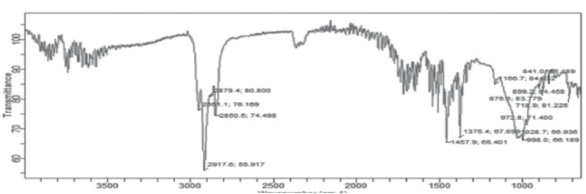
The current study was carried out at the Department of Wildlife and Ecology, University of Veterinary and Animal Science, Ravi Campus Pattoki, Pakistan. The source of PVC-MPs for the study was a mat (locally called ‘chatai’) used in many households in Pakistan for sitting on the floor. These mats gradually disintegrate over time into smaller fragments. These fragments were collected with the help of a brush and passed through sieves with a mesh size of 0.5 mm and 0.3 mm. The largest fragments were found to be in the size range of 0.3 mm and hence selected for this study. The major functional group of the polymer fragments was confirmed through FTIR to be vinyl chloride with absorption bands obtained at 3500-1000 cm-1 (Fig. 1). In FTIR, vibrational absorption bands are used for the identification of specific chemical structures and the quantitative measurement of key chemical compositions [40]. Absorption peaks ranging between 3500-1000 cm-1 support PVC identification.
Healthy adult female albino mice (Mus musculus) were purchased from the animal breeding house at the University of Veterinary and Animal Sciences, Lahore, Pakistan. Female mice were selected for this trial as they were a more relevant model focusing on estrogen-dependent DNA repair mechanisms and investigating reproductive toxicity as estrogen levels influence OGG1 expression. They may exhibit different genotoxic responses to PVC-MPs, particularly in genes involved in DNA repair. OGG1 and HPRT1 are sensitive markers for detecting DNA damage and repair and their relevance to human health. The experiment was run after receiving the approval certificate from the ethical committee for animal use (vide letter no. DR/27/19-01-2023). The trial animals were screened and checked thoroughly for any kind of disease or physical injury, and only healthy mice were selected for experimental work. The experiment was conducted in a controlled environment with room temperature (23+05oC), 50-70% relative humidity, and a regulated light and dark schedule [41]. A total of n=75 mice of similar average body weight (30+05 g) were selected and acclimatized for a period of one week. During this phase, standard feed and distilled water were provided to the experimental animals. During acclimatization, a standard pelleted feed with crude protein 19.05% was given to the mice. The feed was ground into a powder form for the experiment and divided into five batches. Polyvinyl chloride (PVC-MPs) was added to this feed in proportions described below and again shaped into pellets (15 mg each) by adding some water and stored in pre-labeled bags for the experiments. Doses were prepared following [42]. After acclimatization, the mice were randomly distributed into five groups designated as T0, T1, T2, T3, and T4, each with three replicates.
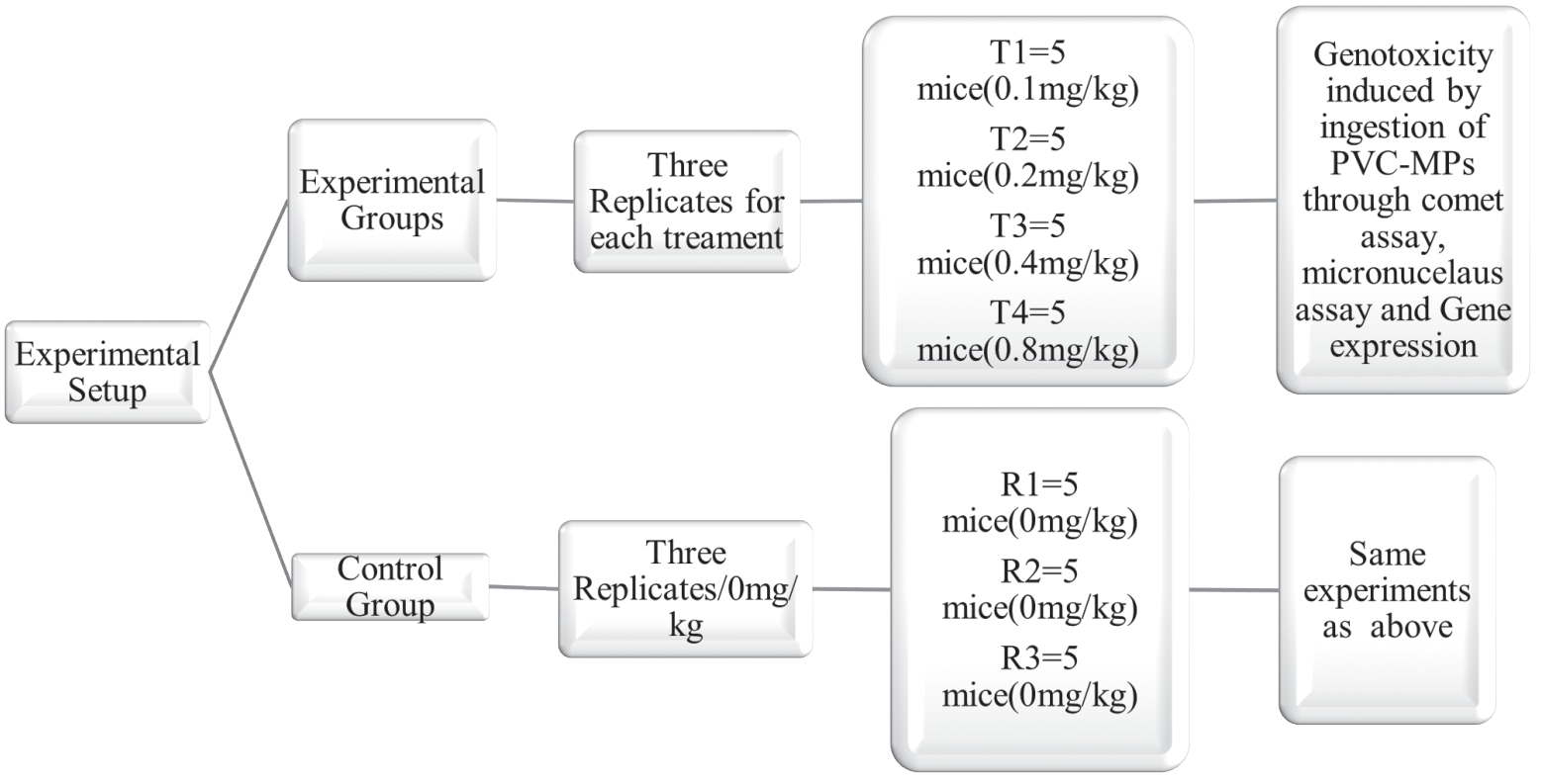
T0 was considered the control group, and no MPs were given. T1, T2, T3, and T4 were fed a basal diet mixed with 0.1, 0.2, 0.4, and 0.8 mg/Kg/BW of MPs, respectively. Body weight and mortality rates were noted regularly, and the mice were maintained for 35 days.
At the end of the trial, the animals were sacrificed, and liver and kidney samples were obtained to determine DNA damage through the comet and micronucleus assays. Gene expression through DNA damage-responsive genes was also determined (Fig. 2).
Micronucleus Assay
The micronucleus (MN) assay is one of the most widely used and reliable genotoxicity assessment tests [43]. The assay was performed following the protocol given by [44]. The treated cells were centrifuged with KCL (1%). The residual pellets were then centrifuged first with 50 mL of fixative (containing glacial acetic acid and methanol at 1:5) and 50 mL NaCl (0.9%) and later with glacial acetic acid (40 mL) and methanol (200 mL). Samples were loaded on pre-chilled slides in triplicate for each treatment and stained with Giemsa stain (10%) to identify and count micronuclei and binuclei under a microscope.
Comet Assay
The alkaline comet assay was performed following the method used by [45] and modified by [46]. Three slides were prepared for each treatment, and DNA damage was scored based on comet tail length. Liver and kidney samples (10 mg each) were taken, chopped finely, and then sieved. Tissue pellets were formed and resuspended in PBS. The treated cells were mixed with 1.5% LMA, and slides (pre-coated with 1% NMA) were prepared. Slides were chilled for five minutes and then subjected to a lysis buffer at 4oC for 1 h. Slides were then kept in an electrophoresis buffer solution for 20 min. Later, electrophoresis was carried out at 4°C for 20 min (25 mV and 300 Amp). After neutralizing with a buffer solution for five minutes, the slides were dyed with ethidium bromide (20 µg/mL) for easy visualization of DNA and observed under a fluorescent microscope at 40X. DNA damage was documented on a scale of 0-4, with 0 being no damage and 4 indicating maximum DNA damage. The following formula was used to calculate the DNA damage:
In this Equation, Ni represents the number of cells in a particular degree of damage, while i represents the degree of damage (0, 1, 2, 3, and 4).
Oxidative Stress
To determine oxidative stress, liver tissues were first homogenized, then centrifuged (8000 rpm, 10 min), and the supernatant was collected to determine physiological indicators such as glutathione and malondialdehyde. All the physiological indicators were determined using commercially available kits (MDA, GSH assay kit) [47].
Table 1.
Pearson’s correlations between the measured parameters for toxicity assessment.
Table 2a).
DNA damage Index and fragmentation % observed in liver cells of experimental groups given different doses of PVC-MPs. Table 2b). DNA Damage Index and fragmentation % observed in kidney cells of experimental groups given different doses of PVC-MPs.
Sample Processing, RNA Extraction, cDNA Synthesis, and Real-time PCR Analysis
Tissue samples of liver and kidney (50 mg) were taken from the treatment and control groups, chopped completely with sterile blades, and transferred into an Eppendorf tube. Trizol reagents (1 ml) and proteinase k (30 µl) were added to the Eppendorf tube, thoroughly mixed in a vortex, and then incubated for 10 min at 37°C. After that, chloroform (200 µl) was added to the Eppendorf tube and inverted for at least 15-30 sec continuously, later incubated for 2 min at room temperature. Samples were then centrifuged at 14,000 rpm for 15 min, resulting in the formation of three layers. The upper transparent layer was taken. An equal amount of isopropanol was added, mixed vigorously, and incubated for 10 minutes at -20°C. The mixture was centrifuged for 15 min at 14,000 rpm at 4°C, and the supernatant was discarded very gently; then 1ml of 75% ethanol was added and centrifuged again (10,000 rpm, 15 min) at 4°C. Ethanol was discarded very gently, and the sample was completely air-dried. An Elution buffer was added and stored at -20°C for further analysis. Complementary DNA (cDNA) was made per the manufacturer’s instructions (Thermo Fisher Scientific Inc.). Briefly, reaction buffer 5x (4 µl), Ribolock (1 µl), 10mM dNTPs (2µl), Revertaid (µl), and reverse primer OGG1 (2 µl) were added to each tube, and RNA (2 µl) was added. The same procedure was repeated for the HPRT1 gene. Four standards and three controls were run (GAPDH was a positive control, no template negative control, and a negative control with no Revertaid). Temperature conditions for cDNA synthesis were 42°C for 60 min, then 70°C for 5 min. The Maxima SYBR Green kit was used for expression analysis; the kit was thawed at room temperature, SYBR Green master mix (12.5 µl), forward primer OGG1 (1 µl), and reverse primer OGG1 (1 µl) were added to each tube of the total sample tubes. Then, a cDNA sample (2 µl) and double-distilled water were added to make the 25 µl reaction mixture. The same procedure was repeated for HPRT1 gene expression analysis [48, 49].
Statistical Analysis
Data were analyzed statistically and represented in terms of mean ± standard deviation using SPSS (v 21.0, IBM, Inc.). Pearson’s correlation was performed to determine the dose–dependency of microplastics with micronuclei formation (α = 0.05).
Table 3.
Pearson’s correlation analysis between doses and measure of DNA damage in both liver and kidney cells.
Table 4.
Effect of different doses of PVC-MPs on oxidative stress enzymes.
Results and Discussion
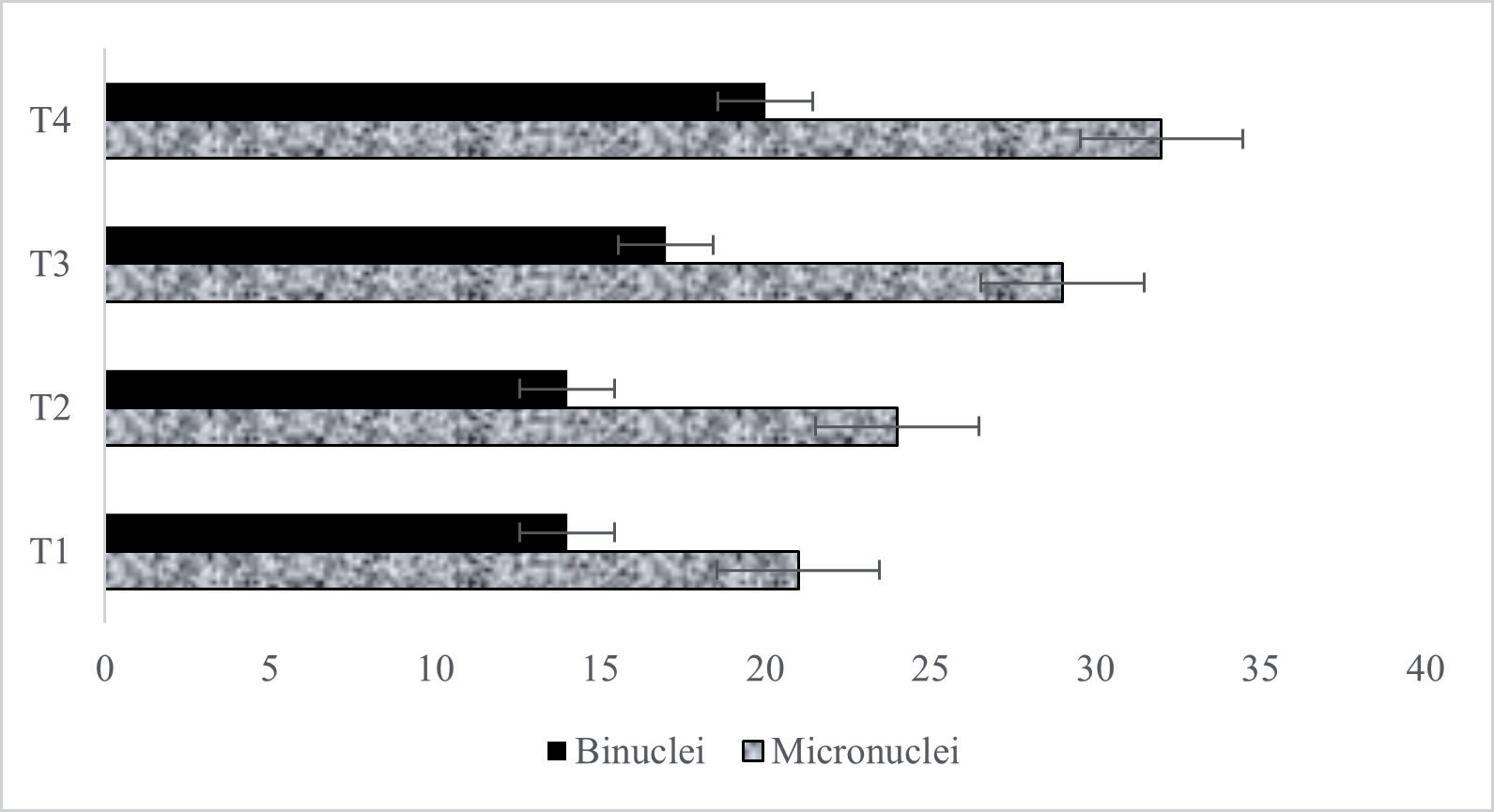
A dose-dependent increase in micronuclei and binuclei formation was observed (Fig. 3), which is indicative of increased genotoxicity with an increase in exposure to microplastics. A strong positive correlation was observed between the doses and BN formation (n = 4; r = 0.950), doses and MN formation (n = 4; r = 0.984), and between BN and MN (n = 4; r = 0.953) at a 0.05 significance level (Table 1).
Similarly, the outcomes of the comet assay indicated that the damage index increased with higher numbers of microplastics added to the feed in both tissues. Fragmentation % was also increased (Tables 2a) and 2b)). A strong positive correlation was observed between the doses and damage index (n = 4; r = 0.987), doses and fragmentation % (n = 4; r = 0.964) in liver cells, and also between fragmentation % in both cells (n = 4; r = 0.963) (Table 3).
When the results were compared with control groups, PVC-MP toxicity inhibited the Glutathione (GSH) levels. PVC-MPs increase the production of reactive oxygen species (ROS), which leads to cell abnormalities and has a negative effect on the activity of glutathione. Table 4 shows a significant (p<0.05) decrease in glutathione (850±31.69) activities in all treated groups and a malondialdehyde increase (1576±23.63) in a dose-dependent manner as compared to the control group.
Gene Expression Analysis of the OGG1 Gene and the HPRT1 Gene
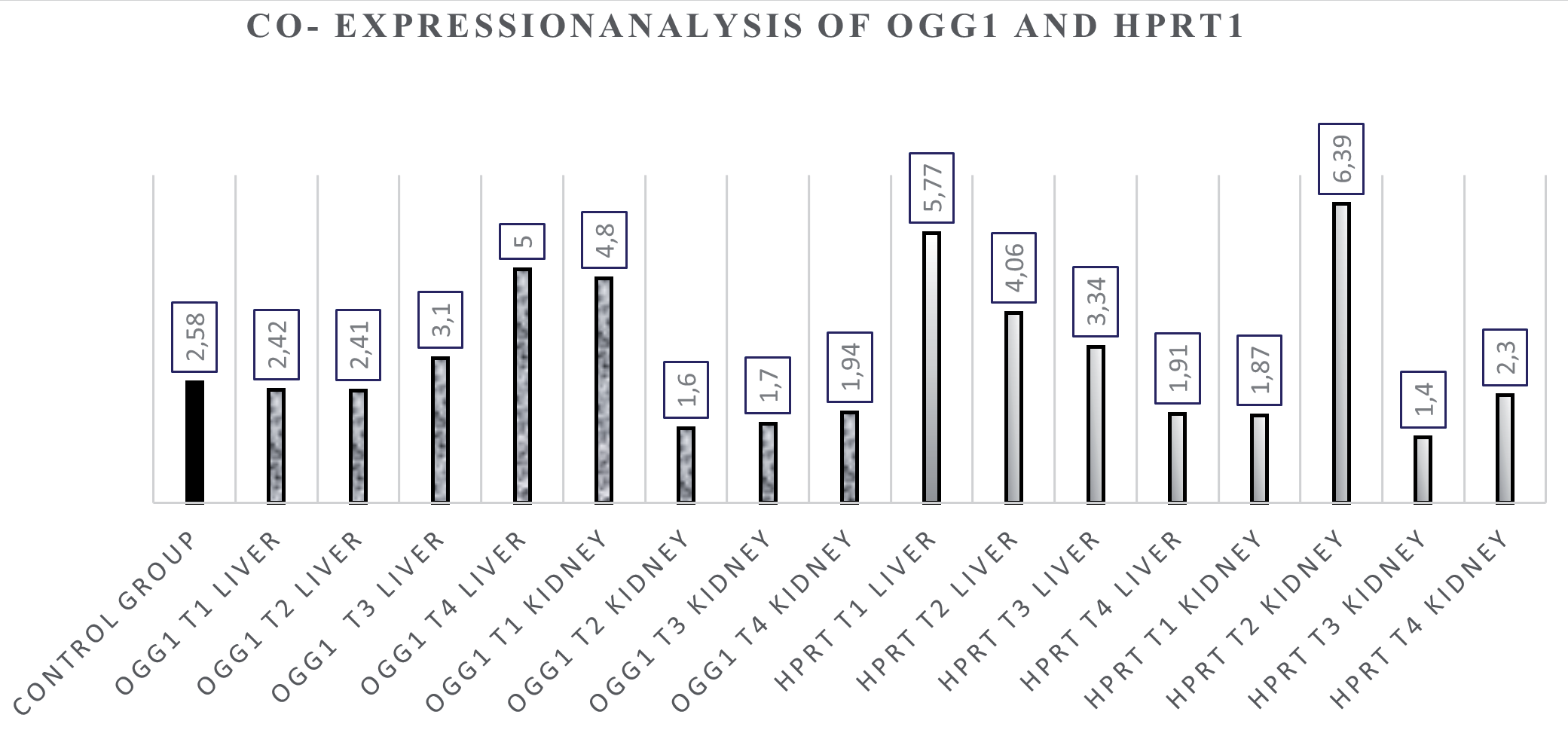
The OGG1 and HPRT1 gene expression levels in mice’s livers and kidneys among four treatment groups were also measured and compared. The co-expression analysis (Fig. 4) of OGG1 and HPRT1 in the liver sample shows an inverse relationship according to doses because OGG1 shows overexpression in high doses, and HPRT1 shows downregulation in high doses. HPRT1 shows a dose-dependent relation, and its expression decreases as the dose increases. Remarkable fold changes were observed between the low- and high-dose groups.
Microplastic exposure has been documented to induce toxicity in numerous aquatic life forms, while studies on terrestrial exposure and toxicity assessment are limited. Moreover, each microplastic type differs from others in terms of its chemical composition and additives added for different purposes, making it difficult to evaluate potential toxicity for each type. Since different plastic products have different chemical compositions, each may impact biological systems differently. PVC was selected for this experiment as it is the second most abundant microplastic type globally [29, 50] and is known to degrade and release toxic compounds such as vinyl and chloride, trimers, dimers, and monomer additives under different conditions [51]. As discussed earlier, the source of polyvinyl chloride for this study was a commercial product, a mat commonly used for sitting on the floor in many households across the country. The major reason for selecting this product was the fact that it keeps on disintegrating into smaller fragments over time and usage, frequently releasing secondary microplastics into the environment.
In the present study, healthy adult female albino mice were exposed to PVC-MPs of different doses (0.1, 0.2, 0.4, and 0.8 mg/Kg) for five weeks to analyze their toxic effects through various parameters including comet assay, micronucleus assay, oxidative stress, and gene expression. The comet assay is a widely accepted technique used for the determination of single and double DNA [52], whereas micronuclei formation also shows a reliable method for the detection of clastogenicity and aneugenicity [53, 54]. While the outcomes of the comet assay may not always correspond to MN formation when the extent of exposure is limited, as MN formation is indicative of damage occurring in at least one mitotic cycle, the comet assay is known to detect more damage than the micronucleus assay. Hence, it is important to employ both parameters as indicators of probable genotoxicity. MNs are formed due to damaged chromosome fragments that fail to incorporate into the nucleus during mitosis and are generally caused by the presence of some genotoxic agent. Hence, their formation is a simple yet effective parameter for genotoxicity screening in any living sample [43, 55].
Our results indicated that prolonged exposure to PVC did induce significant changes in DNA structure as the dose increased. The damage index value for each experimental group increased as the PVC-MP dose increased along with the fragmentation percentage (Tables 2a) and 2b)). As seen in Fig. 3, there was an increase in the number of binuclei and micronuclei formation as the dose increased, indicating increased chromosome instability. A strong positive correlation of 0.94 and 0.98 was found between the doses administered to the experimental groups and the number of MN and BN observed for each respective treatment.
Apart from DNA damage, reactive oxygen species (ROS) formation has also been reported in response to the administration of different types of microplastics in different animals [56-59]. An increase in malondialdehyde (MDA) levels in response to polystyrene exposure in Wistar rats has been observed, along with a decrease in activities of superoxide dismutase, glutathione peroxidase, and catalase [60]. The toxicity of the microplastic type is reflected by the formation of reactive oxygen species, which in turn leads to lipid peroxidation in the cells. MDA is an important product of lipid peroxidation, and its levels indicate the degree of stress caused by the toxic compound in question [59]. Our results also exhibited similar findings as MDA levels increased with an increase in dose, while GSH levels were found to be reduced, which is indicative of oxidative stress in experimental animals.
Toxic impacts of PVC have also been reported in earlier studies but in higher doses. For example, [61] exposed adult male mice to 100 mg/kg of PVC-MPs (2 µm size) for sixty days and observed a marked decrease in mucus secretions in the intestine along with an increase in intestinal permeability as a result of reduced mRNA expression related to the genes responsible for mucus secretions. In another related study, exposure to 0.5 mg/kg of PVC MPs for sixty days resulted in oxidative stress, marked hepatic damage, and changes in the gut microbiota of mice [62]. According to [63], genotoxic and cytotoxic damage was observed in the early juveniles of Oreochromis niloticus when exposed to different doses of microplastic (1 mg/L, 10 mg/L, and 100 mg/L). Likewise, PVC-MPs were reported to alter various metabolic functions in zebrafish larvae, like changes in triglyceride (TG), pyruvate, non-esterified fatty acids (NEFA), total cholesterol (TC), total bile acid (TBA), and glutamic acid (GLU) [64].
The combined effect of PVC-MPs and PS-MPs alters the function of genes that are responsible for lipid metabolism, oxidative stress, and signal transduction in mice. Moreover, PVC-MPs with HFD and ND caused gene expression changes responsible for antioxidant enzymes and immune functions [65, 66]. In addition, disturbances in offspring metabolism and abnormal expression were observed in 674 coding RNAs in blood tissue and other non-coding RNAs in mice, which indicates vascular dysfunction and protein degradation [67]. Recent results have shown that microplastics (MPs) damage the oocyte DNA of mice, including downregulation of action and defects in chromosome morphology [68]. In another study, higher concentrations of microplastics (180 mg/L) increased the DNA damage in fish and caused abnormalities in their gills [69]. Microplastics in combination with copper for 24 to 96 hours caused damage to DNA structure, while polystyrene nanoplastics in combination with carbamazepine significantly affected mussel gene expression. Microplastics with di-(2-ethylhexyl) phthalic acid were reported to increase the production of ROS in mice, which led to DNA oxidative damage, and in another study, the combined effect of microplastics with co-contaminants disturbed the mitochondrial gene function [68, 70-73].
Another parameter included in this study was determining the gene expression of DNA-responsive genes in experimental animals. RT-PCR was carried out to assess the expression of DNA-responsive genes, OGG1 (8-Oxoguanine DNA Glycosylase) and HPRT1 (hypoxanthine phosphoribosyl transferase 1). OGG1 (8-Oxoguanine DNA Glycosylase) gene is the basic gene responsible for DNA repairing function [74], while HPRT1 (hypoxanthine phosphoribosyl transferase 1) is responsible for the production of hypoxanthine phosphoribosyl transferase 1 enzyme, which plays a central part in purine recycling [76]. Our results indicated the expression of the gene’s inverse relation with the given doses. The gene expression of HPRT1 in low-dose (T1, T2) and (T3, T4) high-dose groups was down-regulated when compared with the control group, while upregulation of OGG1 was reported. A similar study conducted by [77] indicated that PVC-MPs induce downregulation in the BCL-2 liver gene of mice and upregulation in the caspase-3 gene. Our results showed similarity to the study conducted by [78], which reported downregulation of BCL-2 gene expression in testicular cells of male albino mice when exposed to different concentrations of PS MPs. A study encompassing the impact of arsenic exposure on different industrial workers also indicated alterations in the expression of OGG1 and HPRT1 genes, which consequently led to oxidative stress and negatively affected purine recycling and DNA damage [77]. While arsenic is a pollutant whose exposure pathways may differ from that of microplastics in our study, the similarity in ultimate outcomes as a result of exposure to potentially hazardous compounds cannot be ignored and needs to be investigated further.
Conclusions
The outcomes of the current experiment conclude that chronic exposure to microplastics can affect animal health and cause genotoxicity. Moreover, while our doses may not correlate with the actual exposure, they still provide insights into how living systems may respond to constant exposure to these persistent and ubiquitous micro-pollutants. However, since microplastic composition, size, and shape are highly variable in the environment, further research is still needed, and investigations should be conducted into important factors that may influence ecosystem dynamics and toxicity assessments.

