

Introduction
Environmental contamination has surfaced as a prominent obstacle for modern society [1], stemming from the swift industrial growth, rising energy needs, and negligent depletion of natural reserves, leading to its escalation over recent decades [2]. Both natural and human-caused sources are constantly releasing different toxic organic and inorganic contaminants into the land and aquatic ecosystem. Heavy metals count among them, and because of their toxic features, perseverance, ability to build up in living organisms, and a tendency to accumulate in the food chain, they have been highlighted as powerful biological poisons that considerably contribute to environmental contamination [3]. The poisoning by heavy metals in aquatic environments is regarded to be harmful to humans and aquatic creatures [4].
Heavy metals are metallic elements that are toxic or dangerous even in small quantities and have a relatively high density (<5 g/cm3) [5]. Heavy metals can easily infiltrate in aquatic environments and eventually reach the bodies of aquatic species [6]. In accordance to [7], cadmium (Cd), lead (Pb), and mercury (Hg) are three of the ten heavy metals that are harmful to human health. Lead is used as a production material for batteries, solder cables, anti-rust coatings, and paint-making materials. Cadmium is widely used as a pigment in the paint industry, stabilizer in the manufacture of PVC, in the manufacture of batteries and ceramics, as well as in iron coatings to be stainless steel [8]. Mercury is a non-essential metal that gets released in the surrounding environment due to natural (earth’s crust) and anthropogenic (fossil fuels combustion and mining) processes [9, 10].
Fish biomarkers represent a valuable approach for monitoring the stress induced by harmful metals. They gauge cellular, bimolecular, and physiological alterations in response to intoxication resulting from exposure to heavy metals [11]. The assessment of blood and its constituents is a key component of hematological analysis, encompassing different blood indices [12]. Biochemical parameters have been widely utilized to evaluate the adverse impact of contaminants in the environment. These factors encompass the levels of electrolytes and metabolites [13]. Heavy metals are also known to be carcinogenic and potentially genotoxic. Reactive oxygen species (ROS) are produced when heavy metals stimulate oxidative stress, which can lead to DNA damage and cell death [14].
Numerous harmful metals are contaminating water and air, endangering the health of hundreds of millions of organisms around the world. Another risk to human and animal health is heavy metal contamination of food [15]. These heavy metals can bioaccumulate and have a wide range of harmful consequences on different bodily tissues and organs. These metals interfere with a variety of cellular functions, such as apoptosis, growth, proliferation, differentiation, and damage-repairing mechanisms [7]. Similar methods of action, such as hemato-biochemical reactions and genotoxic modification in grass carp, are shown by comparing the mechanisms of action of these metals. Severe reactions following high-dose exposure result in increased DNA damage and neuropsychiatric disorders [16]. There is an abundance of information on the influence of heavy metals on the physiology of many fish species, and much of the literature focuses on the effects of certain heavy metals on a small number of physiological characteristics specific to a given fish species [17]. A thorough analysis of every heavy metal is necessary to guarantee a healthy environment for food security and safety, given the detrimental effects of heavy metals on fish and eventually humans. Therefore, it was planned to investigate more about the harmful impacts of specific heavy metals, i.e., Cd, Pb, and Hg on Grass carp physiology, particularly on blood chemistry and molecular changes at the DNA level.
The Grass carp, scientifically known as Ctenopharyngodon idella, is an herbivorous freshwater fish that falls under the Cyprinidae family. It is simple to upkeep in laboratory settings, holds significant commercial value in managing aquatic vegetation, and can serve as an effective indicator for appraising water quality [18]. Our study was devised to examine the hematological, biochemical, and genotoxic consequences triggered by selected heavy metals in Grass carp. As of now, this is the first research work that determined the impacts of cadmium, lead, and mercury salts on Grass Carp.
Materials and Methods
Ethical Approval
The present research was carried out in the Department of Zoology (Fisheries and Aquatic Toxicology Laboratory), Hazara University Mansehra. The ethics of animal handling were strictly followed, as suggested by the Institutional Bioethics Committee (IBC) of Hazara University Mansehra, Pakistan.
Fish Collection
The C. idella specimens, ranging in size from 4 to 5 inches, were collected using a manual net from Hattian fish hatchery Kamra. They were subsequently transported to the zoology laboratory at Hazara University Mansehra in an oxygenated polythene bag for subsequent examination. A 0.2% KMnO4 solution was administered for two minutes to eliminate any external infections, followed by a rinse with tap water.
Acclimatization
Before commencing the experiment, a two-week acclimatization phase was carried out with all the fish placed in a glass aquarium with a water retaining capacity of 60 liters. All the water quality parameters were agreed to international standards of water quality suitable for freshwater fish species. Standard conditions were maintained throughout the experiment. Commercial fish feed of 30% protein was given daily at 2% of their body weight. Oxygen was regulated through aerators, and fecal material was removed regularly to avoid ammonia toxicity. Inspection of each aquarium was done on a daily basis to find any mortality and clinical ailments.
Concentration of Heavy Metals
Analytical grade heavy metals in the form of salts were used in the present study, i.e., mercury as (HgCl2), lead as (Pb(NO3)2), and cadmium as (CdCl2) were purchased from (Merck, Germany) through a local distributor. For the experimentation, the LC50-96h value was initially ascertained for each metal. Over a 96-hour period, toxicity tests for cadmium, lead, and mercury were determined using varied concentrations of Cd (10, 30, 50, 70, 90 mg/l), Pb (20, 40, 60, 80, 100 mg/l), and Hg (02, 04, 06, 08, 10 mg/l).
Experimental Design
After the acclimatization period, fish species were then allocated to four groups (E0-E3) randomly, at which a set of 30 fish were introduced in 3 replicates (10 fish/ replicate). Based on 96h LC50 calculated value for cadmium, lead, and mercury (23.84 mg/l, 38.01 mg/l, and 3.38 mg/l), a sub-lethal concentration of 5.7 mg/l for cadmium (E1), 7.6 mg/l for lead (E2), and 0.6 mg/l for mercury (E3), corresponding to 1/20th of 96-h LC50 used for this study for 1 week. Group E0 was used as a control group with non-exposed healthy fish for the entire experimentation. A semi-static system was employed, with the test solution being replaced every 24 hours. To evaluate the general health, behavior, and any alterations in response to heavy metal toxicity in fish, daily observations were made.
Hemato-Biochemical Analysis
After one week of experiments, the fish from both the control and exposed groups were carefully taken from the aquariums and then exposed to clove oil anesthesia. After that, blood was collected from the caudal vein of the fish with sterilized 2ml syringes containing heparin as an anticoagulant. The sample was taken and placed into the Tubes containing EDTA. Total erythrocyte and leukocyte count, hemoglobin, hematocrit, and platelet values were examined by using a hematology analyzer (Sysmex, Kx21). Packed cell volume (PCV) was determined through the micro-hematocrit technique. Documentation of erythrocyte indices, including mean corpuscular volume (MCV), mean corpuscular hemoglobin (MCH), and mean corpuscular hemoglobin concentration (MCHC), was accomplished through a specific process [19]. The serum has been employed to assess a comprehensive range of biochemical indicators, encompassing electrolytes and metabolites. Metabolites include triglycerides, protein, glucose, cholesterol, serum creatinine, blood urea, and uric acid, and serum electrolytes such as serum calcium, phosphorus, iron, sodium, and potassium were also identified in the bloodstream, utilizing a biochemical analyzer.[20].
Genotoxicity Assessment
The blood was analyzed by Single Cell Gel electrophoresis technique (Comet assay) for DNA damage. Comet assay performed on fish blood was adopted according to the described protocol [21]. Blood samples were tempered by utilizing saline solution. Slides were made using 10 µl of saline solution and 120 ml of low-melting agarose (0.5%) at 37°C. Slides in the lysis solution (1 mL of Triton X-100, 10 mL of DMSO, and 89 mL of lysing solution stock, pH 10.0 – stock solution: 2.5 M of NaCl, 100 mM of EDTA 100, 10 mM to 1 L of Tris) were refrigerated for 60 minutes. After 60 minutes, the slides were retained for 20 minutes at 25 V and 300 mA in a horizontal electrophoresis configuration. Slides were neutralized for approximately 15 minutes with 0.4M Tris (pH 7.5) and then stabilized in ethanol for 10 minutes. Cells without DNA damage exhibit homogeneous movement, but those with DNA damage exhibit fragments of different masses, and smaller cells move more quickly during electrophoresis, like a comet’s tail.
Statistical Analysis
We analyzed the data by using statistical tools, such as SPSS (version-24). A t-test was conducted to compare the control and exposed groups, whereas a one-way ANOVA was performed to analyze all exposed groups. For creating graphs, the software GraphPad Prism (version 9) was used.
Results
This study examined the toxic effects of selected heavy metals Cd, Hg, and Pb on the well-being of C. idella fish by assessing its genotoxic and hemato-biochemical biomarkers. The findings of this current investigation are outlined in this study.
Hematological Responses
In a recent study, we investigated the exposure to specific heavy metals affecting the hematological parameters of the fish C. idella. Alterations in the hematological profiles were observed, indicated by the changes induced by exposure to the toxic substances. Specifically, the blood indices of the experimental groups displayed a significant decline (p < 0.05) in the levels of WBCs, RBCs, platelets, Hb, lymphocytes, and MCHC while there was a significant rise (p < 0.05) in the levels of MCV and MCH in C. idella, in comparison to the control group.
Biochemical Indices
Compared to the untreated or control group, the treated groups exhibited changes in various biological parameters after exposure to specific heavy metals. There was an increase in blood glucose, cholesterol, creatinine, blood urea, and uric acid levels, alongside a decline in proteins across all treated groups in comparison to the untreated group. Additionally, changes in electrolyte values, including phosphorus, iron, calcium, potassium, and sodium were noted.
Fig. 1.
Biochemical Parameters in C. idella exposed to selected heavy metals in contrast with control. Graphs representing hematological parameters with symbols * (p0.05) and ** (p0.00000) denotes significant deviation (p < 0.05) between the control and experimental groups of C. idella, (A) RBCs count (106/µl), (B) Hemoglobin (gd/l), (C) PCV (%), (D) MCH (pg), (E) MCV (fl), (F) MCHC (g/dl), (G) Platelets (103), (H) WBCs count (106/µl), (I) Lymphocytes (%), (J) Neutrophils (%), (K) Monocytes (%).
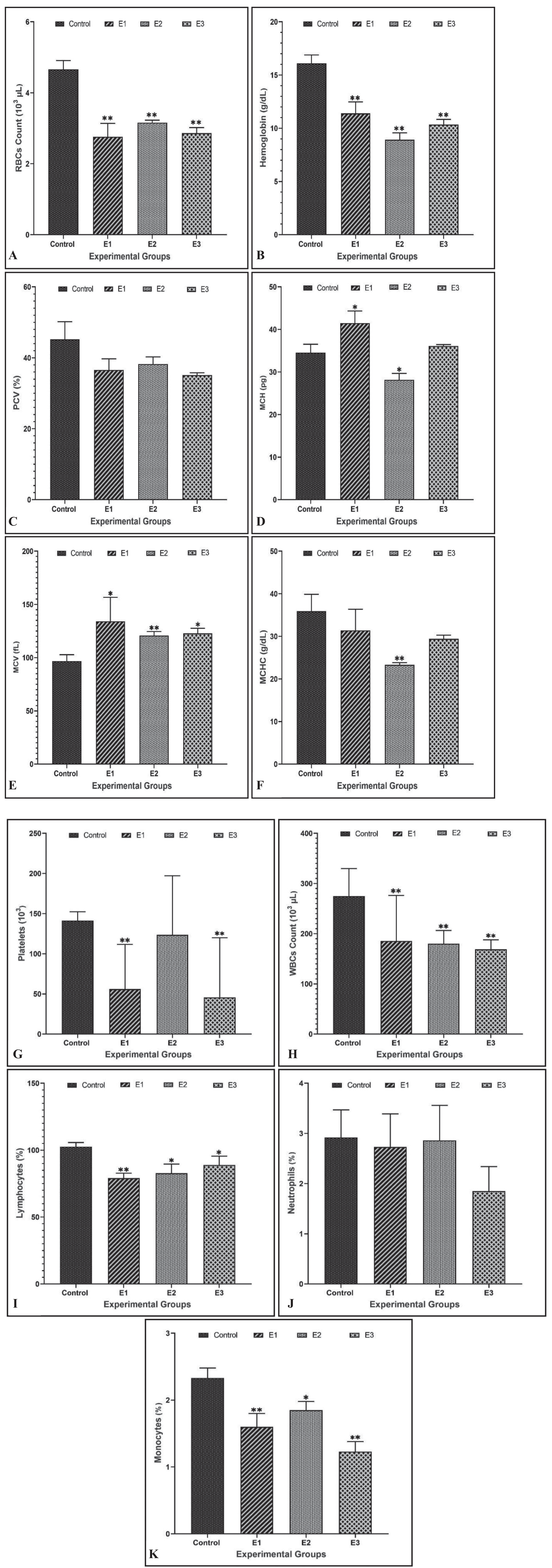
Table 1.
Biochemical Parameters in C. idella exposed to selected heavy metals in contrast with control.
DNA Damage
The intensifying concerns about the toxic effects of heavy metals compelled scientists to investigate the genotoxic effects of selected metal ions on fish. The genotoxicity of cadmium, lead, and mercury in erythrocytes of C. idella was analyzed using single cell gel electrophoresis technique (Comet assay). After the exposure of C. idella to different concentrations of selected metal ions, a significant (p < 0.05) higher frequency of erythrocytic DNA damage was observed in the following trend Pb > Hg > Cd in all treated groups compared to the control group. Details of DNA damage that occurred in treated groups are presented in Fig. 2 and Table 2.
Discussion
In the present study, various hematological indices demonstrated a significant decrease in the number of RBCs, Hct, and Hb in fish when exposed to different metals compared to the control group. The distribution and binding of oxygen in fish blood are reflected in the amounts of Hct and Hb. Our results are consistent with the research of [22], who found that silver carp treated with mercuric chloride had lower hemoglobin, red blood cell counts, and HCT. Similarly, [23] noted that exposure to salts of copper and cadmium caused a drop in red blood cell count, hemoglobin, and hematocrit in C. catla. Zulfahmil I. et al. [24] examined a significant decline in Hb and RBC in Chanos chanos after being subjected to lead nitrate, which concurs with our observations. Hematocrit values typically decline when a fish experiences reduced appetite or stress. The noticeable decrease in hematocrit and hemoglobin values suggests a mechanism of hemodilution resulting from impaired osmoregulation or gill damage. Our results further align with previous research conducted by various scholars, as observed in the works of [25, 26].
The hematological indices, such as MCH, MCV, and MCHC, offer additional insights into the size, relationship, shape, and Hb content of erythrocytes. Moreover, these indices serve as standards for the morphological examination of anemia [27]. The hemoglobin to erythrocyte count ratio is represented by MCH. The MCV value can indicate the efficiency of RBC production throughout the process of erythropoiesis. Our research showed that when grass carp were exposed to particular metals, their total MCH and MCV values rose substantially, while their MCHC values declined. Parallel findings were observed in the milkfish subjected to lead nitrate [24]. Regenerative anemia brought on by erythrocyte hemolysis may be the cause of elevated MCH as well as MCV values. The study on common carp conducted by [28] similarly reported a remarkable decrease in MCHC. The same results in C. batrachus after exposure to arsenic were also reported by [29].
Fig. 2.
DNA damage in peripheral erythrocytes of C. idella due to metals exposure. Representing peripheral erythrocytic cells (A) control-regular cell (B) Cd (5.77 mg/L) exposed (C) Hg (0.67 mg/l) exposed (D) Pb (7.6 mg/l) exposed.
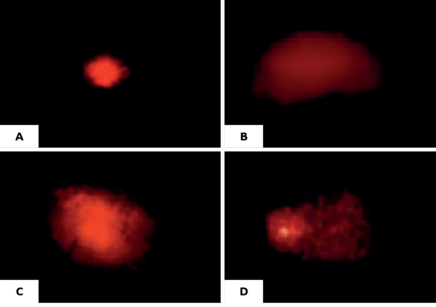
Table. 2.
DNA damage in peripheral erythrocytes of C. idella due to metals exposure.
The white blood cells (WBCs) play a critical role in the cellular and immune systems. Our findings indicated a reduction in WBCs and lymphocyte counts compared to the untreated group. Similarly, [25] observed a significant decrease in WBCs and lymphocyte counts in fish exposed to lead, chromium, and copper, compared to the control. Ergönül M. et al. [30] reported similar results, mirroring our findings. The decline in WBC values may signify an impaired immune system in the fish, as suggested by [31]. In the current investigation, when C. idella was subjected to particular heavy metals, the number of platelets was reduced in the treatment groups relative to the untreated group. The decline in the number of platelets suggested a possible hemodilution mechanism that was probably brought on by gill damage or impaired osmoregulation. Our results confirm those of [32], who subjected C. punctatus to heavy metal exposure.
Blood serves as a comprehensive indicator of the overall body’s health. Changes in different hematological measures signify the response to pollution and can function as a method for the early identification of particular issues related to aquatic pollution [33]. Research on various fish species indicates that heavy metals can impact the biochemical traits present in tissues and blood. Alterations in tissue or blood composition may induce changes, leading to an elevation or reduction in the observed values. Blood glucose emerges as a reliable and sensitive marker for identifying environmental stress caused by pollutants in fish. In the conducted experiment with C. idella, there were significant increases in blood glucose levels. Our study demonstrates similarities to the investigation conducted by [34], in which the Caspian roach was subjected to lead exposure. Islam S.M. et al. [35] documented the occurrence of hyperglycemia in Pangasianodon hypophthalmus exposed to chromium. Cholesterol serves as a crucial structural element in membranes and acts as a precursor to all steroid hormones. The concentration of cholesterol is considered an indicator of environmental stress caused by heavy metals [36]. Our results demonstrated a rise in cholesterol levels across all experimental groups compared to the control. The heightened cholesterol levels induced by heavy metals could potentially be attributed to liver failure, subsequently leading to an elevation in serum concentration. Comparable outcomes were achieved in the case of C. batrachus subjected to arsenic exposure, as documented by [29]. Additionally, Tabat J.L. et al. [37] observed increased levels of glucose and cholesterol in catfish fingerlings when exposed to cadmium.
Triglycerides primarily function as a cellular energy source and can be employed to evaluate nutritional status. A recent study identified a decrease in triglyceride levels, indicating liver damage induced by oxidative stress. Heydarnejad M.S. et al. [38] documented comparable findings, indicating a reduction in triglyceride levels in rainbow trout treated with cadmium. Ali Z. et al. [39] demonstrated a decline in triglyceride levels in C. carpio following exposure to manganese and chromium, exhibiting similarity with our research findings. The levels of tissue proteins serve as an indicator of stress induced by xenobiotics in aquatic organisms. In all groups treated with metals, there was an observed reduction in the entire protein content of the experimental fish’s blood serum. Conditions, including liver cirrhosis, renal nephrosis, or alterations in enzymatic activity necessary for protein synthesis, could be the cause of the drop in plasma protein levels. Similar results in C. batrachus exposed to arsenic trioxide were reported by [40]. The results of our research align with those of [37], who observed similar outcomes. In this study, after being exposed to various heavy metals, the experimental fish C. idella showed higher blood urea, serum creatinine, and uric acid levels than the untreated group. Fluctuations in levels of creatinine are a sign of renal failure. These results are in alignment with the findings reported by [41], who found that C. punctatus exposed to mercuric chloride had higher levels of creatinine. According to research by [42], C. gariepinus subjected to cadmium showed elevated uric acid levels. Ali Z. et al. [39] identified a rise in uric acid and urea after manganese and chromium treatment in C. carpio. Our results are consistent with those of [41], who examined the effects of mercuric chloride on C. punctatus.
The presence of electrolytes in the bloodstream serves as a bioindicator of the health and physiological condition of fish. Exposure of grass carp to cadmium, lead, and mercury resulted in observable changes in electrolyte levels. There was a decrease in calcium and phosphorus levels, accompanied by an increase in sodium, potassium, and iron levels. The exposure of C. idella to selected heavy metals induced hypophosphatemia and hypocalcemia. This result is parallel to the study conducted by [43], who also found hypophosphatemia and hypocalcemia in H. fossilis subjected to lead. Increased sodium and potassium activity were shown in grass carp following exposure to zinc and mercury, as reported by [44]. It was observed that the groups that received heavy metal treatment had higher iron concentrations. The study of [45], which found elevated iron levels in common carp subjected to harmful heavy metals, lends credence to our findings.
Comet test was used to identify metal-induced substantial changes in DNA damage levels. Several genetic pollutants can be efficiently screened using this method in the peripheral blood of various fish species [46]. DNA damage significantly increased in C. idella peripheral blood erythrocytes during the current experiment as metal concentrations increased. In terms of the percentage of erythrocytic damaged comet cells, the different metals’ abilities to damage DNA followed the following sequence Pb>Hg>Cd. Similar results of DNA damage were reported in C. idella induced by metals such as Pb, Cr, and Cu [47]. Kousar S. et al. [48] reported the comparable genotoxic effects of different metals, like Cu, As, and Zn in peripheral erythrocytes of four freshwater fish species viz. L. rohita, C. mrigala, C. catla, and C. idella. Due to their nucleated form, erythrocytes were chosen as the ideal study material for fish DNA damage, even though the damage may also be found in the tissues of the fish’s gills and kidneys [49]. Moreover, metal-induced DNA damage raised severe concerns about the possibility that metals could endanger grass carp’s ability to survive in their native aquatic environments.
Conclusions
In conclusion, anthropogenic activities and manufactur-ing firms are the leading contributors to heavy metal toxicity in aquatic ecosystems. Heavy metals can easily infiltrate aquatic environments and eventually reach the bodies of aquatic species. Fish are a vital source of animal-based protein and come into close contact with heavy metals in the aquatic environment as a result of pollution. The results of our study indicate that grass carp are sensitive to the harmful effects of mercury, lead, and cadmium on their hematological, blood-biochemical, and genotoxic markers. Within the realm of environmental biomonitoring, the factors examined in this research can function as anticipatory markers for evaluating the influence of heavy metals on both fish and other aquatic organisms.

