

Introduction
Arthrospira platensis is a species of cyanobacteria; one of its synonyms is Spirulina platensis [1]. It has czylgz@imau.edu.cn a long history of being used as a food supplement and has gained considerable popularity in the human health food industry due to its therapeutic properties, including antioxidant, anti-inflammatory, immune-modulatory, and anticancer activities [2-4]. More recently, the United States Food and Drug Administration (FDA) has classified A. platensis as “Generally Recognized as Safe (GRAS)” [5], and the National Aeronautics and Space Administration (NASA) has successfully used it as a space travel nutritional supplement for astronauts [6]. A. platensis is both biologically and economically significant due to its numerous applications in the pharmaceutical, biofuel, cosmetics, wastewater treatment, bioremediation, agricultural industries, and biofertilizer industries [7-13]. Because of its nutritional, environmental, and social significance, researchers are campaigning for its extensive production globally [4].
A. platensis was discovered in Lake Bayannur and other nearby alkaline lakes on the Erdos Plateau of China [14]. It had previously been found in tropical and sub-tropical countries in Asia, Africa, North America, and South America [6]. It has been generally accepted that A. platensis is a thermophilic alga, as the optimum temperature for its growth falls between 35ºC and 37ºC [15], and the highest cell dry biomass was obtained at the optimal temperature of 35ºC [16]. In contrast to these conditions, Lake Bayannur and other adjacent alkaline lakes on the Erdos Plateau are situated at latitudes ranging from 39°01 N to 39°23’N in an arid to semi-arid temperate region. The Erdos Plateau experiences four distinct seasons: winters are long and cold, while summers are short and hot. Air temperatures can vary significantly from day to night. The average annual temperature is as low as 6.4ºC, while lows can reach -31ºC and highs can reach 36.7ºC [17]. A. platensis from Lake Bayannur has, over millennia, adapted to these variations in temperature. Gao and Qiao revealed that the optimal temperature for the growth of A. platensis from the Erdos lakes is about 24ºC, far below that reported by Kumaresan [16, 17]. It can survive freezing at -18ºC for 24 h, and slow growth is maintained even when temperature decreases suddenly from 35ºC to 13ºC, meaning that this strain of A. platensis can be regarded as a low-temperature strain [17]. Additionally, the low rainfall, long light exposure periods, and easy availability of carbon in natural alkaline lakes promote the growth of A. platensis [18]. These advantages all make the Erdos Plateau of China a suitable place for A. platensis cultivation.
Knowledge of A. platensis growth is essential for understanding its physiology and yield period. Temperature and light are the main factors affecting growth in outdoor ponds [19]. Tanaka et al. clarified that the low night temperature reduced the net productivity of A. platensis, and night temperature should be managed in outdoor cultivation based on the permissible dark-period temperature range [20]. There have been reports published on the morphology and ultrastructure of A. platensis [6, 11, 21, 22]; in particular, the structural characteristics of A. platensis from Erdos lakes at room temperature (20ºC to 25ºC) were investigated by Tian et al. [21]. Light is known to regulate morphological development and photosynthetic performance. There have been papers published on the influence of low temperatures on the biochemistry and physiology of A. platensis. Growth-temperature stress causes biochemical changes in the cells and a reduction in biomass yield. [20, 23].
It is common knowledge that morphology and anatomical structure are adapted to physiological function. While 4ºC cold-stressed Klebsormidium crenulatum only showed minor ultrastructural alterations, drastic changes to the cell wall and organelle distribution were found in frozen samples (-2ºC and -4ºC) [24]. The morphological and anatomical responses of A. platensis to low-temperature stress have not been adequately elucidated. This study aimed to add to the limited knowledge about the Erdos lakes strain of A. platensis by examining changes in its morphology and anatomical structure as temperature decreased from 5ºC to -5ºC. It was hypothesized that, since A. platensis from Erdos lakes can survive at a latitude of 39°N, the dynamic changes in the morphology and ultrastructure might support its physiological adaptation to cold temperatures. The response of anatomical structure and ultrastructure to temperature stress could be beneficial to exploring low temperature thresholds suitable for production.
Materials and Methods
Materials
A. platensis, a kind of blue-green algae (BGA), belongs to the cyanobacteria phylum (Fig. 1). This cyanobacterium grows as filamentous, helicoidal trichomes, performs oxygenic photosynthesis, and reproduces through binary fission [22]. It prefers tropical or subtropical waterbodies with high carbonate/ bicarbonate content, salinity, and pH, which may not support other plants. Sunlight, CO2, and mineral elements are directly used by the algal cells for growth. Environmental factors affect the growth and biopigment accumulation of microalgae, including nutrient availability, high pH, light, salinity, and temperature [16]. A. platensis is a cyanobacterium with a high economic value and is nowadays one of the most important industrially cultivated microalgae [22].
The strain of A. platensis used in these experiments was obtained from a water bloom in Lake Bayannur, Erdos Plateau, China, then separated and purified.
Methods
Algal Culture
Purified samples were cultured in Zarrouk’s medium with intermittent aeration at room temperature (20ºC to 25ºC) under natural light [25].
Low Temperature Treatment
Algae in the logarithmic growth period were sampled and divided into three groups. Group 1 was placed at 5ºC, group 2 was placed at 0ºC, and group 3 was placed at -5ºC. There were three replicates in each group, and 50 ml of algae samples were allocated. All samples were placed under fluorescent light of 1200 lx intensity with 12 h off and 12 h on. The duration of the experiment was 5 days.
Light Microscope (LM)
After the low-temperature treatments, algae were placed on a microscope slide and immediately observed and photographed using an Olympus BX41 light microscope (Olympus Optical Co. Ltd., Tokyo, Japan) at 100x, 200x, and 400x magnification. For each temperature treatment, trichomes of different morphology were counted in the same field of view (0.95 mm2). A total of 60 observations, the number of replicates, were made at random for each treatment.
Transmission Electron Microscope (TEM)
After low-temperature treatment, cultures were sedimented with a 5 minute centrifuge step at 1500 rpm, and much of the water was removed. A volume of 1 mm3 of the resulting algal pellets was fixed for 2 h at 4ºC in 3% glutaraldehyde-Na phosphoric acid buffer (0.1 mol L–1, pH 7.2). The pellets were then rinsed three times at 15-minute intervals in phosphoric acid buffer (pH 7.2). Post fixation was carried out in 1% osmium tetroxide for 1 h, after which the pellets were again rinsed 3 times at 15-minute intervals in a phosphoric acid buffer (pH 7.2) and dehydrated in an ethanol series starting at 30% and gradually rising to 100% at 10 min intervals. Specimens were then embedded in propylene oxide-Epon 812 (EM bed-812, Electron Microscopy Science, Hatfield, PA, USA) for 24 h at 37ºC, polymerized at 60ºC for 48 h, and sectioned using an ultra-thin microtome (LKB 2088 Ultrascan XL, Bromma, Sweden). The sections (50 nm) were stained using uranyl acetate-lead nitrate. Thin sections and whole mounts were viewed with a transmission electron microscope (TEM, H-700H; Hitachi, Ibaragi, Japan) and photographed.
Results
Morphology
The morphology and quantity of algal filaments were different under the same treatment conditions (Fig. 1). When A. platensis was observed following 5ºC treatment, abnormal as well as normal intact trichomes were detected. Abnormal trichomes took varying forms; some were non-intact, some were arc-shaped with differing lengths, and some consisted of short, straight segments with only a few cells (Fig. 1a, b). In the comparison of A. platensis trichome morphology and quantity in each field of view (0.95 mm2) with an LM following differing temperature treatments (Table 1), the number of intact trichomes was greater than that of other forms (p<0.05). The number of non-intact trichomes was similar to that of arc-shaped trichomes and was significantly greater than that of straight and short segments (p<0.05). Observations about the forms and quantity of trichomes made following treatment at 0ºC were similar to those after treatment at 5ºC (Fig. 1c, d). The number of individuals with different forms in each field of view changed similarly as well. It was noteworthy, however, that the arcs of the arc-shaped trichomes were generally shorter at 0ºC with a lower degree of arc; segments also tended to be shorter in length.
At -5ºC, trichomes became yellow-green, and almost all became either arc-shaped or were broken into short segments with few cells. Arc-shaped trichomes were even shorter in length than at 0ºC. Intact and non-intact trichomes in the process of disintegration were observed (Fig. 1e-h). At this temperature, it was almost impossible to see intact trichomes, and there were a few non-intact trichomes in the field of view. The number of arc-shaped trichomes was not significantly different than that of straight and short segments (p>0.05) and was significantly greater than that of intact and non-intact trichomes (p<0.05).
With the decrease in temperature, the morphology and quantity of A. platensis changed with regularity (Table 1). There was no significant difference in the number of each type of trichome between treatments at 5 and 0ºC (p>0.05). The numbers of intact and non-intact trichomes at -5ºC were much less than those at 5ºC and 0ºC (p<0.05), while the numbers of arc-shaped trichomes and straight and short segments were the highest.
Ultrastructure
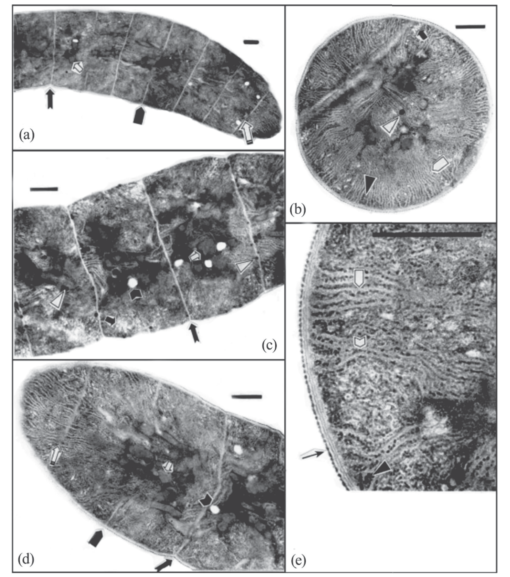
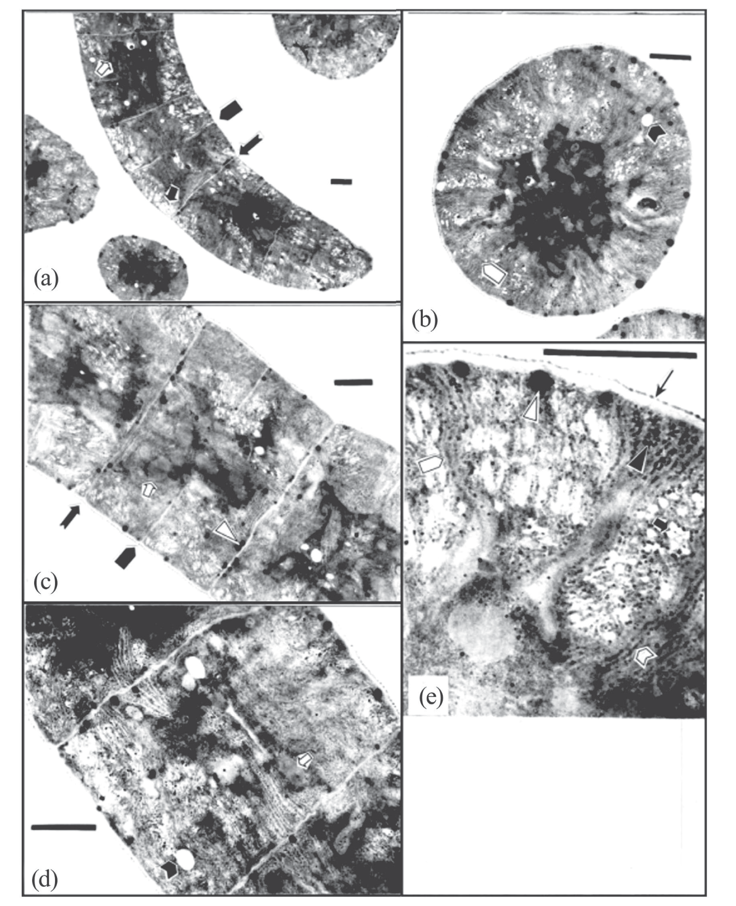
Algae subjected to 5 and 0ºC low-temperature treatment contained abundant, parallel, clearly discernible thylakoids (Fig. 2b, e, and 3b, e) mainly distributed in peripheral areas of the cells; in the cross-section, they were radiate (Fig. 2b and 3b). Phycobilisomes were visible on the outer surface of the thylakoid membranes (Fig. 2e and 3e). Contoured carboxysomes with sharp edges and corners were mainly distributed in the central areas of the cells; some were also present adjacent to the cross walls, but none were detected near the longitudinal walls (Fig. 2a, c, d, and 3a, c, d). In the longitudinal sections, cross walls showed slight constriction (Fig. 2a, c, d, and 3a, c). There were plasmodesmata between adjacent cells (Fig. 2a, d). In the cross sections, longitudinal walls were clear and had demarcated layers (Fig. 2e and 3e). The newly-formed cross walls of cells undergoing division commenced formation at the longitudinal walls, divided the peripheral area into two parts, and gradually stretched to the center (Fig. 2a, d, and 3a, c). There were polyglucan granules in the peripheral areas (Fig. 2b, e, and 3e), and gas vacuoles were mainly distributed in the peripheral areas. They were cylindrical with diameters of 54-67 nm and were arranged in parallel to form a honeycomb-like pattern (Fig. 2b, c, and 3a, e). Vacuoles had no definite structure, and their sizes were random (Fig. 2c, d, and 3b, d). Lipid bodies (droplets) were distributed in both the peripheral and central areas, with concentrations near the plasma membranes (Fig. 2b, c, and 3c, e).
Fig. 1.
LM micrographs of A.platensis at low temperature. (a, b) A.platensis at 5ºC; (c, d) A.platensis at 0ºC; (e-h) A.platensis at -5ºC. Scale bar represents 75 µm.
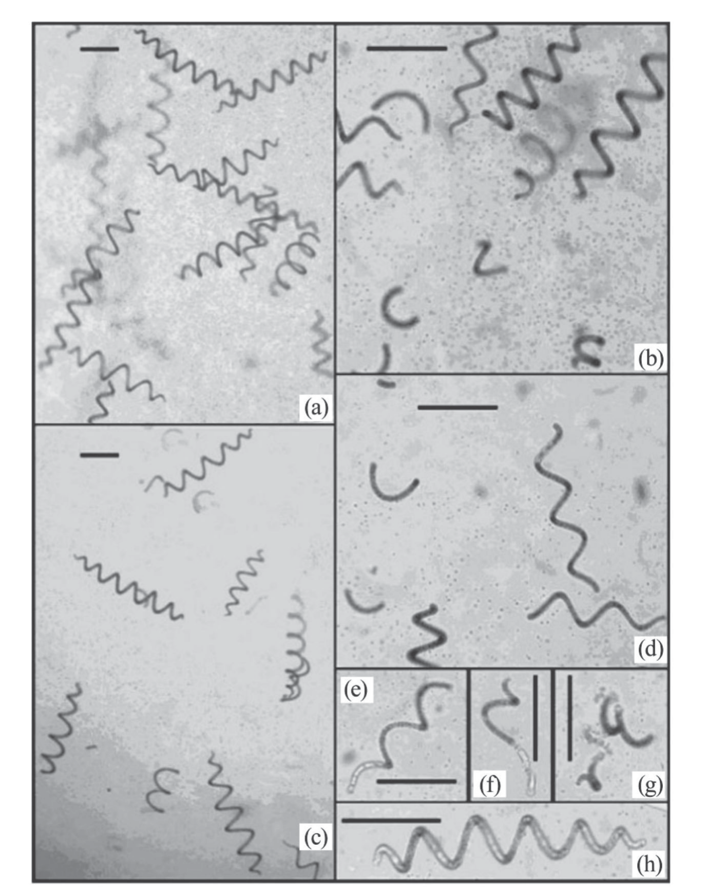
Table 1.
Comparison of A. platensis trichome morphology and quantity in each field of view (0.95 mm2) with LM following differing temperature treatments.
Fig. 2.
TEM micrographs of A. platensis treated at 5ºC. (a, c, d) Longitudinal sections; (b, e) Transverse sections.  : thylakoids,
 : carboxysome,
 : gas vacuoles,
 : vacuole,
 : longitudinal wali,
 : phycobilisomes,
 : plasmodesmata,
 : cross wall,
 : newlyformed cross wall, △ : lipid body (droplet), ▲: polyglucan granules. Bar = 1 μm.
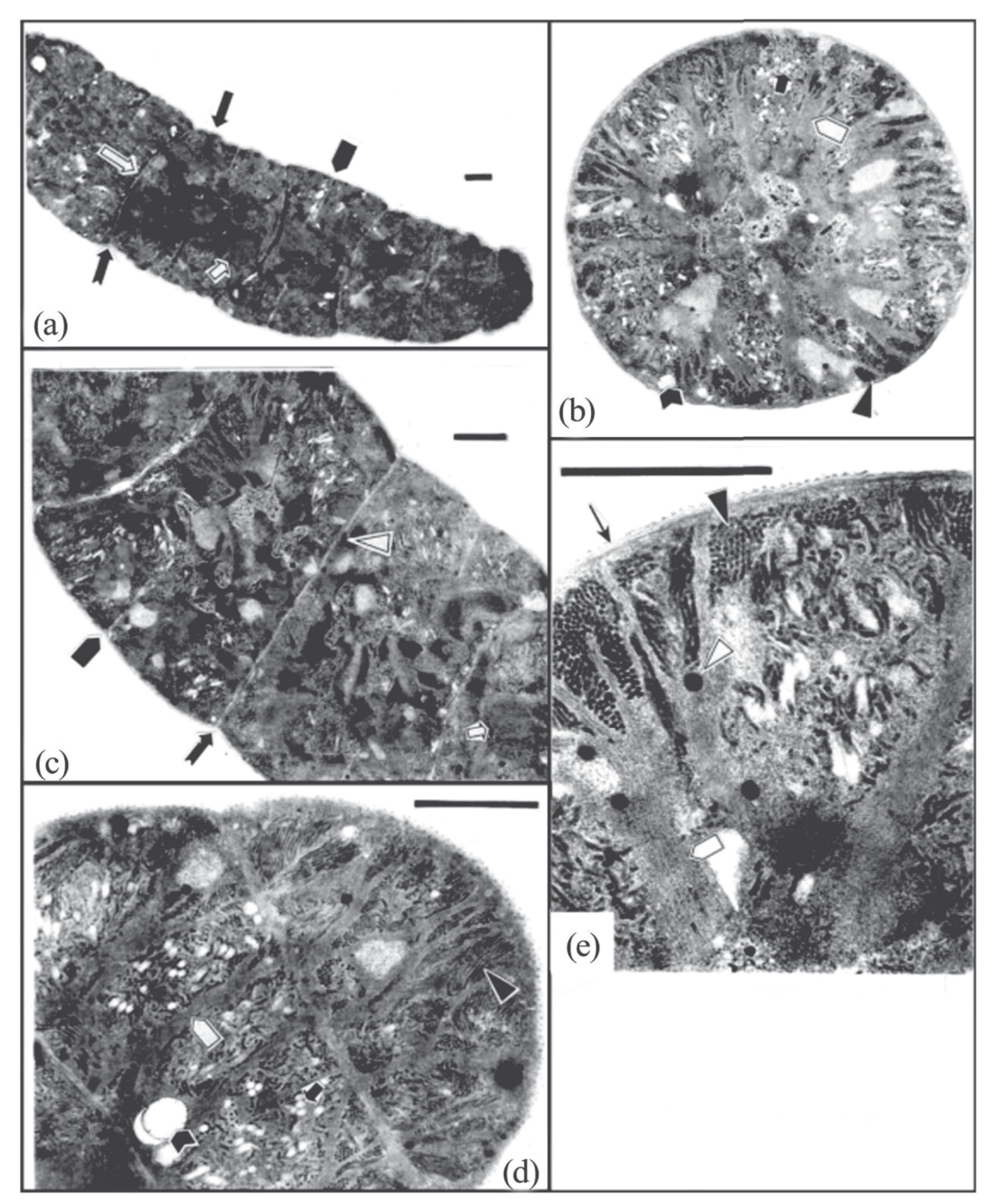
The longitudinal walls of the algal cells subjected to -5ºC were, for the most part, wave-shaped, while the cross walls were arc-shaped (Fig. 4a); newly formed cross walls, however, were nearly straight (Fig. 4a, c). The layers of the longitudinal walls were clear, but their thickness had decreased compared to cells treated with higher temperatures (Fig. 4e). The peripheries of carboxysomes were somewhat indistinct, and their edges and corners were not sharp (Fig. 4a, c). The clarity of the thylakoid membranes was lower, and phycobilisomes on the membranes had disappeared to such an extent that membrane layers were close to each other (Fig. 4b, d, e). There were more polyglucan granules in the cells, which were mostly distributed in peripheral areas; they were rod-shaped and of differing lengths, with diameters of about 27 nm (Fig. 4b, d, e). Honeycomb-like gas vacuoles and unstructured vacuoles were observed (Fig. 4b, d), but in lower quantities than the higher-temperature treatments. Lipid bodies were smaller than those in the 5ºC and 0ºC groups (Fig. 4c, e).
Discussion
All three groups of algae continued to divide during treatments at temperatures of 5, 0, and -5ºC (Fig. 2a, 3a, and 4a). Cells do not generally divide at such low temperatures, but the lowest temperature for the growth and reproduction of A. platensis from Erdos Lakes is about 4ºC [17]. It was hypothesized, therefore, that the fact the algae were collected during a period of logarithmic growth accounts for the observed cell division below the lowest minimum temperature for growth. The aforementioned period witnessed a phase of robust cellular proliferation and reproduction, where the algae continued to undergo cell division despite being exposed to low temperatures.
Fig. 3.
TEM micrographs of A. platensis treated at 0ºC. (a, c, d) Longitudinal sections; (b, e) Transverse sections.  : thylakoids,
 : carboxysome,
 : gas vacuoles,
 : vacuole,
 : longitudinal walil,
 : phycobilisomes,
 : cross wall,
 : newly-formed cross wall, ▲ : polyglucan granules, △ : lipid body (droplet). Bar = 1 μm .
Mühling et al. [26] revealed that subjecting three strains of Arthrospira to a temperature increase from 30ºC to 34ºC for 7 days resulted in a significant alteration in helix orientation. Under specific circumstances, the helical filaments of Spirulina transform atypical morphologies such as irregularly curved and linear shapes [27]. Tian et al. [21] discovered that, as A. platensis’ trichomes became dehydrated, their coils were either constricted in a portion of the spiral or became loose. In the present study, short filaments of algae increased in number as temperature decreased; most of these short filaments were arc-shaped or were quite small in size. At -5ºC, there were some abnormal shapes because of disintegration. The above-mentioned results lead to the conclusion that the morphology of A. platensis’ trichomes changes as environmental conditions change.
Overall, there were no differences in either the anatomic structure or morphology of the Erdos Lakes strain of A. platensis at 5ºC and 0ºC. When the temperature dropped to -5ºC, however, there were distinct changes in cell wall structure, thylakoid membranes, carboxysomes, polyglucan granules, and lipid bodies.
Fig. 4.
TEM micrographs of A. platensis treated at -5ºC. (a, c, d) Longitudinal sections; (b, e) Transverse sections.  : thylakoids,
 : carboxysome,
 : gas vacuoles,
 : arc-shaped cross wall,
 : vacuole,
 : wave-shaped longitudinal wall,
 : longitudinal wall,
 : newly-formed cross wall, ▲ : lipid body (droplet), △ : polyglucan granules. Bar = 1 μm.
The decrease in temperature likely led to alterations in cell walls. A temperature decrease would cause metabolic disorder in trichomes, causing catabolism to take place at a faster rate than anabolism; trichomes would have had to rely on consuming their own material (including cell walls) to survive. A decrease in the quantity of cell wall material, accompanied by a decrease in free water, caused cell walls to thin. When the temperature drops below 0ºC, living tissue freezes, and cells are damaged or die. The observed alterations in the cell walls of A. platensis at -5ºC were likely induced by ice crystallization. The freezing point of extracellular liquid is higher compared to intracellular fluid [28], and the formation of extracellular ice leads to dehydration [24]. Thus, as temperature decreased to -5ºC, liquid outside cells froze before liquid in the cells. As liquid outside cells froze, vapor pressure dropped, and, via exosmosis, water migrated from inside the cells, resulting in cell shrinkage or distortion. Mechanical strain was placed on cell walls, causing them to become wave or arc-shaped.
Transmission electron microscope examination of cells from each temperature treatment did not reveal distinct differences in the ultrastructures of plasma membranes. The work of Zhang on A. platensis from Erdos Lakes demonstrated that plasma membrane H+-ATPase activity as well as fatty acid composition and content changed considerably as temperature decreased [29]. Zhang found that, as temperature decreased from 15 to -5ºC and incubation proceeded from one to seven days, H+-ATPase activity at first increased and later decreased. Zhang compared the change in H+-ATPase activity of A. platensis from Erdos lakes to that of A. platensis from Lake Chad and found the change in H+-ATPase activity of the Erdos Lakes strain to be similar to that observed in Lake Chad A. platensis under the same experimental conditions, with the following exception: at -5ºC, H+-ATPase activity of the Lake Chad A. platensis was zero, while activity in the Erdos Lakes strain was still evident. Plasma membrane saturated fatty acid content of both the Erdos Lakes and Lake Chad. A. platensis strains decreased as temperature decreased, while, conversely, unsaturated fatty acid content increased. In the two strains, the increase in unsaturated fatty acid content was caused by different fatty acids; in the Erdos Lakes strain of A. platensis, flax acid (18:3), and palmitoleic acid (16:1) increased, while in Lake Chad, A. platensis, linoleic acid (18:2), and oleic acid (18:1) increased. Flax acid, with its relatively large number of unsaturated links, and palmitoleic acid with its relatively short carbon chain, have greater resistance to cold and are more effective in maintaining membrane fluidity than linoleic and oleic acid. Zhang concluded that the Erdos Lakes strain of A. platensis has greater resistance to cold than the Lake Chad A. platensis strain.
Phycobilisomes, thylakoids, and carboxysomes are structures that have a bearing on A. platensis photosynthesis. Phycobilisomes are the main collectors of light energy [30]. They transmit captured light energy to chlorophyll on the thylakoid membranes so that photosynthesis can take place. Transmission efficiency is high when phycobilisomes are intact; when phycobilisomes are in a state of disintegration, however, transmission slows and may stop [31]. Thylakoids are not only carriers of chlorophyll and carotenoids; they are also the venue for photosynthetic electron transfer and photophosphorylation. Carboxysomes play a crucial role in the dark reaction of photosynthesis. In cyanobacteria, carboxysomes function as the central CO2-fixing organelles that elevate CO2 levels around encapsulated Rubisco to enhance carboxylation [32]. Abnormalities in the structures of phycobilisomes, thylakoids, or carboxysomes directly affect photosynthesis.
This study showed that, as temperature decreased, A. platensis phycobilisomes disintegrated or disappeared altogether, thylakoid membrane layers became less distinct as the distance between them decreased, and carboxysome shapes became abnormal; because of this, photosynthesis weakened to the point of cessation. The degradation of phycobilisomes occurs rapidly upon nitrogen depletion, following a complex genetic program, as observed in previous studies [31]. Lipid bodies were a mode of fat storage used by A. platensis. Under low-temperature stress, survival was difficult for trichomes, and they were forced to consume stored fat (lipid bodies). Lipid body diameters, therefore, gradually decreased in size. The findings offered cellular-level evidence supporting previous research indicating that the lipid composition and fatty acid profiles of algae exhibit temperature-dependent variations [33]. The present study demonstrated that, as temperature decreased, A. platensis’ polyglucan granules increased in number. The increase was particularly noticeable at -5ºC. Such conversion enables trichomes to better resist cold, and it is one of the reasons why this strain of A. platensis can survive in the alkaline lakes on the Erdos Plateau, an area with a typical temperate arid to semi-arid climate. These observations help confirm that the A. platensis found in the alkaline lakes on the Erdos Plateau is a strain adapted to low temperatures.
In comparison to the ultrastructure at room temperature (20 to 25ºC), lipid bodies in the Erdos Lakes strain of A. platensis decreased in number and size at low temperatures (5 to -5ºC), while the number of polyglucan granules increased. The main storage material of the algae changed with temperature; lipid bodies were the main storage material at room temperature, while polyglucan granules became the main storage material, with lipid bodies becoming subsidiary at low temperatures. This is consistent with the results of a study that showed that the concentration of soluble sugar in A. platensis cells and the rate of sugar synthesis both increased as temperature decreased from 5 to -5ºC. [34]. In general, the overall lipid content of green microalgae tends to decrease when temperature is increased [35].
Algae generally float. This experiment revealed, however, that the Erdos Lakes strain of A. platensis sank to the bottom of its container at low temperatures (5 to -5ºC) over 5 days. It is generally thought that gas vacuoles are mainly responsible for sinking and floating; however, there were no distinct differences observed in the number of gas vacuoles between cells incubated at room temperature and at low temperature. It was hypothesized that the decrease in lipid body number and increase in polyglucan granules as temperature decreased might have played an important role in the sinking or floating of algae.
Conclusions
The findings of this study indicate that the anatomic structure and morphology of the Erdos Lakes strain of A. platensis did not change significantly as temperature decreased from 5ºC to 0ºC. An increase in polyglucan granules as temperature decreased was observed, which likely enabled the Erdos Lakes strain to survive in cold water. This study provides evidence that A. platensis, found in the alkaline lakes of the Erdos Plateau, is a typical low-temperature strain. It also offers valuable insights into the management of criticality at low temperatures in the industrial cultivation of the Erdos lakes strain of A. platensis.