

Introduction
Maize (Zea mays L.) is a vital crop globally, contributing significantly to food security and agricultural economies [1]. Maize is the preferred staple food for over 900 million poor consumers, 120- 140 million poor farm families, and about one-third of malnourished children [2]. Many studies have been conducted to perform and improve maize yield under natural and stressful conditions [3-5]. Maize production faces various challenges from various biotic and abiotic factors [6-9], with late wilt disease (LWD) caused by Magnaporthiopsis maydis being one of the most devastating diseases affecting maize fields worldwide [10]. Late wilt disease was identified in Egypt in 1960 and has since spread to several countries, impacting maize production on a significant scale [11]. It was caused by Magnaporthiopsis maydis (formerly known as Cephalosporium maydis and Harpophora maydis), which caused a serious threat to maize plants due to its ability to reduce crop yield, quality, and grain quantity [12]. The pathogen M. maydis survives in the soil as sclerotia and infects maize seedlings through roots or mesocotyl, causing root necrosis and impairing sprout development [13]. The disease is characterized by rapid wilting of maize plants, particularly after tasseling and shortly before maturity, leading to substantial yield losses [14]. Symptoms include dull green leaves, yellow-brown discoloration of vascular bundles, and hollow lower stems [15]. Managing LWD involves understanding host-pathogen interactions, environmental factors, and disease prevention strategies. While chemical and biological fungicides offer control options [16], genetic resistance and tolerant maize varieties are effective long-term solutions [17]. However, the presence of aggressive M. maydis strains and environmental influences can affect resistance levels [18]. Efforts to manage LWD include genetic resistance, cultural practices, and biological control methods. Genetic resistance through the development of resistant germplasm and tolerant maize varieties has shown promise in reducing disease impact [17]. Cultural practices such as balanced soil fertility and adjusted tillage systems also play a role in disease prevention. Numerous research initiatives are underway to reduce M. maydis infection through various control measures, including early planting, regular irrigation, stroking, avoiding the use of contaminated seeds, planting on diseased lands, and using pesticides [15].
Understanding the interactions between maize plants and pathogens like M. maydis is crucial for developing effective disease management strategies. Enzymes play a pivotal role in these interactions, as they are involved in host-pathogen interactions, plant defense mechanisms, and disease progression in several plants. Enzymes play a vital role in plant tolerance to diseases and pests; the majority of plant pathogens secrete themselves either naturally or as a result of interaction. The first contact between the disease and the pathogenic host occurs at the surface of the plant, a surface that is primarily made of cellulose or cellulose plus cutin in the aerial parts of the plant, as may be present (pectin and lectins) in the walls of epidermal cells [19]. Plant pathogenic fungi are characterized by their ability to secrete a wide spectrum of decomposing enzymes such as (protease - peroxidase - tyrosinase - phenol oxidase - amylase - lipase - cellulase). Not all pathogenic fungi have the ability to produce all these enzymes; some pathogenic fungi differ in their ability to produce degrading enzymes [19]. The current study attempted to explain the relationship between the maize plant and the fungus that causes late wilt disease. This fungus delays wilt symptoms until the flowering stage and prevents the infected plant from forming the female fruit called the ear. Therefore, this study aimed to follow M. maydis fungi in maize roots and stems by performing cross-sections and determining the enzymatic activity, total chlorophyll, total phenol, total carbohydrate, and total protein content in the different growth stages of maize plants.
Materials and Methods
Isolation, Identification, and Infection of Maize Plants with the Cause of Late Maize Wilt
About 15 samples were collected from maize plants grown in the Sakha Agricultural Research Farm, which showed symptoms of late wilt disease. Then, the roots were separated, washed well with tap water, and cut into small pieces with a maximum thickness of 1 mm and a length of 1 cm. The surface was sterilized by submersion for 3 minutes in a 0.5% sodium hypochlorite solution, followed by numerous washes with distilled water. Samples were put into Petri dishes containing potato dextrose agar (PDA) medium under sterile conditions to isolate the late wilt pathogen. The plates were kept at 28°C for 3 to 7 days, with daily inspections to check M.maydis growth. Isolates were further purified using the hyphal tip culture technique after microscopic inspection. The pathogen of late wilt disease was identified according to the techniques outlined by Samra [20].
To prepare the inoculum, 150 g of grain sorghum seeds were soaked in water overnight in a 500 ml glass bottle. The excess water was then decanted, and the seeds were autoclaved for 1 hour to ensure sterility. Subsequently, a 5 mm agar mycelia disc from a 7-day-old purified culture of M. maydis, which had been grown on a PDA medium supplemented with 0.2% yeast extract, was transferred to each autoclaved bottle containing the sorghum seeds. The bottles were then incubated at room temperature (approximately 27±2°C) for 15 days until sufficient fungus growth was observed, following the methodology outlined by El-Shabrawy and Shehata [17]. Then, the contents of the bottles of each fungal isolate were poured out, and the inoculum was used for soil infestation.
Field experiments were conducted at the Sakha Agricultural Research Station, Agricultural Research Center (ARC), Egypt, on the Research Farm during two growing seasons, 2020 and 2021. The experiment was arranged in a randomized complete block design with three replicates. Each replicate consisted of four rows, each 6 m long and spaced 80 cm apart, with a plant distance of 20 cm within each row. Maize seeds of 3 varieties were sown in the first and second seasons on 15th June, and samples were taken at 40, 60, 80, 90, and 100 days from sowing in both seasons. To conduct the row infestation, the inoculum prepared from M. maydis was mixed with seeds of three maize varieties with a predetermined degree of susceptibility to late wilt disease: These cultivars, SC10 (V1), TWC 324 (V2), and SC 128 (V3), were obtained from the Egyptian Agricultural Research Centre (ARC). Within rows, the initial seeding rate is 2 seeds per hill; the plants were then thinned to 1 plant per hill after 3 weeks. Throughout the experiment, fields were irrigated and treated with pesticides and fertilizers according to a predetermined protocol. This protocol is designed to support the growth and development of maize plants while maintaining a controlled environment for study. The presence of the fungus causing late wilt of maize in root and stem was traced by making cross-sections and examining them microscopically from 40, 60, 80, 90, and 100 days after sowing. The plants that were selected for sampling were those that showed pale green leaves, symptoms of drying upward from the lower leaves, or stems that showed a yellowish-brown color, as these are the symptoms that are primarily known for late wilt disease in maize [11, 21]. The maize varieties used in the study were categorized as follows: V1 was a resistant variety with infection rates of less than 10%, V2 was a sensitive variety, and V3 was a moderate variety exhibiting moderate resistance.
Anatomical Studies
Anatomical studies were conducted in the second season in order to follow up on the changes that occurred in the roots and stems of maize plants under infection by the pathogen. Samples (0.5 cm) were taken from the second internode of the stem from the bottom and the roots of three cultivars. The samples were taken at 40 and 100 days from the sowing date; root and stem samples were kept in a glass container with a fixing and killing solution (distilled water: glycerol: ethanol (1:1:1)) for 24 hours, then the samples were kept in 70% ethanol. Samples were embedded in paraffin wax, and the cross-sections were done at 10-15 μm with a microtome, fixed on glass slides through Haupt’s adhesive, and kept to dry for 12 hours. The sections were cleared by xylene and mounted in Canada balsam after the wax had been removed. The double staining was done with Safranin-Fast Green, the images of stems and roots were done using light microscopy with a digital camera, and the anatomical characteristics were recorded [22, 23].
Studying the Relationship between Enzymatic Changes in Maize Plants and the Development of Late Wilt Infection
Polyphenol oxidase and peroxidase extracts were prepared according to the methods recommended by Matta and Dimond [24].
Assay of Polyphenol Oxidase (PPO)
One ml of extraction solution containing 0.1 M sodium phosphate buffer (pH 6.5) was used to extract 1 g of maize fresh weight, which was ground to a fine powder in liquid. Using the supernatant as an enzyme source, the homogenate was centrifuged at 15,000 rpm for 15 min at 4°C. When 0.2 ml of 0.01 M catechol was added, the reaction mixer got going, and the polyphenol oxidase (PPO) activity was spectrophotometrically estimated at 495 nm after 5 min using the same spectrophotometer recommended by Maxwell and Batman [25].
Assay of Peroxidase (POX)
0.1 cm3 enzyme extract, 0.5 cm3 of 1% H2O2, and 3 cm3 of pyrogallol phosphate buffer were included in the reaction mixture. One unit of peroxidase is defined as one unit per mg protein per min; this unit will form 1.0 mg of purpurogallin from pyrogallol in 20 s. The activity of POX was determined at 420 nm as u/mg protein/min. [26].
Determination of Phenylalanine Ammonia Lyase (PAL) Activity
For each maize variety, leaf tissues (300 mg) were homogenized in an ice bath with 0.25 M borate buffer (pH 8.7). At 4°C, the homogenate was centrifuged for 15 min at 5000 rpm. Following this, the supernatant was centrifuged at 15000 rpm for 15 min at 4°C. As a result, the clear, yellowish-green supernatant that was produced was employed as a basic enzyme extract. The reaction mixture comprised 1 ml of enzyme extract, 0.5 ml of 0.2 M borate buffer (pH 8.7), 1.3 ml of distilled water, and 0.2 ml of 1 ml of phenylalanine. A spectrophotometer was used to track changes in absorbance at 290 nm (Hitachi, Japan, 2000). One unit of enzyme activity produces 3.37 nm of cinnamic acid/hour in a reaction mixture without substrate [27].
Determination of Total Phenols
One gram of fresh sample was homogenized with 10 ml of 80% methanol and agitated for 15 ml at 70°C [28], while 1 ml of the methanolic extract was added to 5 ml of distilled water and 250 l of Folin-Ciocalteau reagent (1N). Consequently, the solution was kept at 25°C. The absorbance of the blue solution was measured using a spectrophotometer (Hitachi, 2000, Japan) at 725 nm, and catechol was used as the standard.
Determination of Total Protein
Total protein in maize plants has also been estimated to be one of the important compounds used by pathogens. Determination of total protein using Coomassie brilliant blue G250 is based on the observation that Coomassie brilliant blue G250 exists in two different color forms, red and blue. The red forms are converted to the blue form upon binding the dye with protein; the protein-dye complex has a high extinction coefficient, thus leading to great sensitivity in the measurement of the protein. Total protein content was determined according to Bradford [29].
Chlorophyll Determination
For total chlorophyll determination, 1 g of fresh maize leaves was taken and placed in 5 nm N-dimethylformamide for 24 hours in the dark in the refrigerator. The samples were then taken and measured at wavelengths of 470, 647, and 663 nm [30].
Chl. A = 22.7 A663 - 2.79 A647
Chl. B = 20.76 A647 - 4.62 A663
Total Chl. = 17.9 A647 + 8.08 A663
Determination of Total Carbohydrates
Total carbohydrates were determined calorimetrically by the method of Dubois et al. [31]. The samples were taken in tubes and allowed to stand for 10 min. Then, they were shaken and placed in a water bath for 10 to 20 min at 25°C to 30°C. before readings were taken. The color is stable for several hours, and readings may be retaken later. The absorbance of the characteristic yellow-orange color is measured at 490 nm for hexoses and 480 nm for pentoses and uronic acids. Blanks are prepared by substituting distilled water for the sugar solution. The amount of sugar may then be determined by reference to a standard curve previously constructed for the particular sugar under examination.
Results
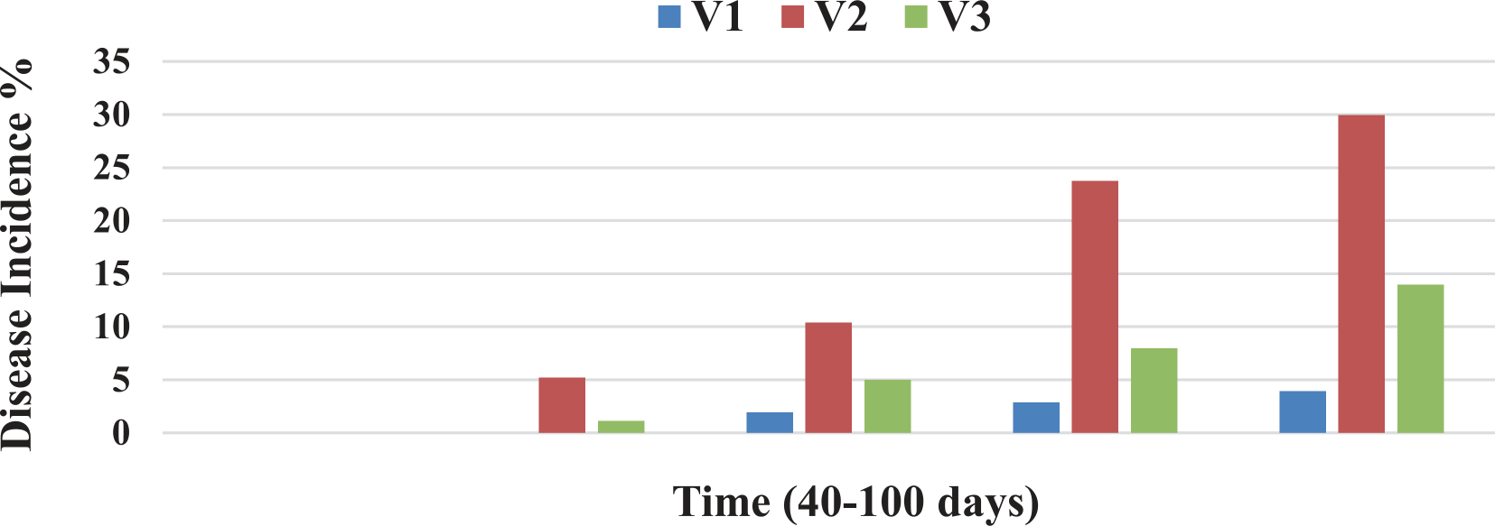
Fifty samples of growing maize roots of the Sakha Agricultural Research Station research farm, which showed symptoms of late wilt, were selected, and many fungal isolates were obtained. Among these fungal isolates, the focus was on the fungal isolates belonging to the fungus under study, which was identified in the Central Laboratory for Fungal Identification at the Agricultural Research Center in Giza as Cephalosporium maydis based on morphological and microscopic characteristics. The presence and progression of late wilt disease (LWD) were observed and characterized by the development of brown necrotic bands starting from the first internodes and extending to adjacent internodes until the entire plant exhibited signs of dehydration. The development of late wilt disease in three maize cultivars was investigated at the Sakha Agricultural Research Station farm. Among the three cultivars studied, SC10 exhibited slower disease incidence and acted as a resistant variety (V1); the disease incidence (DI%) for this variety remained below 4% (Fig. 1). Disease incidence increased, and the highest level of DI% was recorded at 100 days from sowing and at the grain-filling stage of the studied varieties (Fig. 1). Moreover, the sensitive variety TWC 324 (V1) exhibited the fastest disease development rate, and the disease incidence rate of this variety increased from 5.20% to 29.94% (Fig. 1); meanwhile, the moderately sensitive variety SC 128 (V2) exhibited a moderate progression rate of late wilt disease with a DI % around 14% (Fig. 1).
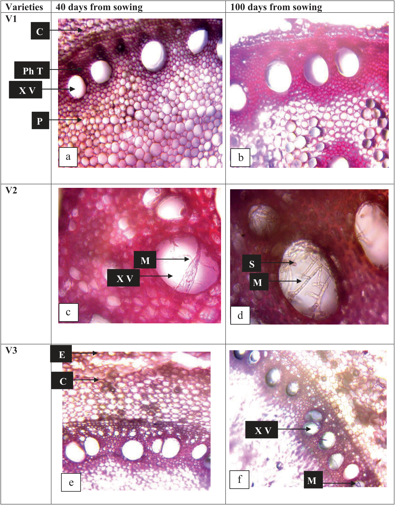
Fig. 2.
Cross sections in roots of the three maize varieties (SC10 (V1), TWC 324 (V2), and SC 128 (V3)) at 40 and 100 days from sowing in the second season. V1: resistant variety SC10, V2: sensitive hybrid TWC324, and V3: moderately sensitive variety SC128.
Anatomical Changes in the Roots and Stems of Maize Varieties (SC10 (V1), TWC 324 (V2), and SC 128 (V3)) under Infection with Late Wilt Disease
Anatomical studies were conducted in order to follow up on the changes that occurred in the roots and stems of maize plants and follow up on the growth of the pathogen. Samples were taken from the second internode of the stem and the roots at 40 and 100 days from the planting date. The illustrated results in Fig. 2 showed that the infection with late wilt disease harmfully affected the anatomical characteristics of maize roots. The infection decreased the root diameter and vascular cylinder diameter of infected maize plants (Fig. 2). Also, cortex thickness and xylem vessel diameter were negatively affected by the infection with M. maydis. Results in Fig. 2 showed that the variation in the fungal growth was recorded in the three studied varieties (SC10 (V1), TWC 324 (V2), and SC 128 (V3)). The cross sections in roots of the variety SC10 (V1) showed no growth of M. maydis recorded at 40 and 100 days from the sowing date (Fig. 2a) and b)). However, the results obtained from Fig. 2c) and d) showed that the growth of M. maydis, especially mycelium, was observed in the xylem vessels of the variety TWC 324 (V2) at 40 and 100 days from the sowing date; mycelium growth was greater at 100 days than at 40 days from sowing.
Fig. 3.
Cross sections in the stems of the three maize varieties (SC10 (V1), TWC 324 (V2), and SC 128 (V3)) at 40 and 100 days from sowing in the second season. V1: resistant variety SC10, V2: sensitive hybrid TWC324, and V3: moderately sensitive variety SC128.
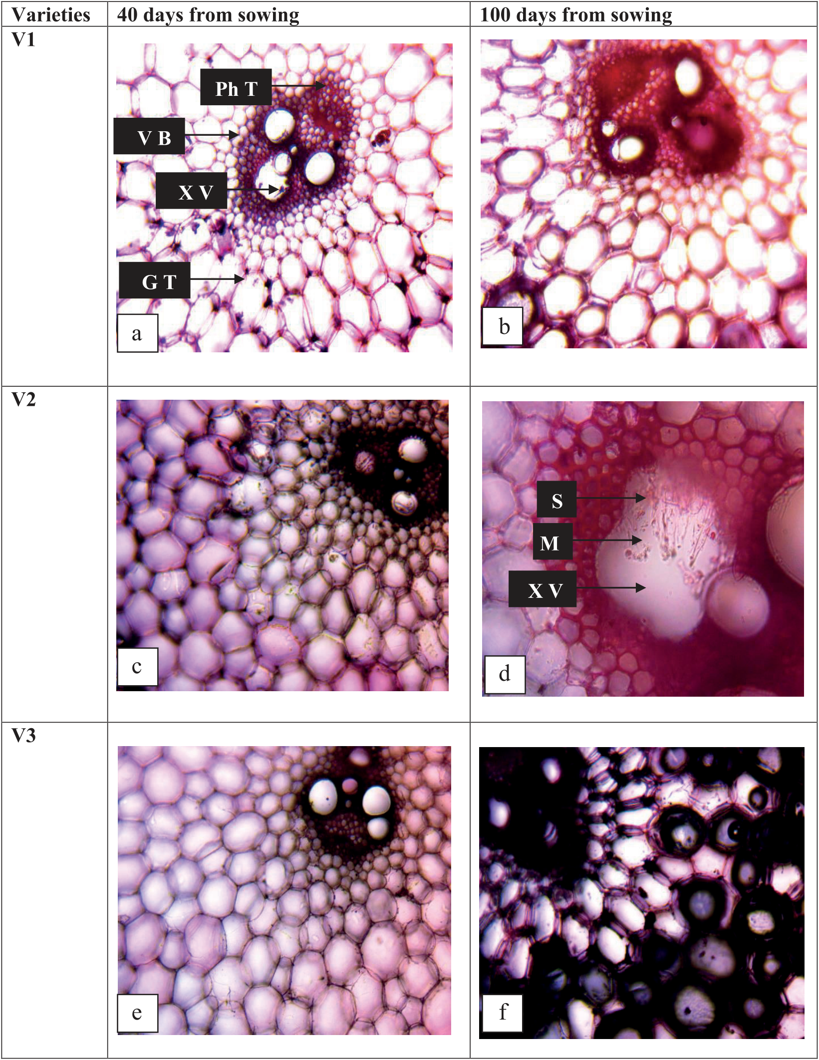
On the other hand, the illustrated photos in Fig. 2e) and f) showed that the mycelium growth of M. maydis was observed only in the transfer sections of the maize root variety SC 128 (V3) at 100 days from sowing; however, mycelium growth was not observed in the transfer sections of the maize root at 40 days from sowing. According to the obtained results in Fig. 3, the illustrated photos showed that the anatomical characteristics of maize stems were negatively affected by the infection with late wilt disease. The results showed that the lower internodes were reduced and dried, associated with brown vascular bundles (Cimmyt, 2004), and finally, the plant died at a late stage, leading to huge economic losses. The anatomical characteristics of the stem, such as stem diameter, length and width of the vascular bundle, phloem thickness, and xylem vessel diameter, were decreased owing to the infection with late wilt disease (Fig. 3). These results were observed in the sensitive variety TWC 324 (V2) (Fig. 3b) and c)) more than in other varieties (SC10 (V1) and SC 128 (V3)). Additionally, the transfer sections of the sensitive variety TWC 324 (V2) showed more mycelium growth of M. maydis in the xylem vessels than the moderately sensitive variety (SC 128 (V3)) at 40 and 100 days from the sowing date. However, no mycelium growth of M. maydis was observed in the stems of the resistant variety (SC10 (V1)) at 40 and 100 days from the sowing date (Fig. 3a) and b)). It was also noted that infection with this fungus led to the destruction of the plant’s internal tissues, especially the vascular tissue.
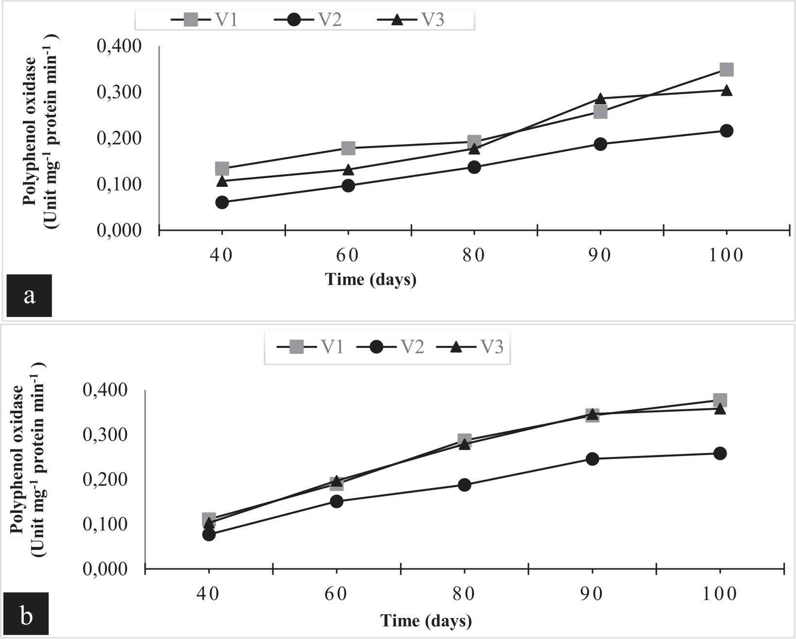
Fig. 4.
Effect of infection with M. maydis on polyphenol oxidase enzyme activity (PPO) in three maize varieties (SC10 (V1), TWC 324 (V2), and SC 128 (V3)) after 40, 60, 80, 90, and 100 days from sowing in the two seasons. V1: resistant variety SC10, V2: sensitive hybrid TWC324, and V3: moderately sensitive variety SC128.
Studying the Relationship between Enzymatic Changes in Maize Plants and the Development of Late Wilt Infection
The results presented in Fig. 4 show that the activity of the polyphenol oxidase enzyme increases gradually with the age of maize plants. The highest value of the enzyme was in the resistant cultivar, SC10 (V1), at 100 days from sowing, and the lowest value was in the sensitive cultivar, TWC324 (V2). It was also noted that there is a correlation between this enzyme activity and the degree of resistance of the cultivated maize variety to infection with late wilt disease. The results indicated that the degree of enzyme activity in the resistant variety intersects with the degree of enzyme activity in the moderately resistant variety at some stages of plant growth in the first season (Fig. 4a)). In the second season, however, there was a close convergence in the activity of this enzyme in both the resistant and moderately sensitive varieties to late wilt (Fig. 4b)).
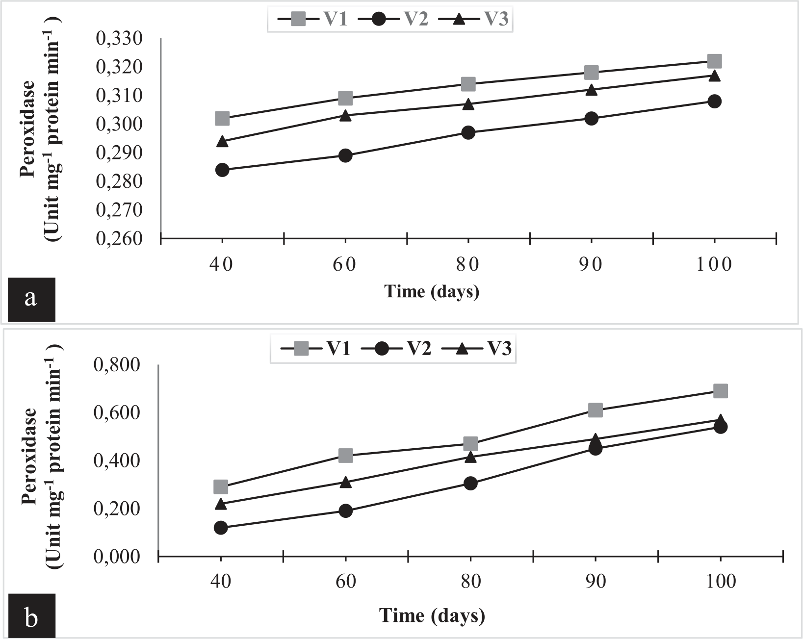
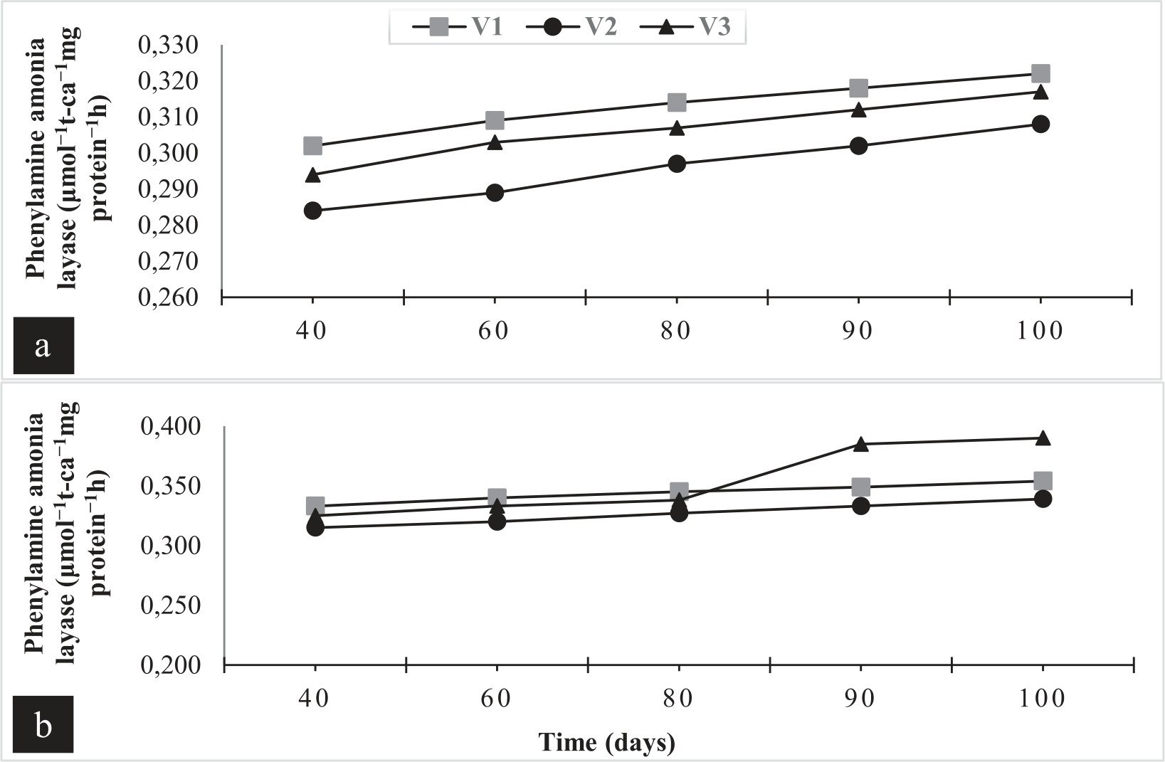
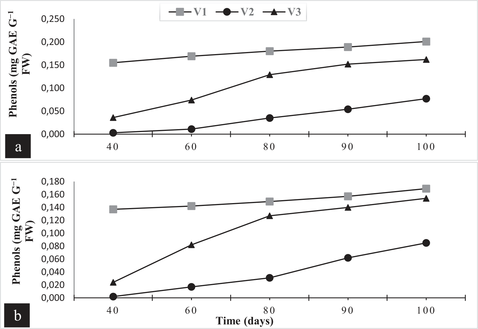
Regarding the activity of the peroxidase enzyme, Fig. 5 showed the activity of the peroxidase enzyme for the three maize varieties under study in both seasons; it was found that it was almost identical to the results of the activity of the polyphenol oxidase enzyme, with very slight differences. The higher degree of resistance of the maize variety to late wilt was correlated with the higher activity of the polyphenol oxidase enzyme. Furthermore, the activity of the phenylalanine ammonia lyase enzyme is similar, especially in the first season of the study, with slight differences in the activity of this enzyme in the second season (Fig. 6a)). A higher activity of this enzyme was recorded in the moderately sensitive variety than the resistant variety, starting at 80 days after planting (Fig. 6b)). Phenol content was determined in the three maize varieties under this study; it was observed that the phenolic content increases with the age of the plants and is strongly correlated with the degree of resistance of the plants to late wilt disease (Fig. 7a) and b)).
Fig. 5.
Effects of infection with M. maydis on peroxidase enzyme activity (POX) in three maize varieties (SC10 (V1), TWC 324 (V2), and SC 128 (V3)) after 40, 60, 80, 90, and 100 days from sowing in both seasons.
Effect of M. maydis Infection on Total Protein, Carbohydrate, and Total Chlorophyll Content in Maize Varieties (V1, V2, and V3) at 40, 60, 80, 90, and 100 Days from Sowing
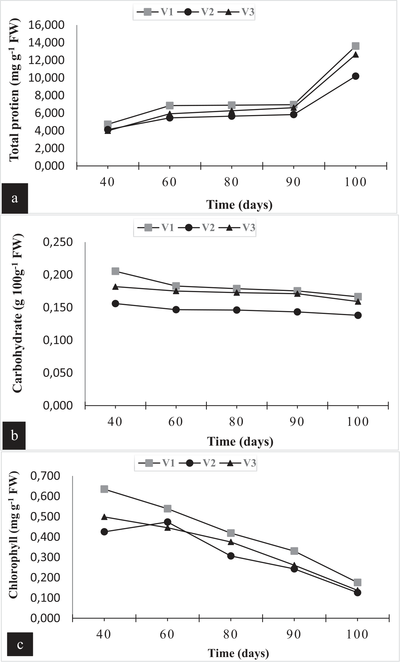
In the current study, total protein, carbohydrate, and total chlorophyll content were determined in the infected maize varieties (V1, V2, and V3) with M. maydis at 40, 60, 80, 90, and 100 days after sowing (Fig. 8). The results indicated that the total protein content was significantly decreased under infection with late wilt disease in the sensitive and moderately sensitive varieties (V2 and V3) compared with the resistant variety (V1) at 40, 60, 80, 90, and 100 days from sowing (Fig. 8a)). Additionally, the infection with M. maydis led to a significant decrease in carbohydrate levels in the sensitive and moderately sensitive maize varieties (V2 and V3) compared with the resistant variety (V1) at all time sampling (Fig. 8b)).
Total chlorophyll content was measured in the three studied varieties (SC10 (V1), TWC 324 (V2), and SC 128 (V3)); the results showed that the total chlorophyll content was significantly reduced according to the infection with M. maydis (Fig. 8c)). The lowest level of total chlorophyll was recorded in the sensitive variety (V2), followed by the moderately sensitive variety (V3) compared with the resistant variety (V1) at 40, 60, 80, 90, and 100 days after sowing (Fig. 8c)).
Discussion
Late-wilt disease caused by the fungus M. maydis is an important fungal disease of maize, especially in Egypt; it causes severe economic losses in maize growth [15, 20]. Three varieties of maize were used in this study according to their susceptibility to this pathogen (resistant variety, moderately sensitive variety, and sensitive variety) to determine their relationship with the fungus M. maydis. To track the stages of the appearance of the somatic structures of M. maydis in the xylem vessels of the stems and roots of maize plants, anatomical studies were conducted on the roots and stems of the 3 varieties at 40 and 100 days from the sowing date. The results from cross-sections of the resistant variety (SC10) at 40 and 100 days from sowing showed the absence of the fungus M. maydis, whether in the form of mycelium, spores, or fungal structures. However, cross sections of the roots and stems of the sensitive variety (TWC 324) showed the presence of the somatic structures of M. maydis starting at 40 days and increasing at 100 days from sowing in the xylem vessels of the stems and roots. Also, the anatomical characteristics of the moderately sensitive variety (SC128) showed the presence of fungal mycelium at 100 days from sowing in the xylem vessels. It was also observed that the fungus led to the destruction of the internal tissues of the maize plant, especially the vascular tissue, resulting in the prevention of the translocation of nutrients and water from the root to the top part of the plant, which led to a decrease in the morphological characters and consequently decreased the anatomical characteristics of the maize plant, such as decreased root and stem diameters. However, the growth and development of fungi was not observed in the resistant variety (SC10); this variety has the ability to withstand the infection with M. maydis. The negative effects of fungal infection on anatomical characteristics have been recorded in many plants, such as wheat [32- 34].
Fig. 8.
Effect of infection with M. maydis on total protein (a), carbohydrate (b), and chlorophyll content (c) in three maize varieties (V1, V2, and V3) at 40, 60, 80, 90, and 100 days from sowing in the second season. V1: resistant variety SC10, V2: sensitive hybrid TWC324, and V3: moderately sensitive variety SC128.
Enzyme activity is a very important indicator for the infection with fungi; the activity of polyphenol oxidase, peroxidase, and phenylalanine ammonia lyase was increased in the resistant variety (V1), followed by the moderately sensitive compared with the sensitive variety (V3). These results may be due to the important role of these enzymes in the defense system against the infection with M. maydis, so the enzyme activity in the resistant variety was higher than in the sensitive and moderately sensitive varieties. These results agree with many researchers under biotic [32, 35] and abiotic stress factors [36-38]. Phenols were also estimated to be one of the important compounds used as a defense system against pathogen infections and to demonstrate the plant’s ability to reduce the progression of infection.
In the current study, total protein content was significantly decreased under infection with late wilt disease at 40, 60, 80, 90, and 100 days from sowing in the maize-sensitive variety (V2), followed by the moderately sensitive variety (V3) compared with the resistant variety (V1). This result may be due to the negative effect of M. maydis on the nutrient uptake that participates in the protein synthesis in maize plants, consequently decreasing the total protein content. Infection with M. maydis was ineffective in the resistant variety (V1) because the growth of M. maydis was not developed, and the mycelium was not produced in the vascular tissue, especially in xylem vessels. Some researchers also recorded this negative effect on the growth characteristics and total protein content [12, 39, 40]. Furthermore, carbohydrate and total chlorophyll content were negatively affected and decreased in the infected maize plants with M. maydis. The decrease in carbohydrate and total chlorophyll content was significant in the sensitive variety (V2), followed by the moderately sensitive one (V3) at 100 days from sowing. This reduction reflects the harmful effect of fungal infection on the morphological characteristics and growth of maize plants, decreasing the physiological and biochemical characteristics. These harmful effects were associated with rapid wilting of the upper part of the maize plant, which happens at 60-80 days from sowing [30]; total dehydration and yield loss can be observed under severe infection [40]. These negative effects of fungal infection on the physiological and biochemical characteristics were recorded in some studies [32, 33, 41].
Conclusions
In conclusion, our study concluded that the sensitive maize variety TWC 324 (V2) to late wilt disease showed that disease incidence was increased compared with the moderately sensitive SC 128 (V3), while the resistant variety SC10 (V1) showed the lowest disease incidence. Likewise, protein, total carbohydrates, total chlorophyll, total phenols, and enzyme activities were augmented in the resistant variety (V1) compared with the sensitive variety (V2). Additionally, the anatomical characteristics of the stems and roots were decreased in the sensitive variety (V2) compared with the resistant variety (V1). However, more studies are needed to determine the best strategy to control Magnaporthiopsis maydis in maize plants and improve crop productivity.

