

Introduction
Khanspur is located in the Ayubia National Park and Reserve Forest (34.0227690, 73.4209476). The vegetation of the park is dominated by conifers, mainly pines and firs. MHB are beneficial because they promote the health and growth of tree seedlings [1, 2]. Some soil microorganisms, such as bacteria and fungi, have an influence on symbiosis formation. Ectomycorrhizae are symbiotic organs formed during interactions between a soil fungus and most tree species or perennials. The plant partner can select bacterial strains that are helpful for ECM symbiosis in nature [3, 4]. Plant growth-promoting rhizobacteria (PGPR) can support plant development through a variety of processes [5-8]. They can act directly by producing chemicals that stimulate growth and improve nutrient availability in the soil or indirectly by suppressing plant diseases in the rhizosphere [9-11]. Although research on PGPR in agriculture settings has progressed, much more research is needed on this group of bacteria in forest habitats [12]. Most antibiotics are derived from terrestrial fungal and bacterial strains that produce inhibitory chemicals such as azoles and quinolone derivatives [11]. Researchers are focusing their research on natural chemicals derived from microbes or herbal extracts to find novel and safe lead compounds. The first step in the development of novel antibiotic compounds is the screening of fungal/bacterial strains capable of producing inhibitory chemicals [13]. The genus Bacillus is one of the most frequently discovered bacterial strains in soil and can produce a wide range of antibiotic compounds [14]. Peptides, such as phospholipid derivatives (i.e. Bacilysocin), have been discovered as antibacterial compounds produced by this terrestrial genus. Numerous studies have been conducted to isolate different strains of terrestrial Bacillus sp. and determine their inhibitory components [15]. Approximately one-third to one-half of prescribed antibiotics are considered unnecessary, and up to one-half are administered unnecessarily [16]. Bacterial resistance to drugs is mediated by four main pathways [17]. Bacteria produce enzymes that inactivate drugs, such as beta-lactamases that cleave the ring of penicillins and cephalosporins [18, 19]. Bacteria produce altered targets against which the drug is ineffective, e.g. a mutated protein in the 30s ribosomal subunit causing streptomycin resistance or methylation of the 23s rRNA causing erythromycin resistance [20]. The bacteria reduce their permeability to such an extent that an effective intracellular concentration of the drug cannot be achieved [21]. Due to widespread bacterial and fungal resistance to commonly used bioactive secondary metabolites, new antifungal and antibacterial chemicals need to be developed [22, 23]. For many years, researchers have been working on the project of microorganism screening for the development of novel antibiotics. A number of industries, including agriculture, veterinary medicine, and the pharmaceutical industry, use antibiotics [24]. Fourier transform infrared spectroscopy (FTIR) is a common technique for identifying the functional groups of materials (solids, liquids, and gases) by measuring their infrared absorption or emission spectra [25]. An FTIR spectrometer simultaneously collects high-resolution spectral data over a broad spectral range. The spectrum is usually plotted as a percentage transmittance versus wavenumber (cm-1). The infrared spectrum is influenced by the type of bonds in a molecule, which can absorb infrared radiation if they have an electric dipole [26]. The infrared region has a higher energy and shorter wavelength than microwaves, but a lower energy and longer wavelength than UV-visible light. When a covalent bond with a dipole moment interacts with infrared radiation, it absorbs energy and vibrates back and forth. The vibrations change the net dipole moment of the molecule, which leads to infrared absorption [25]. To obtain the actual spectrum from the raw data, a mathematical process called Fourier transformation is required. This process converts a range (specular displacement in cm) into its inverse range (wavenumbers in cm-1).
Material and Methods
Site and Sampling
Soil cores with Ectomycorrhizae were taken with soil cores with Ectomycorrhizae from different locations at a depth of 15 cm and a diameter of 10 cm. The soil digger was rotated to extract the soil, and we obtained about 450- 500 g of soil from the root zones of Abies pindrow (Royle ex D. Don) Royle and Pinus wallichiana (P. griffithii) from Helipad forest and Green Spot in Khanaspur KP, Pakistan. The soil samples were placed in polythene bags and labeled with codes according to the method of [27].
Isolation of Bacteria
LB medium was prepared according to [28] in order to observe different properties of the culture medium. The medium and Petri dishes were autoclaved at 121°C at 15 lb/inch2 and then cooled at room temperature before use. We used the serial dilution method to count the bacteria in the soil and made dilutions of (10-1, 10-2, 10-3 10-4, 10-5, 10-6). We poured 15-20mL of melted, cooled (45-50 ̊C) LB medium into 90mm autoclaved Petri dishes and allowed it to solidify. We then poured 100μL of the dilution from each labeled test tube into the respectively labeled Petri dishes and spread evenly. We wrapped and incubated these for 24 h at 28 ± 2 ̊C in an incubator [27].
Isolation of Different Microbial Communities
Different microbial communities were isolated by taking an inoculum of the bacterial colony using a cooled, flame-sterilized inoculation loop. This was spread on the surface of the LB medium in horizontal and vertical lines on the Petri plates. A similar method was repeated for other microbial communities to isolate them. Pure strains were obtained and stored at 4 °C to prevent overgrowth of bacterial colonies [29].
Macro Morphological Characters
Different bacterial species form colonies with different shapes and textures depending on the conditions of the substrate. Colony patterns can be useful for identifying the species or monitoring the growth process, but they are also very sensitive to environmental factors. However, there are some common rules for the selection of patterns for each species, such as color, shape, size, etc.
Morphological Characteristics of Bacteria
The Gram staining technique was used to differentiate between Gram-positive and Gram-negative bacteria, and the prepared slides were examined under a microscope [30]. The heat-fixed slides were placed on one side of the staining tray. They were flooded with crystal violet for 45˗50 sec and carefully rinsed with water. The slide was then flooded with Gram’s iodine solution for 45 sec to one min and carefully rinsed with water. The slide was decolorized with 95% ethanol until a bluish color or no color appeared on the washed-off alcohol, and then the side was rinsed with water. For counterstaining, the slide was carefully flooded with safranin for 35-45 sec and rinsed with water. The slide was allowed to air dry for 15 min. The slide was then ready for observation under the compound light microscope.
SMAN Production
It was carried out using shake flask fermentation. The medium of choice (L-Broth) was used to cultivate the bacterial isolates in the shaking incubator for 3 days. After 3 days, the fermented culture broth was aseptically transferred into sterilized 50 mL falcon tubes in the laminar air flow hood and labeled according to the respective strains. After decanting the culture broth into sterilized falcon tubes, these were centrifuged at 6000 rpm for 10 min, and the supernatant was separated into another falcon tube and labeled accordingly [31, 32].
For the isolation of SMAN, the supernatant was treated with 20 mL ethyl acetate in the separatory funnel and mixed vigorously to mix the 2 layers evenly. The funnel was then placed on the stand for 1 h and waited for complete separation of the two layers in the funnel. After one hour, both layers were collected separately, and the ethyl acetate layer was kept in the Petri dish where the solvent evaporated overnight. After 12 h, the dried layer of secondary metabolites of antibiotic nature was scraped off of the Petri dish with the help of sterile surgical blades. The extracted compound was weighed on a balance and stored in clean, dried, and autoclaved Eppendorf tubes with the appropriate labels.
Antimicrobial Activity Test of Isolated Bacterial Strains
To ensure confluent growth of the organism, LB plates were densely inoculated with standardized inoculum by spreading 100 µL of the bacterial dilution. Standardized 6 mm filter paper discs were used to determine the sensitivity of secondary metabolites of antibiotic nature (SMAN) of bacteria by the agar diffusion method, following [33, 34]. The discs were aseptically impregnated with specific concentrations of different SMAN and placed on the surface of an agar plate that had been colonized with the organism to be tested. Each disc was carefully touched with sterile forceps to ensure that it was firmly attached to the surface of the medium. After incubation at 28 °C for 24 h, the plates were examined for the presence of an inhibition zone [22, 34, 35], which was characterized by a distinct zone around each disc. The size of this zone in mm determined the susceptibility of an organism to SMAN. The antimicrobial activity of crude extracts obtained from selected strains was determined against a range of test organisms, including Gram-positive bacteria (Bacillus meurellus and Bacillus subtilis) and Gram-negative bacteria (Acinetobacter rhizosphaerae and Escherichia coli).
FTIR Analysis
The FTIR analysis technique was applied to the materials to examine their organic, inorganic, and polymeric components using infrared light. The change in material composition can be seen by the change in the pattern of the transmission band, which is characteristic of each compound. The unknown chemicals were also described and identified using FTIR [36]. Moreover, FTIR was also used to detect impurities and contaminants in the sample. This method is based on the measurement of wavelength transmission in the range of 500-3500 cm [37-39].
Results
Isolation and Enumeration
For the isolation of MHB from the Ectomycorrhizosphere of A. pindrow and P. wallichiana, soil blocks were collected from Helipad Forest and Green Spot, Khanaspur, KP, Pakistan. The samples were processed within a week to isolate the bacterial colonies. Twenty bacterial strains were obtained from sample AP-10, namely AP10-1-1, AP10-1-2, AP10-1-3, AP10-1-4, AP10-2-1, AP10-2-2, AP10-2-3, AP10-2-4, AP10-3-1, AP10-3-2, AP10-3-3, AP10-3-4, AP10-4-1, AP10-4-2, AP10-4-3, AP10-5-1, AP10-1-1, AP10-1-1, AP10-1-1 and AP10-1-1 (Table1). The size of these colonies ranged from punctate to large, and the color of the colonies was white, off-white, dull-white, creamy, and matte. The shape of the colonies was circular, filamentous, irregular, and pulvinate. The margin of the colonies was entire, filamentous, lobate, and opaque, and the colonies were convex, crateriform, flat, elevate, raised, and umbonate. Five bacterial strains were obtained from the sample PW-2, namely PW2-1-1, PW2-2-1, PW2- 3-1, PW2-3-2, PW2-4-1, and PW2-5-1. The size of these colonies ranged from small to medium, and the color of the colonies was white, off-white, and cream. The shape of the colonies was circular and regular. The margin of the colonies was entire, and the colonies were convex, flat, and raised (Table 1).
Table 1.
Description of characteristics of Bacterial Culture Isolated from Dilution Plates (10-1, 10-2, 10-3, 10-4, 10-5 and 10-6) From Ectomycorrhizosphere of A. pindrow (Sample AP-10) and P. walichiana (Sample PW-2)
Antibacterial Activity
The antibacterial activity of selected bacterial strains was determined by solid media bioassay against Bacillus meurellus, Bacillus subtilis, and Escherichia coli, and only potentially active isolates were selected for further work, while the inactive strains were discarded (Table 2, 3).
FTIR Analysis
FTIR Analysis of Selected SMAN from Bacterial Strains
The FTIR spectrum with a scan range of 500 to 3500 wavenumbers cm-1 was used for the spectra obtained, which gave a comprehensive metabolic fingerprint of the ethyl acetate extract of strain PW2-3-1 with characteristic peaks at 3272.13, 1658.11, 1408.61, 1018.11, and 560.12 cm-1 (Fig. 1). The ethyl acetate extract of strain AP10-2-4 showed characteristic peaks at 3271.25, 1659.03, 1408.47, and 1017.67 cm-1 (Fig. 1). The extract of strain AP10- 4-2 showed characteristic peaks centered at 3272.52, 1650.64, 1408.26, and 1017.97 cm-1 (Fig.1). The extract of strain AP10-6-2 showed characteristic peaks centered at 3278.58, 1651.23, 1408.60, and 1019.67 cm-1 (Fig. 1). The ethyl acetate extract of strain AP10-1-1 showed characteristic peaks centered at 3272.57, 2923.41, 1651.40, 1408.02, 1017.80, and 519.93 cm-1 as shown in Fig. 1. The peak value at 3272.52 cm-1 showed the presence of an alcoholic compound due to O-H stretching vibrations. The peak values at the wavelength of 1650.64 cm-1 indicate the presence of amide due to C=H bending. The peak values at 1408.26 cm-1 indicated the presence of alkanes (methyl group) due to C-H bending. The peak values at 1017.97 cm-1 are due to the presence of C-O stretching and indicate the presence of vinyl ether.
Table 2.
Zones of Inhibition for Antibacterial Activity of Supernatant of Selected Bacterial Strains.
Table 3.
Zones of Inhibition for Antibacterial Activity of some quantitative extract SMAN in water and Standard Antibiotics against Four bacterial strains, their shape and type after Grams’ staining.
Discussion
The study was conducted in Khanaspur, Helipad Forest, and Green Spot, KP, Pakistan, to explore its microflora. The study focused on the Ectomycorrhizosphere of two Pine species, A. pindrow and P. wallichiana, for the isolation of different microbial communities. These colonies were characterized using morphology and Gram’s staining. The FTIR spectroscopic analysis was also used to identify different functional groups present in the extract of secondary metabolites of antibiotic nature. Additionally, biological screening was done to investigate the antimicrobial potential of bacterial strains. Current research work focused on the isolation of 26 different bacterial strains belonging to four genera: Bacillus, Coccus, Streptococcus, and Staphylococcus. These findings are in line with previous reports by [40] and [41] on the role of bacteria in the ectomycorrhizosphere of plants and their impact on fungal and soil bacterial communities [12, 42, 43]. Ectomycorrhizae occur in about 10% of the world flora, mainly in Pinaceae (pine, fir, larch, and spruce hemlock), Fagaceae (oak, chestnut, beech), Betulaceae (alder, birch), Salicaceae (poplar, willow), Juglandeceae (American walnut, pecan), Myrotaceae (eucalyptus), and some other trees. Many fungi have been identified as ectomycorrhizal producers [44]. Oh and Lim [45] identified thirteen species of microbes in pine roots and reported that they were all members of Bacillales or Burkholderiales. Alibrandi et al. [46] made a taxonomic identification of bacterial endophytes (fifty unduplicated bacterial isolates) obtained from three terrestrial Mediterranean orchid species based on the 16S rRNA gene and revealed that they belonged to the genera Pseudomonas, Pantoea, Rahnella, Staphylococcus, Sphingomonas, Microbacterium, Streptomyces, Fictibacillus, and Bacillus. Another study, Gonzales-Escobedo et al. [47] described a total of seven bacterial phyla, 14 classes, 26 orders, 43 families, and 51 genera from Pinus arizonica and P. mavigensis. They found that Enterobacteriaceae was the most abundant family in all samples, followed by Acetobacteraceae and Acidobacteriaceae, which is consistent with previous studies in other pine and conifer trees. MHB can be found in many bacterial species, both Gram-negative and Gram-positive. Most of them belong to Proteobacteria such as Agrobacterium, Azospirillum, Azobacter, Burkholderia, Brayrhizobium, Enterobacter, Pseudomonas, Klebsiella, or Rhizobium. But others are found in the phyla Firmicutes (Bacillus, Brevibacillus, Paenibacillus) and Actinomycetes (Rhodococcus, Streptomyces, Arthrobacter) [1]. It is clearly understood that bacterial isolates and plants with various activities can be considered as potential sources of environmentally friendly bioactive metabolites [48]. It has been reported that some MHBs may also have additional beneficial effects, such as direct stimulation of plant growth and protection against pathogens or adverse conditions. These bacteria have applications because they indirectly improve the health and growth of seedlings. For example, the use of plantations or short-rotation coppices has been suggested to reduce fertilizer use in tree nurseries and fast-growing tree plantings such as poplar [49].
Fig. 1.
FTIR spectrum of Bacteria showing different peaks at specific wavenumber cm-1 (a) Strain PW 2–3–1 (b) Strain AP 10–2–4 (c) AP 10–4–2 (d) AP 10–6–2 (e) AP 10–1–1
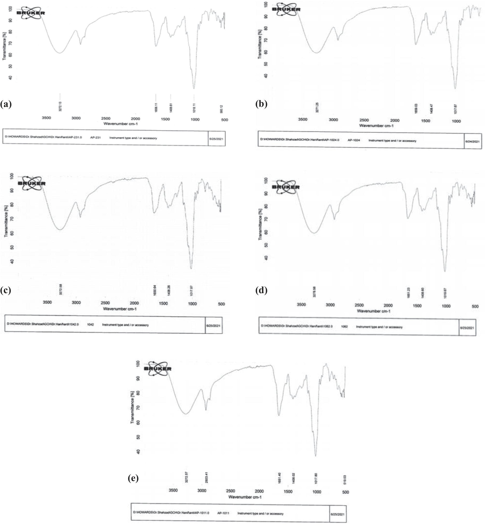
In this research, different colonies of bacteria were isolated and characterized morpho-anatomically based on their colonial shape, size, color, elevation, and texture using Gram’s staining [50], as followed by [51]. Among members of the plant microbiota, mycorrhizal fungi (MF) and plant growth-promoting bacteria (PGPB) (which facilitate root penetration through their ability to promote the growth of AMF hyphae) interact in rhizospheric environments, leading to additive and/or synergistic effects on plant growth and health [52-55]. Selvakumar et al. [56] observed that Gram-positive bacteria were more associated with AMF spores than Gram-negative bacteria, hence the name spore-associated bacteria (SAB). The susceptibility of an organism to a secondary metabolite was determined by the size of the zone of inhibition, which is dependent on various factors [57]. Antibacterial activity was performed against four identified bacterial strains: Bacillus meurellus, B. subtilis, A. rhizospherensis, and E. coli. Antimicrobial activity was performed using the well diffusion method against selected strains, which were processed for further work [58]. An antimicrobial assay was also done using the disc diffusion method. Antibiotics have been used for decades to fight bacterial infections in general. In this research, some bacteria with rapid growth rates were selected for the production of antibiotics. These strains have high metabolic activity and produce more secondary metabolites into the culture medium. The highest yield was detected by the strain AP 10-2-4, which produced 0.119 mg of antibiotics per 50 mL of culture medium. The findings are in line with the work of [59] and [60]. On the same pattern, Gislin et al. [61] assessed the antimicrobial activity of soil bacteria isolated from 10 different rhizosphere locations and various cultivation areas in Kochi, Kerala, India. They evaluated that both isolates (S1A1 and S7A3) showed positive results against S. aureus and Enterococcus sp. Frey-Klett et al. [62] showed that the proportion of Pseudomonas inhibiting the growth of seven fungal root pathogens in ectomycorrhizae of L. bicolor was significantly higher than in the surrounding soil. Many Pseudomonas strains produce antimicrobial metabolites such as phloroglucinols, phenazines, pyoluteorin, and pyrrolnitrine [63, 64]. The data from FTIR analysis revealed the presence of secondary metabolites of antibiotic nature in the crude extract. FTIR spectroscopy has become a standard method for analyzing extracted bacterial metabolites [65]. The spectral values of FTIR analysis provided comprehensive fingerprints that allowed for the prediction of possible functional groups associated with some popular antibiotics. Most of the bacterial isolates exhibited secondary metabolites containing compounds of antibiotic nature. Damavandi et al. [66] evaluated the anticancer and antibacterial potential of bioactive secondary substances derived from bacterial endophytes associated with Artemisia absinthium. The researchers of this article examined various endophytic bacteria for P. aeruginosa SD01 and found discernible activity against both bacterial pathogens and malignancies. The crude ethyl acetate extract of P. aeruginosa SD01 showed MIC values of 32 and 128 µg/mL for S. aureus and MRSA, respectively. Furthermore, they evaluated 2 aminoacetophenone, 1,2 apyrazine-1,4-dione, phenazine, and 2-phenyl-4-cyanopyridine as the main bioactive secondary metabolites (via FTIR and GC-MS analysis). They concluded that their findings indicate that bacteria derived from A. absinthium plants, and in particular from P. aeruginosa SD01, are a remarkable source of untapped therapeutic compounds, i.e. antimicrobial and anticancer compounds. 2-aminoacetophenone is a single-benzene-ring volatile molecule with a grape-like odor from P. aeruginosa cultures [66, 67]. Consequently, Damavandi et al. [66] noted that the peaks presented are similar to the presence of aromatic structures typical for favonoids. They considered that there are various types of favonoids, but all share the general structure of C6-C3-C6 phenyl benzopyran, consisting of aromatic rings. They noted that this effector molecule can promote persistent phenotypes through its effects on both the bacterial cell and the host, leading to long-term bacterial survival in a stationary phase and a reduction in bacterial virulence in a variety of hosts. Moreover, it likely helps bacteria survive within plant tissue and has been reported to protect bacteria from the plant’s defenses. Phenazines, one of the most widely used bacterial secondary metabolites, have broad-spectrum antibiotic properties against a wide range of bacterial and fungal pathogens. Pseudomonas and Streptomyces are among the most common bacterial species that produce phenazine compounds [66-68]. Significantly, Lee et al. [19] studied Sophora koreensis, an endemic species of the Gangwon-do region of Korea. They analyzed and compared the compounds found in the leaves, stems, and roots of S. koreensis collected from three different habitats in Chuncheon, Inje, and Yanggu in South Korea. This research also benefited from the analysis of soil microorganisms in the three habitats to determine the relationship between the compound and microorganisms. Notably, they found that N-methylcytisine was the most common compound in all three habitats, but the amounts varied, with Chuncheon having the highest amount (509 mg/L), followed by Yanggu and Inje (102 mg/L and 39 mg/L, respectively). As a result of their important findings, they revealed that soil microorganisms in different habitats affect the variations of N-methylcytisine. Soil biodiversity is a crucial factor in controlling and sustaining ecosystems. Microflora creates diversity that maintains soil functionality and drives ecosystem processes. Ecosystem management can be achieved by conserving soil microflora, which varies according to different environments.
Conclusions
Mycorrhizal fungal hyphae improve soil structure and soil porosity by binding the soil into aggregates, producing humic compounds and organic sticky substances. Such fungal-plant associations lead to increased tolerance of plants to salinity and heavy metals, increased resistance to insects feeding on plant green parts, and increased uptake of nutrients including P, Zn, Fe, N, K, and Mg by plants. Furthermore, mycorrhizal associations play a role in the biological control of plant diseases and in the growth and development of plants. In the current investigation, it was found that MHB produce metabolites that inhibit the growth of soil microflora and facilitate the growth of soil mycorrhizae. Plants associated with mycorrhizal associations are resistant to various environmental factors, such as drought or flooding, and showed significant growth in comparison to non-mycorrhizal plants. Some isolated strains showed significant inhibition against grams’ negative and positive bacteria that can be used for further investigation in the field of medicine and research work for improved and modified drugs. FTIR analysis confirmed the presence of certain functional groups in the crude extract obtained from bacterial strains in nature, providing comprehensive fingerprints that allowed for the prediction of possible functional groups associated with antibiotics. Soil biodiversity, driven by microflora, is crucial for controlling and sustaining ecosystems. This research supports previous work on the production of antibiotics by bacterial strains and the importance of soil biodiversity in ecosystem management.

