

Introduction
Herbal therapy has its roots in ancient times in human history. The therapeutic effects of plants have been learned through trial and error through the most effective learning method and have been transferred from the past to the present [1, 2]. Plants are a valuable source of medicines and play a significant role in the healthcare system. Around 4.22 million flowering plants have been identified globally, with over 50,000 being used for therapeutic purposes [3]. Plants are essential for population health because of their curative potential, without them, life would be impossible. Medicinal plants are plants with curative characteristics or that have a significant pharmacological impact on individuals [4]. Medicinal plants have always been regarded as a vital source of medicines and treatments and have developed to play a significant part in healthcare systems all around the world. This includes using medicinal plants not just to cure diseases but also as a potential material for maintaining better health and the environment [3]. The uses of ethnomedicinal plants should be confirmed by the experimental and phytochemical studies to increase the safety and effectiveness of these plants and witness to develop modern drug development [5]. In folk medicine, health is considered holistically by incorporating environmental and spiritual elements into the healing process [6]. The use of medicinal plants is seen to be quite safe, as there are no or very few side effects [7]. Medicinal plants produce a huge number of complicated and valuable compounds, which are referred to as secondary metabolites. Secondary metabolites are bioactive chemicals found as resin, tannins, volatile oils, glycosides, flavonoids, and saponins, and are utilized in a variety of pharmaceuticals [8, 9]. In recent years, natural compounds produced from plants, such as alkaloids, flavonoids, and terpenoids have gained a lot of interest due to their wide range of medicinal properties including anticancer, antifungal, and antibacterial capabilities [8, 10].
The interest in nanotechnology, which is one of the determining factors in shaping the future world, has increased considerably in recent years. Nanotechnology presents a solution for environmental, health, and technological challenges, including agricultural science [11]. With nanotechnology applications, it has become possible to investigate the antibacterial, anticarcinogenic, or antibiofilm effects of nanoparticles produced by chemical or biological methods. In the treatment of diseases, these effects can be investigated and used to learn which treatments are effective (biological or chemical). These studies have revealed the necessity of researching a less toxic and waste-generating system. In the following years, the term “Green Nanotechnology”, which is based on nanoparticles with less toxic effects and using living organisms in nature, has emerged [12]. It has been reported in many studies that plants and herbal products are cheap and renewable resources for nanoparticle production, and that the use of plant extracts is an alternative to physical and chemical methods in recent years and is widely used in the field of health [13]. For these reasons, interest in performing phytonanoparticles synthesis with the use of plant extracts has increased in recent years. Phytonanoparticles which are used in many areas, draw attention in the field of health with their antimicrobial, antifungal properties. Silver nanoparticles (AgNP), has drawn attention with their antimicrobial properties, which provide an advantage in the fact that there is almost no bacterial resistance in silver and, as mentioned before, it is not toxic at low concentrations. In particular, AgNPs produced by plants reduce the side effects of drugs used in treatment at lower doses due to their properties [14, 15]. The suitability of the shape and size of the silver nanoparticles is characteristic of both its antimicrobial activity and its effect on reducing toxic tissue in the cell. Their size is very well defined to reduce metal ions. This green silver nanoparticle has many advantages such as easy processing, measurement, and economic accessibility for synthesizing, this method of other medical and electronic applications is increasing its potential attractiveness and its usage area is expanding and more new inorganic nanoparticle syntheses will be discovered. Toxic studies of silver nanoparticles on human pathogenic cells open the door for the formation of a new range of antibacterial agents [16]. Nanoparticles have distinct physicochemical features that are used for a variety of applications in biological sciences, chemical engineering, medicine, and agriculture [17].
In the current work, silver nanoparticles (AgNP), which have a very important place in nanobiotechnology research in line with their advantages, were produced using the A. euchroma plant, and their effects on some biological activities were investigated. A. euchroma, also known as Pink Arnebia is a medicinal plant belonging to the family Boraginaceae [18]. The plant can be used to treat toothaches, ear infections, and eye infections. Cuts and burns are treated using the plant’s antibacterial and anti-inflammatory qualities. The plant aids in urination and phlegm expulsion, which aids in the relief of persistent cough and cold symptoms [19]. A clinical investigation using A. euchroma extracts revealed that it has the potential to treat diabetes by suppressing adipocyte signaling and increasing glucose sensitivity. The roots of A. euchroma are antipyretic and anticancer, inhibiting cancer cell development on the chorion membrane. The root also includes shikonin, an anticancer and antibacterial, antimicrobial, chemical that inhibits E. coli growth. S. aureus, Bacillus typhi, and P. aeruginosa [20, 21]. A. euchroma roots produce many secondary metabolites and purified from the roots shikonin, acetylshikonin, isobutyrylshikonin, β,βdimethylacrylshikonin, isovalerylshikonin, β-hydroxyisovalerylshikonin, deoxshikonin, isobutylshikonin. Important phytochemicals such as arnebinone, arnebin-7, stigmasterol are used to treat various harmful diseases [21, 22]. The root part of the plant contains biologically active compounds. In addition, in this study, silver nanoparticles were added to the root extract, and root extract was to be obtained, the effect of the root alone and the synergistic effect that it will form together with the silver nanoparticle were analyzed. To the best of our knowledge, there is no study examining some biological properties of AgNPs added to extracts of A. euchroma roots. Hence, in the present research we aimed to determine the phytochemical component and the biological activities such as (i) antibacterial, (ii) antifungal (iii) antipellicle as well as (iv) antibiofilm activity of different extracts of A. euchroma collected from Chitral, Khyber Pakhtunkhwa Pakistan.
Material and Methods
Collection and Processing of Plant Specimen
Fresh roots of A. euchroma were collected from Chitral, Khyber Pakhtunkhwa Pakistan, and subjected to shade drying for 25 to 30 days at room temperature. The roots were then pulverized into fine powder through an electric grinder.
Extraction and Fractionation
500g of root powder was mixed in 2 L of ethanol in a bottle and kept in a water bath until it was completely evaporated. The extract was then used for phytochemical screening and biological activities [23]. 10 g of crude extracts were dissolved in 300 mL of distilled water, shaken for 20 min, and added 100 mL of chloroform. Allow it to stand in the separating funnel for 1-2 hr. Two distinct strata were observed. The chloroform layer was separated in a beaker, and the remaining solvent was mixed with 200 mL of methyl acetate and poured into a separating funnel in the same manner. After the two layers were visible, the methyl acetate layer and any remaining aqueous were removed, and the fractions were put in a water bath at 55ºC to dry. These extracts were used for a variety of activities [24].
Phytochemical Screening
Screening of phytochemical constituents of the plant was done both qualitatively and quantitatively using standard procedures described by Edeoga et al. [25], Evans, [26], Kareemi & Jaafar, [27], Nazir et al. [28], Ajmal et al. [29].
Extracts Preparation for HPLC-UV Characterization
HPLC-UV characterization and quantification were carried out according to the reported method [30]. To prepare the extract for HPLC analysis 1 gm powdered sample was mixed in methanol and water (1: 1; 20 mL; v/v). The mixture was heated at 70ºC for 1 hr in the water bath and centrifuged for 10 min at 4000 rpm. After that sample (2 mL) was filtered into HPLC vials through Whattman filter paper. For the identification of phenolic compounds, the high performance liquid chromatography (HPLC) Agilent-1260 infinity system was used, with basic parts like UVAD (ultraviolet array detector), a quaternary pump, a degasser, and an auto-sampler. The separation was carried out by the Agilent-Zorbax-Eclipse column (XDB-C18). Column gradients system consists of solvent B and solvent C. Solvent B composed of deionized water: methanol: acetic acid in the ratio of 180: 100: 20; v/v and solvent C composed of deionized water: methanol: acetic acid in the ratio of 80: 900: 20; v/v. The gradient system was started with solvent B 100 %, 85 %, 50 %, and 30 % at 0, 5, 20, and 25 min, and finally, solvent C (100 %) started from 30-40 min. Elution occurred after 25 min. The ultraviolet array detector (UVAD) was set at 280 nm for the analysis of phenolic compounds and the chromatogram was recorded from 190-500 n. Identification of phenolic compounds was done using their retention times, UV spectra, and available standards while quantification was done taking the percent peak area. All these phenolic compounds were identified with standard phenolic compounds in Malaysian plant methanolic extract. Quantification of antioxidants was measured by the formula:
Cx = Sample concentration; As = Standard peak area; Ax = Sample peak area; Cs = Standard concentration (0.09 µg/mL).
Bacterial and fungal strains collection
Four Gram-negative bacteria (E. coli, S. typhi, P. aeruginosa, P. vulgaris) and two Gram- positive bacteria (Enterococcus, S. aureus). Five fungal strains (Rhizoctonia, F. oxysporum, C. auris, A. niger, and C. fasciculus) was collected. Fungal strain collected from the Department of plant pathology, the University of Agriculture, Peshawar, and the bacterial strain was collected from Khyber Teaching Hospital, Peshawar.
Antibacterial activity assay
For bacterial culture, nutrient agar medium was prepared by dissolving 28 g nutritional agar in 800 mL distilled water. The media was made in a bottle and then dissolved with a stirrer. To avoid contamination, a sterile environment was maintained during pouring. In the test tube, the medium was used as a stock for bacterial growth. Four wells were formed in the medium of the Petri dishes using a sterilized cork borer. With the help of a cotton swab, bacterial strains were streaked over the medium surface. Then, extract solutions of different concentrations were poured into the wells. (2, 4, 6, and 8 µg/mL). The standard antibacterial was co-amoxiclav. The Petri dishes were then incubated for 24 hr at 37ºC. After 24 hr, the reading was taken in mm using a ruler and measuring the inhibitory zone [31, 32].
Antifungal activity
For fungal activity, 39 g PDA (potato dextrose agar) was dissolved in 1000 mL of distilled water. The medium was prepared in a bottle and autoclaved for 15 min at 121ºC. The standard antifungal was fluconazole, which was used at a dosage of 2 mg/mL. For 24 hr the Petri plates were incubated, and then a reading in mm was taken using a ruler to measure the zone of inhibition [32, 33].
Antipellicle activity
The anti-pellicle activity was assessed using the technique of Khan et al. [34] and Joshua et al. [35], with slight modifications. Descriptions of the provided literature were used to compare results with controls. 5mL of nutrient broth was added to sterilized and labeled test tubes, followed by 60l of tested bacterial and fungal inoculum. After using the bacterial inoculum, different amounts of ethanolic extracts (12, 14, and 16 µg/mL) were added to test tubes. These test tubes remained undisturbed in an incubator at 370ºC for seven days. The strong (+), moderate (++), weak (+++), and no inhibition (-) pellicle layers were used to compare the results to the controls.
Antibiofilm activity
Cultured bacteria and fungi were grown on a 96- well polystyrene microtiter plate. Media was dissolved and sterilized in an autoclave using a Thermo Magnetic Stirrer. Different concentrations of the stock solution were taken, i.e. 12, 14, 16 µg/mL and 80 µL nutrient broth, 20 µL cultured bacteria, and fungi were poured into each well of a sterile 96-well polystyrene microtiter plate and were incubated at 37ºC for 24 hr [36].
Synthesis of AgNPs
AgNPs were produced using the procedure of Ikram et al. [37], Ullah et al. [38] with slight modifications. 10 g powdered plant root components were mixed with 100 mL distilled water and shaken for 24 hr. Whatman filter paper was used to filter the final solution twice. For further use, the filtrate was stored in the refrigerator at 4ºC. The A. euchroma extract and AgNO3 salt were used to make AgNPs. In 400 mL distilled water, a 1 mM AgNO3 salt solution was prepared. On a hotplate, the solution was heated and continually stirred while adding plant extract until the color turned light yellow. After that, the mixture was centrifuged for 15 min at 12000 rpm. The supernatant was discarded, and the lower pellet was suspended in distilled water again before being centrifuged for 5 min. To eliminate impurities, the procedure was done three times. The production of AgNPs was determined by scanning the UV-visible spectrum [37].
Results
Plants naturally contain a variety of phytochemicals with important biological activities. Medicinal plants have traditionally been used in various forms in the processes from when these properties were not known to the present day. It is used in the form of herbal teas, extracts, poultices, or ointments. Owing to nanotechnology, which has gained momentum in the last two decades, its use in the form of phytonanoparticles, which is a new form of use, has been presented to the scientific world. In particular, a synergistic effect is created by combining the antimicrobial effect of silver (due to its non-toxicity at low doses) with the effect of the plant, and many effects, especially antimicrobial and antifungal studies, have been studied. Therefore, the phytochemical component and some biological abilities of the A. euchroma were investigated to better understand the effect of AgNPs on various activities.
Table 1.
Phytochemical screening of root of A. Euchroma.
| S. No | Constituents | Results |
|---|---|---|
| 1 | Carbohydrates | + |
| 2 | Amino acids | + |
| 3 | Alkaloids | + |
| 4 | Phenol | + |
| 5 | Terpenoids | _ |
| 6 | Saponin | + |
| 7 | Tannins | + |
| 8 | Flavonoids | + |
Phytochemical screening
Firstly, to improve our understanding of the biological activities of plants it is important to know their molecular composition. Thus, the qualitatively phytochemical screening of A. euchroma extract was performed. Table 1 shows the presence of bioactive components of the extract such as carbohydrates, alkaloids, amino acids, flavonoids, phenols, saponins, and tannins.
On the other hand, the quantitative phytochemical screening was done through GC-MS and HPLC methods. The GC-MS results of the phytochemical contents of the plant are shown in Table 2 and Fig. 1, and the HPLC results are shown in Table 3 and Fig. 2. GC-MS method was used to evaluate different fractions of the root. As seen in Table 2, 10 compounds were detected. In addition to that, typical HPLC-UV chromatograms of plant methanolic extracts are presented in Fig. 2.
Table 2.
GC-MS analysis of root of A. Euchroma.
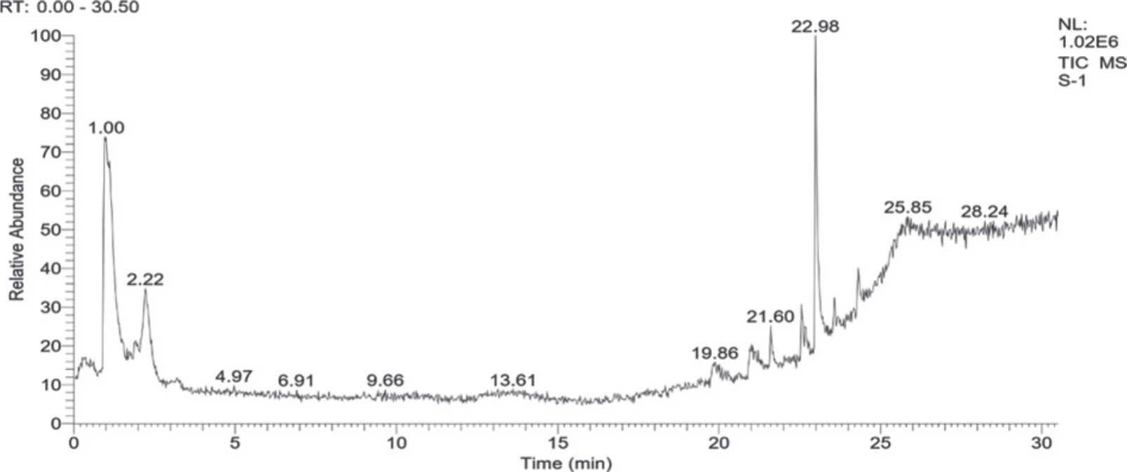
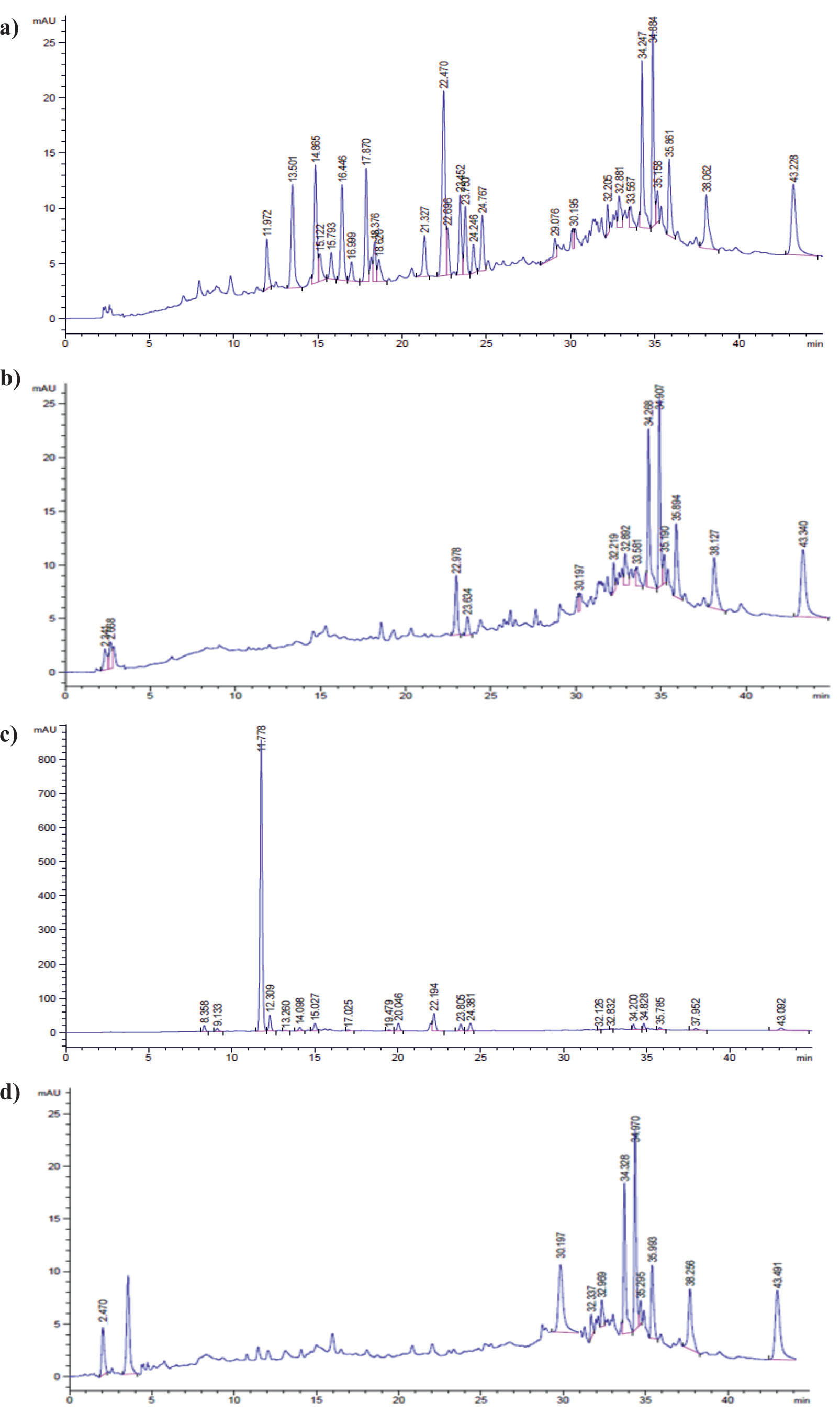
Table 3.
HPLC analysis of phenolic compounds of root of A. Euchroma.
A total of three phenolic compounds ellagic acid, rutin, and mandelic acid were identified in the Met. Ext-ZDL (Fig. 2a) and malic acid, rutin, and mandelic acid were identified in the Met. Ext-2D (Fig. 2b). In Met. Ext-MNP a total of four phytochemicals like epigallocatechin gallate, morin, catechin hydrate, and rutin were identified (Fig. 2c), while two (malic and mandelic acid) phenolic compounds were identified in the Met. Ext-1D (Fig. 2d). The quantification and identification of each phenolic compound with its particular peak position and retention time (Rt) in the chromatogram are presented in Table 3. If antimicrobial activity is added to this feature, it shows that it will be a sought-after main product in the phytocosmetic industry. It is seen that the root of the A. euchroma plant contains compounds with a wide spectrum of biological effects and uses. These contents suggest that the plant can be used in the treatment of diseases, in drug design in pharmacology.
AgNPs Specification
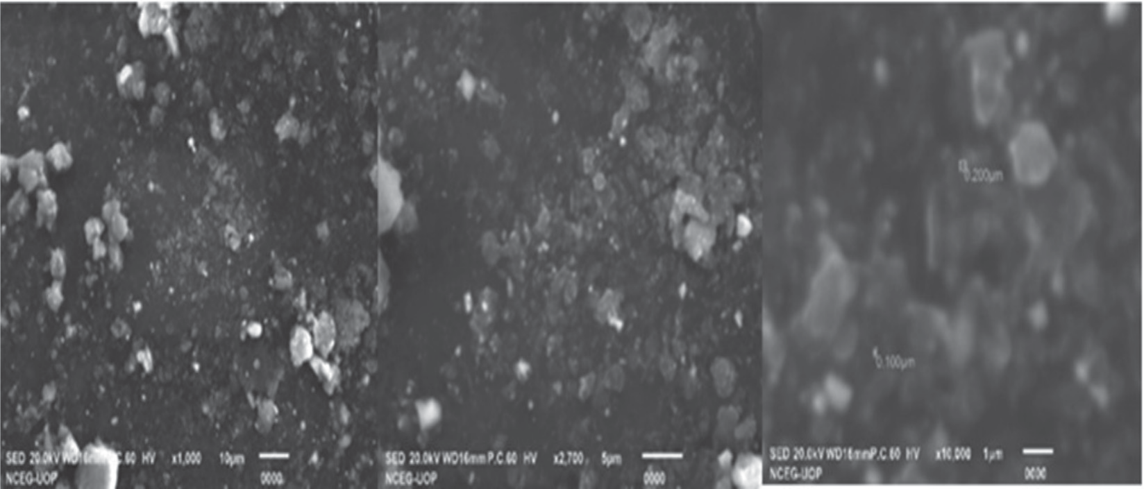
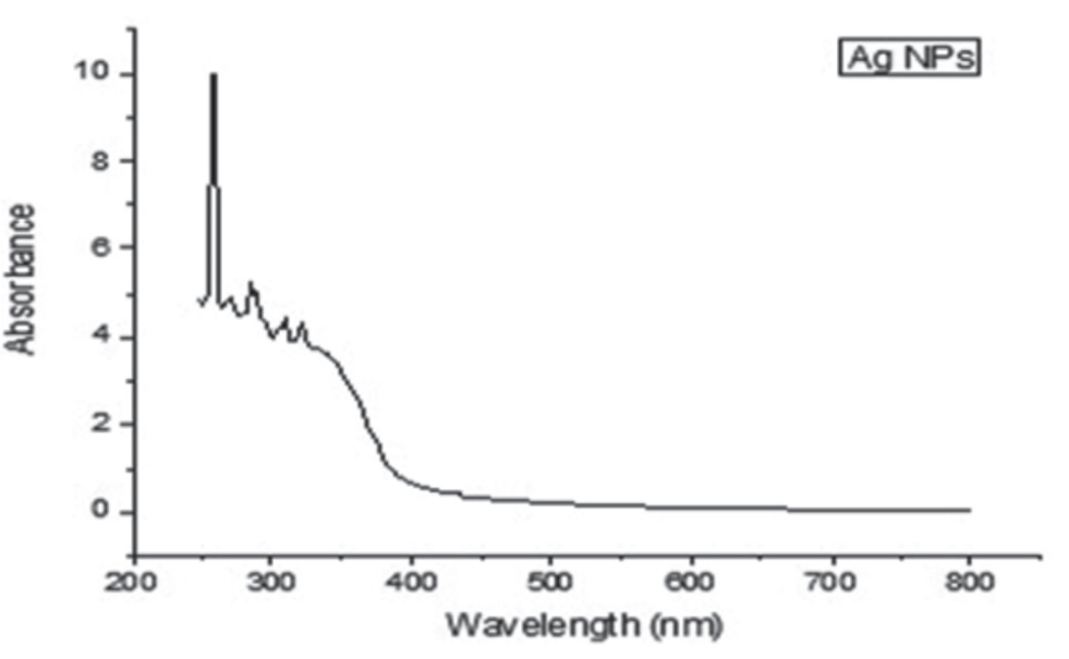
At this stage, the specification of AgNPs was carried out. SEM images (SEM analysis gives information about the sizes and morphological properties of the synthesized nanoparticles) of AgNPs are shown in Fig. 3 and Fig. 4. AgNPs are synthesized by A. euchroma plant. The UV-vis spectrum diagram showing its formation is presented in Fig. 3. Silver nanoparticles were synthesized from the fresh aqueous extract of root of the A. euchroma using 1mM of AgNO3 solution. By adding more silver nitrate solution, the intensity of the peak was increased, suggesting a higher concentration of AgNPs. In addition, as the concentration of AgNO3 was increased, the brown color of the solution became thicker, indicating the presence of more AgNPs. Solution of 2:1 and 3:1 used for UV spectroscopy at the wavelength of 200-800 nm for root. The UV showed a maximum wavelength of 250 nm.
Table 4.
Antibacterial activity of plant extracts and AgNPs.
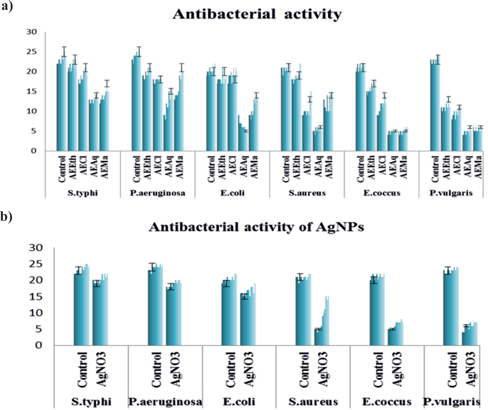
Antibacterial Activity of Plant Extracts and AgNPs
Here, we investigate the effects of various extracts of A. euchroma root with inhibitory potential (Ethanolic crude), methyl acetate, chloroform, and aqueous, selected bacterial strains (P. aeruginosa, P. vulgaris, E. coli, S typhi, S. aureus and Enterococcus) were added to the agar investigated using the diffusion method. Against bacteria, co-amoxiclav was used as a control. The results are summarized in Table 4 and Fig. 5. This makes A. euchroma root extract more valuable for study. In this work, different bacterial strains showed different zones of inhibition toward different extracts; the most susceptible strain was S. typhi and P. aeruginosa, which showed higher inhibitory zones, followed by E. coli, S. aureus, Enterococcus, and P. vulgaris, which showed the least inhibitory zone toward all extracts. The ethanolic crude and chloroform extracts were the most active of all the extracts. It can also be noted that as the concentration was increased, a considerable zone of inhibition was found. A. euchroma root extract appears to have greater inhibitory potential against Gram-positive bacteria. Contrary to these studies, in our study, different plant extracts showed activity on both Gram-positive and Gram-negative bacteria, albeit with varying degrees of inhibition. This situation can be explained by the biodiversity of the plant source and the difference in the extraction method. Because it is known that plants can produce an unlimited number and amount of aromatic compounds. Besides, plant components with antimicrobial properties, natural compounds directly or indirectly affect the biochemical processes of cells in cells, disrupting their physicochemical integrity. Chain reactions that may occur when any natural component affects the target region can also cause similar cell destruction in another part of the cell. Antimicrobial components are also known to affect proteins found in the cell wall. In this regard, A. eurochroma contains many secondary compounds that are biologically active. It can be said that the plant extracts may have exerted an antibacterial effect through these mechanisms, with the effects of these plants alone or in combination. Since the antimicrobial effect of silver is known, studies have generally focused in this direction. Thus, in this work, AgNPs were obtained from the plant root extract, and their antimicrobial effect was investigated. The results are summarized in Table 4 and Fig. 4. Hence, the antibacterial activity of AgNPs was also dose-dependent, zone of inhibition was increased by increasing the concentrations. S. typhi was the most sensitive of the bacteria tested. As a result, the strongest inhibition zones against S. typhi were detected. In our study, it was determined that the plant root contains very valuable compounds, and we can conclude that these compounds make a good combination and this situation positively affects the biological activity.
Table 5.
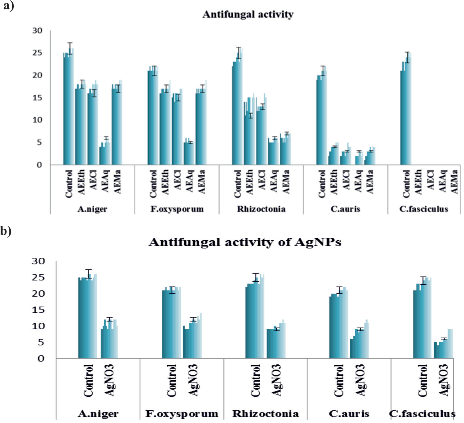
Antifungal activity of plant extracts and AgNPs.
Antifungal Activity of Plant Extracts and AgNPs
The antifungal effectiveness of ethanolic (crude), chloroform, aqueous, and methyl acetate extracts of A. euchroma root against fungal strains Rhizoctonia, C. fasciculus, A. niger, F. oxysporum, and C. auris was tested. Results of our study are depicted in Table 5 and Fig. 6. Remarkably, the various fungal strains displayed varied zones of inhibition towards different extracts. The most susceptible fungi were A. niger and F. oxysporum, which showed the highest inhibitory zones against ethanolic extract, chloroform, and methyl acetate while in aqueous extract all the strains showed less inhibition as compared to other extracts. Rhizoctonia showed moderate inhibition, while C. fasciculus was completely inactive against ethanolic extract, chloroform, aqueous, and methyl acetate. It’s also important to note that when the concentration was raised, a significant zone of inhibition was found. Moreover, the MIC values of the analyzed plant extract were still higher than those reported for other species. Furthermore, a secondary metabolite of saponin was detected in the studied plant. It is known that the saponin substance found in plants is mostly found in plants that are resistant to environmental conditions, but it is a compound with antifungal and antibacterial properties. On the other hand, we also analyzed the antifungal activities of AgNPs. Interestingly, AgNPs were active towards all the selected fungal strains at all the concentrations but at higher concentrations more inhibition was observed. The most susceptible fungi were F. oxysporum followed by A. niger, Rhizoctonia, C. auris while C. fasciculus showed less zone of inhibition among all the strains. This result showed a synergistic effect when combined with the plant’s antifungal activity capacity and AgNPs. At the end of this study, it can be suggested that plant extracts can be used safely with AgNPs in the treatment of these fungi that cause various diseases.
Antipellicle Activity of Bacterial and Fungal Strains
Another important biological activity parameter that constitutes our research is the evaluation of antipellicle activity. As is known, immediately after the removal of bacteria and all organic material from the tooth surface, new organic material begins to accumulate. In a short time, a cell-free, structureless organic film called a pellicle can completely cover the tooth structure. The pellicle is primarily formed by the selective precipitation of various components of saliva. In the structure of the pellicle, the components coming from the gingival groove fluid, dead cell wall residues of bacteria, and other microbial products were also found. The pellicle is an organic enamel cover composed mostly of proline-rich protein-containing saliva components. The pellicle contains receptors specific for the adhesion of different bacterial species. Recently, research on this subject has been intensified and especially studies on the prevention of its occurrence maintain the importance and up-to-dateness of the subject. For this purpose, initially, the antipellicle activity was tested against S. aureus, S. typhi, E. coli, P. vulgaris, Enterococcus, and P. aeruginosa bacteria using crude ethanolic extracts, chloroform, aqueous, and methyl acetate from the root of A. euchroma. The results 12.5 mg/mL and 16 mg/mL concentrations were used. Strong (+), moderate (++), weak (+++), and no inhibition (-) were used to indicate pellicle layer disruption. S. typhi all extracts showed strong inhibition except aqueous. At both concentrations (12.5 mg/mL and 16 mg/mL) ethanolic extract, chloroform, and methyl acetate showed strong inhibition however aqueous extract showed weak inhibition. P. vulgaris showed weak inhibition against ethanolic extract, chloroform, aqueous, and methyl acetate at both concentrations (12.5 mg/mL and 16 mg/mL). Secondly, the antipellicle activity was tested against Rhizoctonia, C. fasciculus, A. niger, F. oxysporum, C. auris fungi using crude ethanolic extracts, chloroform, aqueous, and methyl acetate from the root of A. euchroma. Rhizoctonia and A. niger showed strong antipellicle activity against all extracts, including ethanolic extract, chloroform, aqueous, and methyl acetate, at both concentrations (12.5 mg/mL and 16 mg/mL). To the best of our knowledge, the present study is the first report of plant extract’s antipellicle activity against bacterial and fungal strains.
Table 6.
Antipellicle activity against bacterial and fungal strains of root of A. Euchroma.
Antipellicle Activity of Bacterial and Fungal Strains
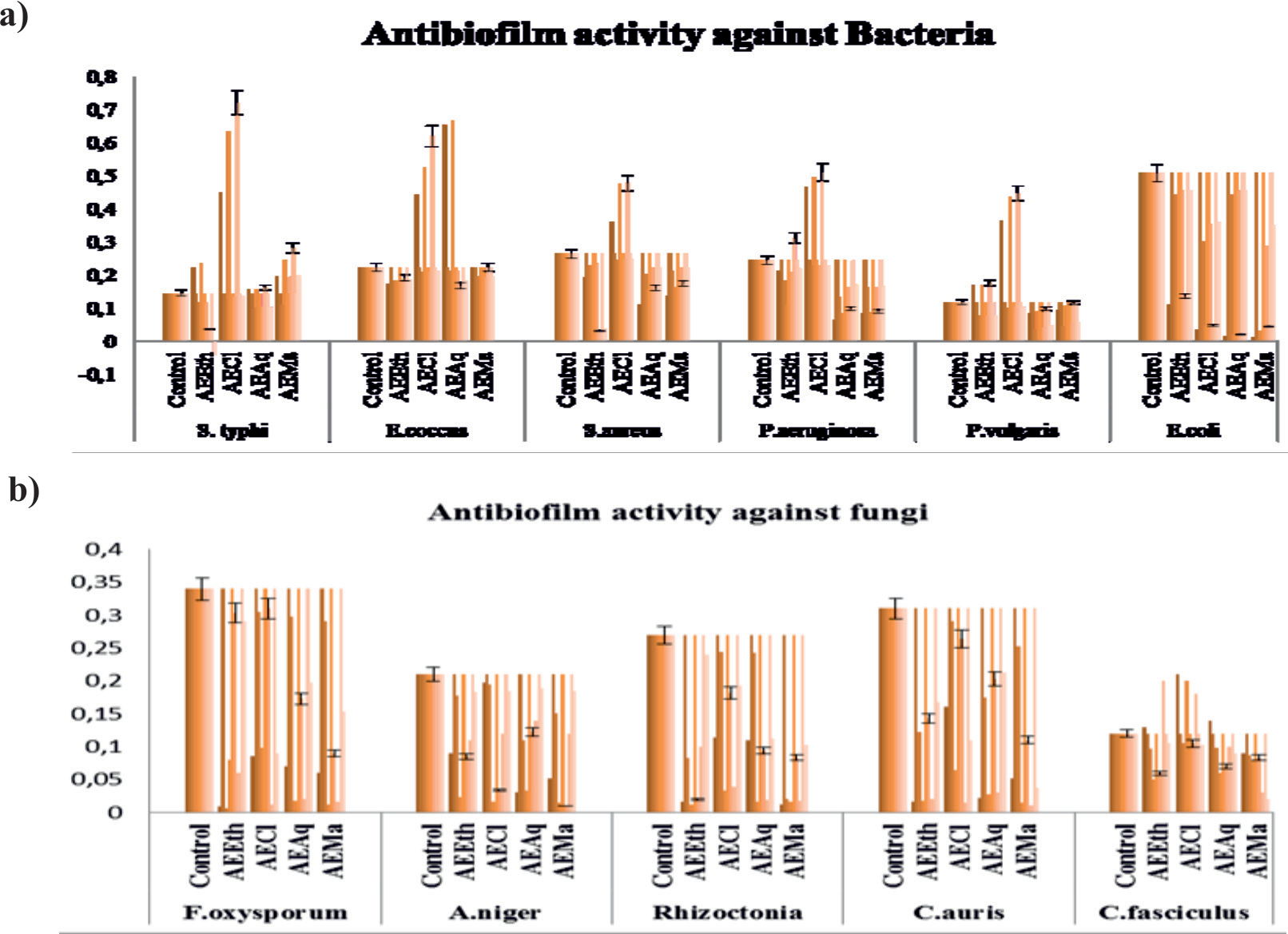
Since it is known that some plant extracts have bactericidal and antifungal activities as well as antibiofilm (biofilm inhibition) properties, we investigated the antibofilm activities of the A. eurochroma plant extracts we used in this study. For this, firstly, using four extracts (ethanolic (crude) chloroform, aqueous, and methyl acetate), the antibiofilm test was performed on six bacterial (P. aeruginosa, E. coli, Enterococcus, S. aureus, S. typhi, and P. vulgaris) and five fungal (Rhizoctonia, C.fasciculus, A. niger, F. oxysporum, C. auris) strains. The results of the antibiofilm activity showed in Table 7 and Fig. 7. There was also a negative and positive control, the positive control contained nutrient broth media and bacterial inoculum, while the negative control contained co-amoxiclav antibiotic, nutrient broth medium, and bacterial inoculum. The following formula was used to determine the biofilm formation of the extracts in comparison to a variety of bacteria and fungi.
More than one represents strong biofilm formation (no inhibition), 0.5 represents moderate biofilm formation (less inhibition), and less than 0.5 represents weak biofilm formation (strong inhibition). Against selected bacterial strains, all of the plant extracts showed a significant level of biofilm inhibition. S. typhi and Enterococcus were the most susceptible bacteria, as biofilm formation by these bacteria was greatly disrupted by all of the plant extracts, indicating that the plant extracts are significantly active to strongly inhibit biofilm formation, as the resultant SBF values towards bacteria were less than 0.5. The SBF values at higher concentrations are similarly higher than those at lower concentrations. Against selected fungal strains, all of the plant extracts showed various levels of biofilm inhibition. All of the plant extracts used greatly disrupted the biofilm formed by these fungi. Among the fungal strains, the most susceptible fungus was F. oxysporum followed by A. niger and Rhizoctonia because biofilm formed by these fungi was greatly inhibited by all the plant extracts, as all the resultant SBF values were less than 0.5 which show high inhibition. All the extracts which are used methyl acetate showed a higher extent of inhibition with the higher dose of concentration. In the literature review, no study was found showing the antibiofilm activity of the extracts obtained from the Arnebia species. In light of all these studies, it can be deduced that the root of A. euchroma contains very valuable biochemical molecules and that they greatly affect the biological activities of the plant. Without exception, all of the plant extracts tested were found to be quite effective against both bacterial and fungal strains, albeit at various rates. This study is the first to reveal the antipellicle and antibiofilm properties of the plant extracts used. Therewithal, the formation of plant extracts with AgNPs added a synergistic effect on the plant’s biological activities, and our study also provided unique information to the world of nanoscience. Considering these results, the extracts of the studied plant species and the active substances to be obtained from them can be evaluated as alternative drugs in the treatment of various diseases, especially bacterial and fungal. In addition, these plants can be an alternative drug source in the treatment of infections caused by biofilms formed by bacteria and fungi.
Discussion
Firstly, to improve our understanding of the biological activities of plants it is important to know their molecular composition. Thus, the qualitatively phytochemical screening of A. euchroma extract was performed. Table 1 shows the presence of bioactive components of the extract such as carbohydrates, alkaloids, amino acids, flavonoids, phenols, saponins, and tannins. Kala, [39] stated that different types of secondary metabolites found in A. euchroma correspond to their medicinal value worldwide. Xin et al. [40] reported that the medicinal parts, namely the roots, of A. euchroma contain naphthoquinones, phenols, benzoquinones, phenolic acids, alkaloids, and sterols. Especially, many studies have shown that naphthoquinones (phenolic compounds), the most prominent compound of A. euchroma, have various physiological activities such as anti-inflammatory, anticancer, and antibacterial, and are also widely used as natural pigments in the pharmaceutical, cosmetic, and printing industries [41]. The dried roots of the plant are used in industry to extract natural compounds for the preparation of medicines [39]. Triethyl borate (TEB) is used in industrial applications [42] as a solvent and/or catalyst in the preparation of synthetic waxes, resins, paints, and varnishes, as a component of some flame retardants and some welding fluxes in the textile industry. Octadecanoic acid, (2-phenyl-1,3-dioxolan-4- yl)methyl ester, cishas known for anti-tumourogenic properties [43]. Octadecanoic acid, (2-phenyl-1,3- dioxolan-4-yl) methyl ester, cis-have antioxidant and anti-inflammatory activity (Ganesh and Mohankumar). Siswadi and Saragih, [44] have reported that hexadecanoic acid, and ethyl ester have antioxidant, antimicrobial, and anti-inflammatory activity. Heptadecane, 9-hexyl- and 1,2-benzenedicarboxylic acid, and diisooctyl ester have antifungal activity [45- 47]. Hexasiloxane, 1,1,3,3,5,5,7,7,9,9,11, 11-dodecamethyl have antimicrobial, antibacterial, antiseptic, hair conditioning agent, and emollient properties [48]. Cyclotrisiloxane, hexamethyl-showed an anti-microbial activity [49]. The phenolic compounds identified in the methanolic extracts of Arnebia densiflora (Nordm.) Ledeb plant was also reported by Zengin et al. [50]. According to Zengin et al. [50], gallic acid, rutin, rosmarinic acid, quercetin, luteolin, and apigenin phytochemicals were identified from the methanolic extract of A. densiflora. Wang et al. [51], reported that shikonin, acetylshikonin, and β,β’ -dimethylacrylshikonin in root of A. euchroma. In this study, biologically active compounds were found in the root of the plant. Especially, malic acid and mandelic acid demonstrated antimicrobial activity [52, 53]. Moreover, malic acid is also a commonly used additive in skin care products. It has long been used in skin products due to its exfoliating properties. In addition, due to its chemical composition, it is able to penetrate deeper into the skin [54]. The synthesized AgNO3 was characterized by ultraviolet (UV)-visible spectroscopy [37]. As known, in addition to the shikalkin derivatives, the A. euchroma root contains some new compounds [55]. The antimicrobial effect of A. euchroma has been reported in many previous studies [56, 57]. Haghbeen et al. [20] reported that A. euchroma root THF-extract grown in Iran showed antimicrobial activity (shikalkin pigment) and demonstrated that the highest control yield of the extract was obtained on M. luteus. Sasaki et al. [58] stated in their study that acetyl shikonin obtained from A. euchroma inhibits C. albicans. Huang et al. [59] described shikonin substances obtained from the root of Arnebia that it has antibacterial effects at various rates against bacteria such as S. aureus (128 μg/μL), Streptococcus agalactiae (128 μg/μL), E. coli (256 μg/μL), Salmonella isolates (128 μg/μL), P. aeruginosa (512 μg/μL). Considering previous scientific reports. Due to the need for new antimicrobial agents that can kill or inhibit the growth of a wide variety of microbes, the use of nanotechnology in the development of effective antimicrobials has taken its place on the scientific scene as a promising new alternative [59]. The most important physicochemical parameters affecting the antimicrobial potential of AgNPs are size, shape, surface charge, concentration, and colloidal state. Its mode of action can be associated with various mechanisms such as adhesion to microbial cells, penetration into cells, generation of ROS (reactive oxygen species) and free radicals, or modulation of microbial signal transduction pathways [60, 61]. It is known that flavonoids, alkaloids, polyphenols, and terpenoids are the main metabolites used in phytonanoparticle production due to their strong reducing properties [62]. Doulah et al. [63] confirmed that A. fimbriopetala, A. linearifolia, A. garandiflora, and A. tubata species growing wild in Iran have a slightly significant effect on fungi. Other study, [64] also showed that the MIC and MFC values of commercially purchased shikonin had a 30% greater effect on fungi [dermatophytic fungal strains; Trichophyton mentagrophytes (PTCC5054), Trichophyton rubrum (PTCC5143), Microsporum canis (PTCC5069), and C. albicans (PTCC5027), saprophytic strains; Aspergillus fumigatus (PTCC5009) and Penicillium chrysogenum (PTCC5076)] than the alcoholic extraction of the root. Shen et al. [65] reported that the A. euchroma extract had little effect on fungi strain. In contrast to these studies, Haghbeen et al. [20] demonstrated that A. euchroma extracts were ineffective against the fungi (C. albicans and A. niger) they tested. The previous studies are evaluated, and it is clear that different plant extracts have an inhibitory effect on the fungi we tested. Saponins act as a chemical barrier against fungi [66]. It can be said that the antifungal property of the plant is due to the valuable secondary metabolites it contains. The effect of different plant extracts and secondary metabolites on pellicle inhibition has been reported previously. Khan et al. [34] reported that both Mirabilis jalapa and Ajuga bracteosa extracts showed moderate (+++) and weak (++++) effects on pellicle inhibition against the tested P. aeruginosa strains. Pattiyathanee et al. [67] examined the effect of curcumin on Helicobacter pylori biofilm both qualitatively by pellicle test and quantitatively by crystal violet staining, making it a potential complementary drug for the treatment of H. pylori biofilm-related infections. We believe that the valuable findings of our study will contribute to the scientific world. Biofilm is a layer consisting of colonies of microorganisms attached to a surface and the extracellular polysaccharides (EPS) they produce, proteins, and organic and inorganic substances. Many gram-positive, gram-negative bacteria and fungi of clinical importance form biofilms. Approximately 65 % of infectious diseases are associated with biofilm-forming bacteria [68]. For such reasons, the use of molecules and plant extracts that prevent or inhibit biofilm formation has become promising for the treatment of agents in recent years [69]. Therefore, our study represents a first and therefore has importance. However, Wan et al. [70], it was found that shikonin showed antibacterial activity against both planktonic and biofilm forms of S. aureus. They mentioned that shikonin metabolite destroys the integrity of the cell membrane of S. aureus, depolarizes the cell membrane, decreases the intracellular ATP concentration, changes the cell morphology and shows strong antibacterial activity by inhibiting the transcription, and expression of virulence genes. Furthermore, they said that since shikonin has strong antibacterial activity against S. aureus, it can be used in food production and processing environments to effectively control S. aureus. Vysakh et al. [71] reported that the ethyl acetate fraction (EFRA) of Rotula aquatica was able to inhibit bacterial growth and biofilm formation in their study evaluating the anti-bacterial and anti-biofilm activity against clinically isolated uropathogenic E. coli BRL-17. Based on their current study, they suggested the potential use of EFRA as an anti-bacterial drug.
Table 7.
Antibiofilm activity of root of A. euchroma against bacterial and fungal strains.
Conclusions and Recommendations
It is concluded that the root of A. euchroma has some active constituents as revealed by qualitative (using chemical reagents) and quantitative (GC-MS and HPLC analysis) screening. With the simple, fast, cheap, and environmentally friendly biological synthesis method, it can synthesize new generation nanomaterials that can be easily used in different application areas. Pharmacological evaluation revealed that both extracts and AgNPs of the root of A. euchroma have significant antibacterial, antifungal agents against selected bacterial and fungal strains. It has been a promising study for the synthesis of more efficient nanostructures and the expansion of their use in biomedical and bioanalytical applications by bringing together materials with different properties and providing a synergistic effect between them. With detailed studies on this subject, the usage areas of nanoparticles can be further expanded. The plant could be used in developing modern antimicrobial drugs and therapeutic purposes for various diseases in the future.

