

Introduction
Thyroid diseases are the second largest disease in the endocrine field and the incidence of thyroid cancer is increasing worldwide [1, 2]. At present, the causes of thyroid diseases are still unclear. Studies have confirmed that environmental endocrine disruptors (EDCs) are one of the risk factors for thyroid diseases [3, 4]. Nonylphenol (NP) as a typical representative of EDCs is widely used in industry and daily life and led to serious environmental pollution [5]. The previous studies on NP of the research group focused on its influence on the stomach [6], endocrine immunity [7- 9], nerves [10], cardiovascular disease [11, 12], etc. There are no specific reports of NP effects on thyroid diseases around the world. Regarding the interference influence of EDCs on thyroid function, most of the current studies focus on the detection of hormones in animals at short-term exposure [13, 14], and the study of the influence of chronic NP exposure on thyroid function has not been reported.
Guizhou zinc-selenium tea, which originated in Fenggang County, Zunyi City, Guizhou Province, China, is a green tea (Camellia sinensis) that is rich in zinc and selenium, two trace elements required by the human body due to the local special geographical environment, as well as contains normal levels of tea polyphenols [15-17]. Current studies have indicated that tea polyphenols, catechins, flavonols, lignans, minerals, and other bioactive substances in tea act as antioxidants [18,19], and polyphenols may prevent cancers by affecting cancer cell proliferation, differentiation, apoptosis, inflammation, and signal transduction pathways, and by regulating enzymatic activity [20, 21]. It has been reported that the biosynthesis of thyroid hormones requires assistance from hydrogen peroxide [22]. Tea polyphenols can promote central nervous system excitation and metabolism [23]. It has also been proven that tea effectively reduces the risk of thyroid cancer [24], and protects the impaired thyroid tissues [25]. Thus, we speculated that zinc-selenium green tea may inhibit the development and progress of thyroid diseases.
However, most of the studies on tea focused on tea intake and basic metabolism, antioxidant action, and cardiovascular diseases, but few have examined the protective mechanism governing thyroid-related diseases. Therefore, to further investigate the effect of NP exposure on thyroid function and subsequent tissue structure damage in rats, the protective effect of zinc-selenium green tea on damage to thyroid function induced by NP was studied. This study explored 1) whether chronic exposure to NP at environmental concentration would induce the imbalance of thyroid hormone levels and thyroid dysfunction, and explore the alterations in the expressions of estrogen receptors and thyroxine receptors in NP-induced thyroid hyperplasia disease. 2) To examine the protective effect of zinc selenium tea on thyroid function in NP-induced thyroid hyperplasia disease, and explore the protective mechanism of zinc selenium tea on NP-induced thyroid diseases.
Materials and Methods
Experimental Animal Treatment
Four-week-old male SPF SD rats (60-80 g) were housed at temperature-controlled (24±2ºC) and humidity-controlled (50%±5%) conditions in a 12-hour light/dark cycle with ad libitum access to food and water. Squirrel cages made of polyacrylamide were selected to avoid the extra NP intake of experimental rats. The rats were obtained from the Animal Center of Tianqin Biotechnology Co., Ltd. (License No: SCXK2014- 0011, Changsha, China). This animal experiment was approved by the Animal Experiment Ethics Committee of Zunyi Medical University (No. 2018-2-166).
Grouping and Exposure Dose
Relevant research shows that the highest content of NP in the Pearl River waters is about 3 µg/L. The highest content is about 12 mg/kg, while the NP content in carnivorous fish is about 9 mg/kg [26]. Danish Institute of Safety and Toxicology proposes that the tolerable daily intake (TDI) of NP in the human body is 5 µg/kg/d [27], comprehensively considering that NP is difficult to degrade and its bioaccumulation, as well as related diseases caused by low concentration exposure reported in recent years. In this study, rats were weighed daily and the dose of toxicity was determined by gavage based on their body weight. The gavage volume was 5ml/kg/d and a high dose NP (40 mg/kg/d) similar to the environmental exposure concentration was selected for the toxicity experiment with continuous exposure during the treatment.
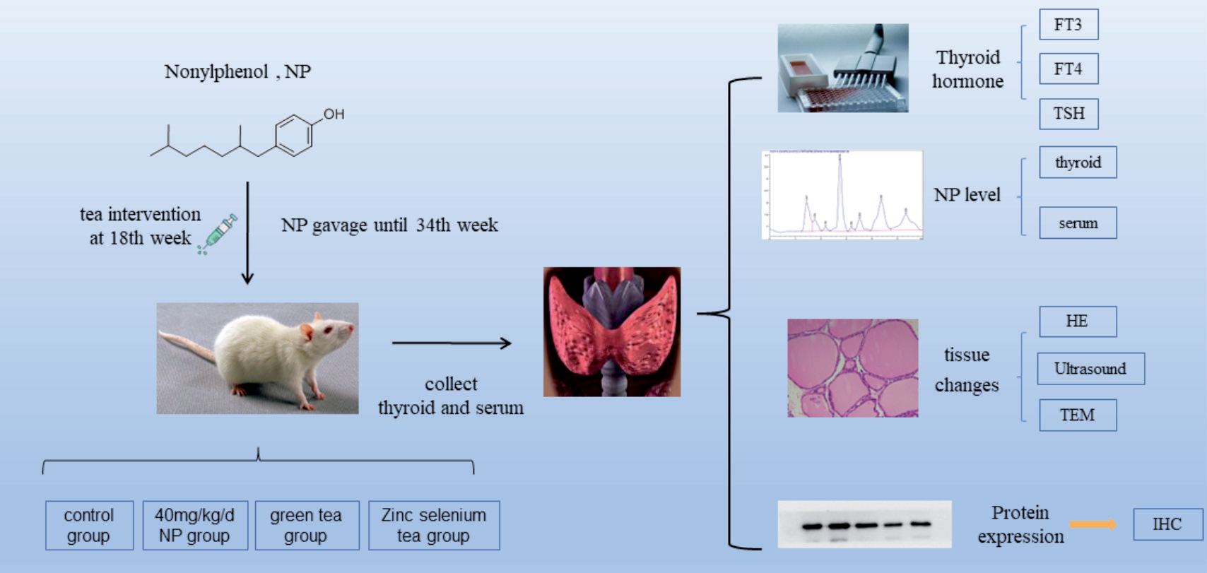
Forty rats were divided into four groups (each with 10 rats): control group (corn oil), NP group (40 mg/kg/d), NP (40 mg/kg) + green tea group (1 g/kg/d), NP (40 mg/kg) + zinc-selenium tea group (1 g/kg/d). The rats were treated by gavage for 34 weeks. Tea intervention was initiated in the 18th week with a dose of 5 ml/kg/d by gavage at a concentration of 0.2 g/ml. A flowchart of exposure protocols for the rats and the duration of exposure is shown in Fig. 1.
Tea preparation: tea soups for gavage were prepared with zinc-selenium green tea (Guizhou Wanhuyuan Tea Co., Ltd, China. Zinc content: 40-100 mg/kg, selenium content: 0.05-2.5 mg/kg) or common green tea (Guizhou Weidao Tea Co., Ltd, China). Zinc-selenium green tea/green tea was weighed and decocted in boiling double-steamed water (weight of water:weight of tea = 3:1), for 3 consecutive times and 10 minutes each time. The tea decoctions were combined together, concentrated to 0.2 g/ml under reduced pressure, and stored at 4ºC.
Experimental Process and Methods
Animal Tissues and Blood Collection
The rats were fasted overnight before execution. They were anesthetized by intraperitoneal injection of 20% ethyl carbamate (5 ml/kg). After anesthesia, the blood was taken from the abdominal aorta, and then placed in a refrigerator at 4ºC for 12 hours and centrifuged. The serum was collected and placed in a refrigerator at -80ºC. The left thyroid tissues were taken and fixed in 4% paraformaldehyde and the right thyroid tissues were placed directly in Eppendorf (EP) tubes and frozen in refrigerator at -80ºC.
B-Ultrasound Examination
One week before the dissection, the rats were subjected to a B-ultrasound examination of the thyroids at the State Key Laboratory of Pharmacology of Zunyi Medical University. The animals were anesthetized by intraperitoneal injection of 20% urethane (5 ml/kg). After anesthesia, the rats were shaved and taken to a supine position to acquire B-ultrasound images of thyroids.
FT3, FT4, and TSH Tests by ELISA
Levels of FT3, FT4, and TSH in serum were detected by ELISA kits, and the procedures were carried out by the manufacturer’s instructions. (FT3 kit, product code: JL21437, Jianglai Biotechnology Co., Ltd, Shanghai, China), (FT4 kit, product code: JL21436, Jianglai Biotechnology Co., Ltd, Shanghai, China), (TSH kit, product code: JL11830, Jianglai Biotechnology Co., Ltd, Shanghai, China).
Immunohistochemistry of Thyroid Tissues
After dewaxing, antigen retrieval, inactivation of peroxidase, blocking, primary antibody incubation (anti-estrogen receptor antibody, anti-thyroxine receptor 1:100), secondary antibody incubation, hematoxylin counterstaining, dehydration, mounting, etc., immunohistochemical sections were made and photographed under an optical microscope. Image Pro Plus 6 (Media Cybernetics, USA) was used for the analysis of immunohistochemical images.
Histopathology of Thyroid Tissues
After weighing, the left thyroid tissues were quickly placed in EP tubes filled with 4% paraformaldehyde and fixed for 24 h. Samples were immersed in xylene and alcohol (100%,95%,80%,70%), stained with 1% hematoxylin for 5 min, stained with 0.5% Eosin for 3 min, and re-immersed in alcohol (70%, 80%, 95%, 100%) and xylene, and sealed with neutral gum to make HE slices. Slices were observed and photographed under an optical microscope. Images were analyzed by Image J software (National Institutes of Health, USA).
ICP-MS Detection of Zinc and Selenium in Tea Water
After mixing the tea water by vertexing, an appropriate amount of tea water was put in a digestion jar containing 5 ml of nitric acid (HNO3) and was then heated at 110ºC for 30 min. After slightly cooling, an additional 1 ml HNO3 was added, and the digestion jar was then sealed and placed into a microwave apparatus for digestion. Following digestion, the digestion jar was removed from the apparatus, and the acid was removed by evaporative heating. After the solution had cooled, the solution was transferred to a 50 ml volumetric flask. The inner wall of the digestion jar was cleaned with HNO3 solution, and the washing solution was collected and transferred into the volumetric flask. Gradually, the HNO3 solution was further added until reaching the scale line on the volumetric flask. Then the concentration of zinc and selenium in tea water was determined by inductively coupled plasma mass spectrometry (ICP-MS) [28].
Statistical Methods
SPSS software (v18.0; SPSS Inc., Chicago, IL, USA) was used to perform statistical analysis. All descriptive statistics for variables in this study were reported as mean ± standard deviation. When analyzing the difference between different dose groups at the same exposure time, if the data obeys a normal distribution, the method of single-factor analysis of variance was applied, a multiple comparison in ANOV analysis was then performed on the groups with significant differences; if the variances are uniform, the Tukey method was used for pairwise comparison; if the variances were not uniform, the Dunnett’s T3 method was used for pairwise comparison. If the data did not obey the normal distribution, Kruskal Wallis was used for group comparison. Independent sample t-test was used in the comparison of different exposure durations at the same dose; the statistical chart was made by GraphPad Prism 6, and the test level was α = 0.05, and P<0.05 indicated that the difference was statistically significant.
Results
Concentrations of Zinc and Selenium in Tea Water
The concentrations of zinc and selenium were 3,959±51.32 ng/ml and 27.74±0.36 ng/ml in zinc-selenium tea water, and 2,106±27.31 ng/ml and 23.34±0.30 ng/ml in common green tea respectively. Both the concentrations of zinc and selenium were higher in zinc-selenium tea water than those in common green tea (P<0.01, Fig. 2).
Effects of Zinc-Selenium Green Tea on Thyroid Function-Related Serum Hormone Levels
The level of FT3 in the NP group was significantly higher than that of the control group (H = 8.64, P = 0.03). Although there was no statistical difference between the two tea groups and the NP group, the FT3 level in the two tea groups was still slightly lower than that of the control group (Fig. 3a). After exposure to the dose of NP, the FT4 level of serum showed a significant decrease, which was statistically different from that of the control group (F = 4.21, P = 0.02) (Fig. 3b). However, after tea treatment, the FT4 level increased slightly, and there was no statistical difference compared with the control group. Simultaneously, the TSH level of serum showed no statistical difference among groups (Fig. 3c).
Effects of Zinc-Selenium Green Tea on the Thyroid by Cardiac Doppler Ultrasound
There were thyroid nodules in both the zinc-selenium green tea group and NP group rather than the normal green tea group and control group. There was no hyperechoic point in the hypoechoic area of the right thyroid parenchyma in the zinc-selenium green tea group, and the symptoms were milder than those in the NP group (Fig. 4).
Pathological Alterations of Thyroid in Rats
The morphological structure of thyroid follicular cells in the control group was normal, while the group exposed to NP showed varying degrees of morphological and structural damage, manifested as irregular follicle shapes, increased number of small follicles, and increased follicular epithelial thickness. Although both the green tea group and the zinc-selenium green tea group exhibited damage compared with the blank group, the damage was decreased compared with the NP group and was mainly manifested by thinner epithelial thickness (Fig. 5a and b).
Fig. 2.
Concentrations of zinc and selenium in tea water (n = 5 - 7). a) Concentration of zinc in different tea treatment groups. b) Concentration of selenium in different tea treatment groups. * vs General tea treatment group, P<0.01.
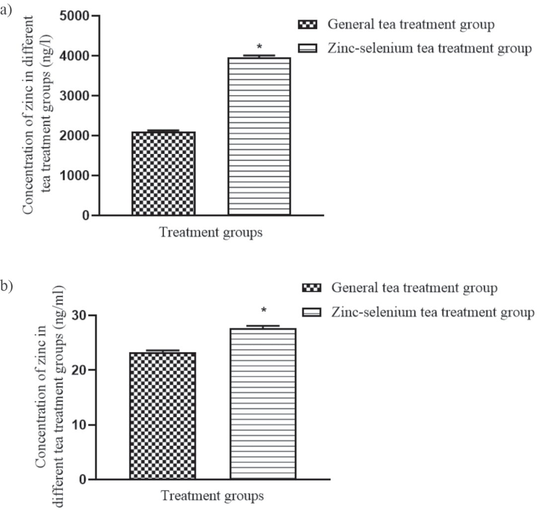
Fig. 3.
Changes in serum-related hormones in rats (n = 5 - 7). a) Serum FT3 level in rats. b)Serum FT4 level in rats. C: Serum TSH level in rats. * vs Control group, P<0.05,† vs Low NP group, P<0.05.
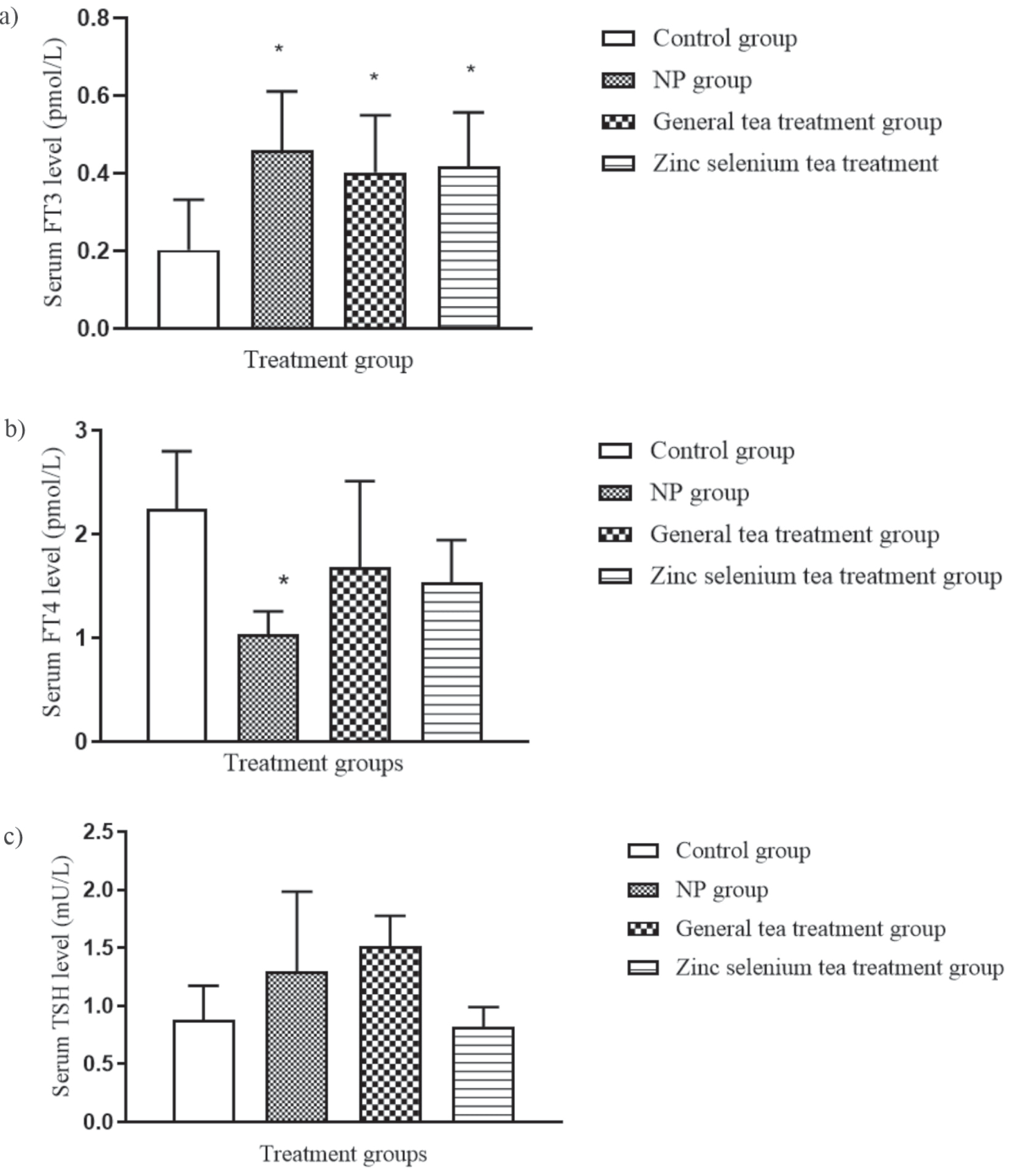
Fig. 4.
Effects of zinc-selenium green tea on right thyroid parenchyma in the treatment groups. (Red arrow: hypoechoic area). C: Control group. NP: NP group. G: Green tea treatment group. Z: Zinc-selenium green tea treatment group.
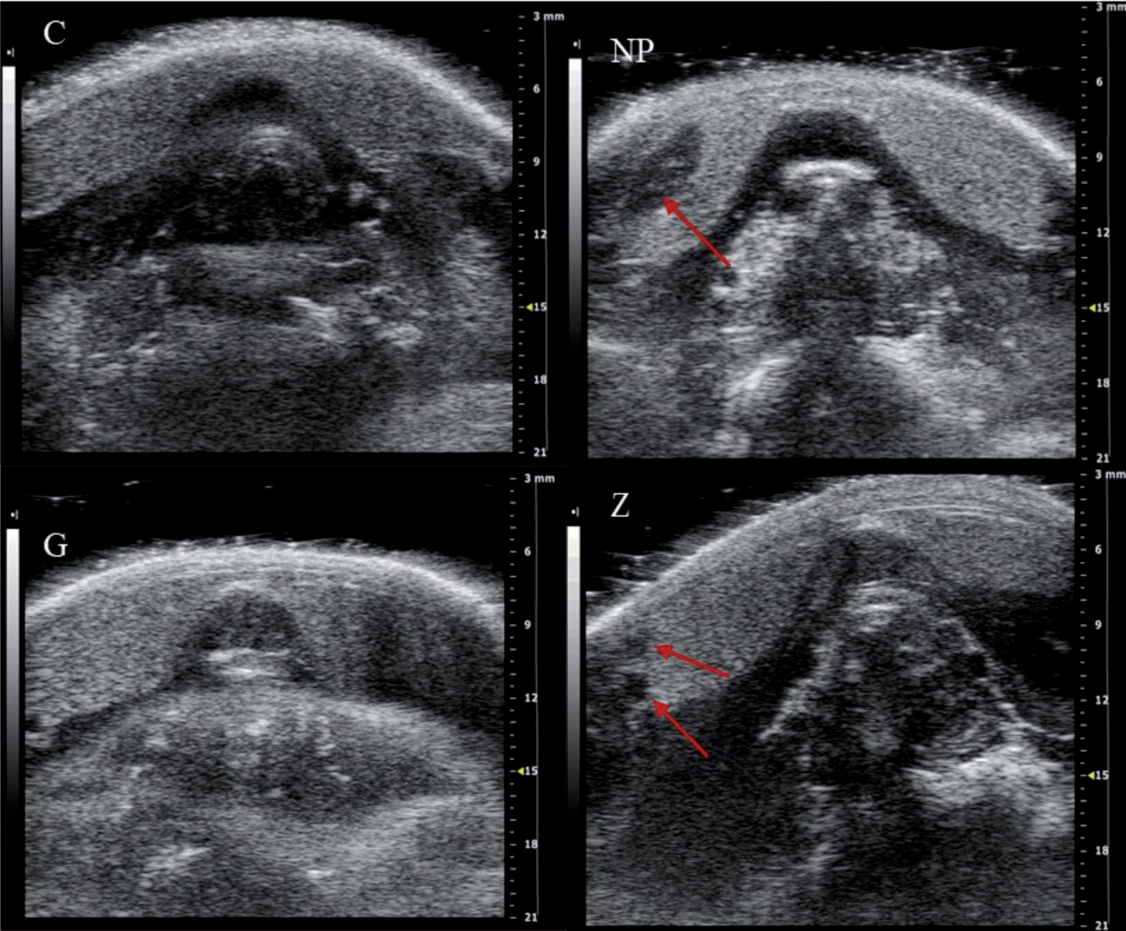
Fig. 5.
Pathological alterations of thyroid follicular cells in rats. (Black arrow: epithelial thickness of thyroid follicular cells. Red arrow: follicular size of thyroid follicular cells). C: Control group, NP: NP group, G: General green tea treatment group, Z: Zinc-selenium green tea treatment group. A: Morphological changes of thyroid follicular cells in rats (400×). B: Morphological changes of thyroid follicular cells in rats (200×). C: Changes in the single follicular area and epithelial thickness of thyroid cells in each group under light microscopy. * vs the Control group, P<0.05,† vs the NP group, P<0.05.
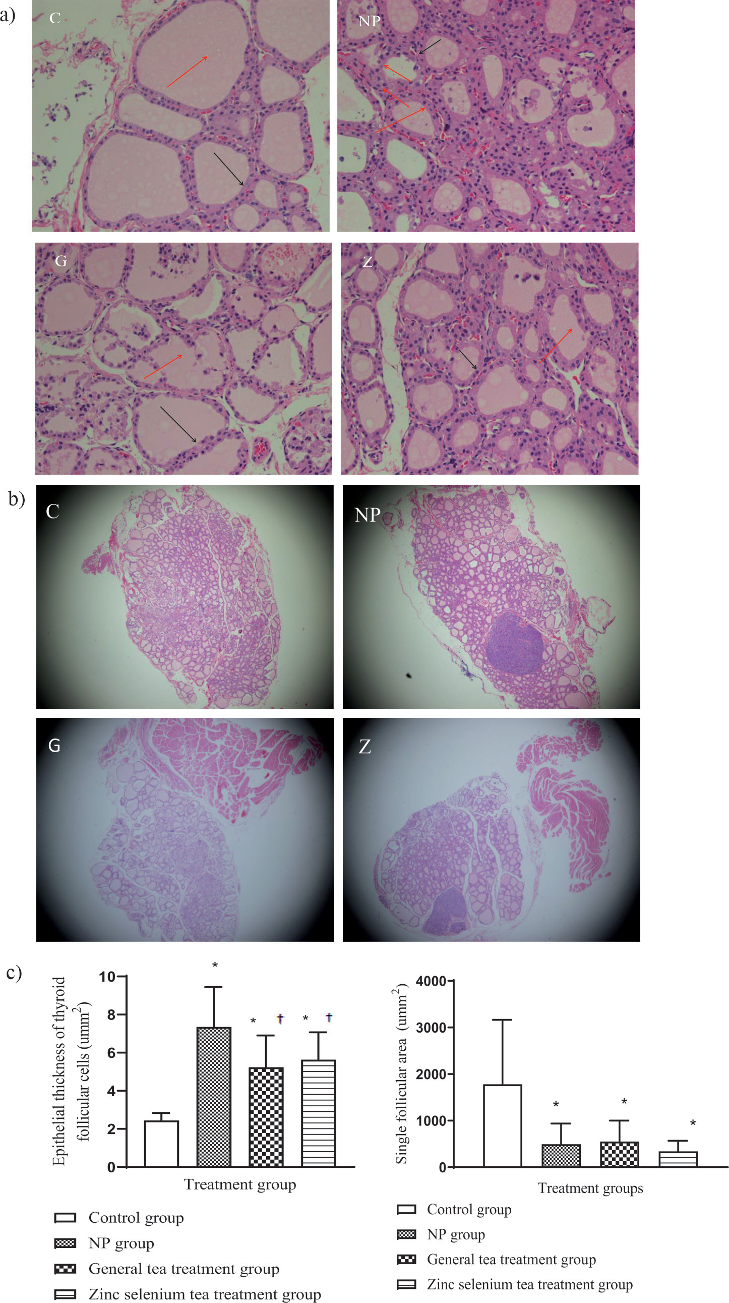
Quantitative analysis of thyroid follicular cells in each group under light microscopy can be observed. There were statistical differences in the follicular epithelial thickness (H = 38.85, P≤0.001) and single follicular area (H = 14.55, P≤0.001) between NP and tea groups with the control group. The epithelial thickness of the tea group was significantly lower than that of the NP group. The single follicular area in the tea group was similar to that in the NP group (Fig. 5c).
Effects of Zinc-Selenium Green Tea on the Expression of Proteins Related to Thyroid Tissue Injury
Exposure to NP could induce the up-expression of ERα, ERβ, TRα, and TRβ proteins in thyroid tissues. After the intervention of tea, the expression of ERα (F = 20.75, P≤0.001), ERβ (F = 32.32, P≤0.001), TRα (F = 13.81, P≤0.001), and TRβ (F = 13.92, P≤0.001) in the tea group was decreased compared with that in the NP group, and there was a statistical difference between the zinc-selenium green tea group and the NP group (Fig. 6).
Discussion
In the current study, the protective effects of zinc-selenium tea on thyroid damage induced by long-term exposure to NP were first explored by the long-term intragastric administration of zinc-selenium tea in rats. The results of the study showed that the damage of NP to thyroid tissue structure was reduced, the expression of hormone receptors increased by NP exposure in thyroid tissues was significantly reduced, and the thyroid hormone levels tended to be normal after long-term NP exposure rats ingested zinc-selenium tea.
Green tea contains tea polyphenols, catechins, chlorophyll, vitamins, and other nutrients, and is widely cultivated and drunk by the Chinese [29]. Guizhou zinc-selenium tea, as a kind of green tea, is richer in zinc and selenium elements and tea polyphenols [30]. Zinc and selenium are essential trace elements in the human body that act as important cofactors for multiple mammalian proteins, thereby performing a vital role in reducing oxidative stress and preventing the resulting DNA damage caused by reactive oxygen species [31]. It has been shown that both zinc and selenium are implicated in the pathogenesis of diabetes. Zinc has been shown to activate key molecules involved in various cell signaling events that maintain glucose homeostasis, modulate insulin receptors, and prolong the action of insulin [32]. Selenium is a vital trace element for maintaining the proper functioning of the immune system and the thyroid. Animal studies have demonstrated that moderate selenium supplementation can decrease adverse events due to iodine overdose and prevent destructive inflammatory lesions of the thyroid [33]. In the present study, ICP-MS revealed that the content of both zinc and selenium in zinc-selenium tea was significantly higher than that measured in regular green tea drinks. Moreover, we observed that rats consuming zinc-selenium tea displayed less severe pathological lesions of the thyroid than those drinking regular green tea. This discrepancy may be attributed to the protective effects of the zinc and selenium present in the tea on thyroid damage caused by the medication NP-Thyroid. Studies have shown that tea polyphenols are the main functional components of green tea, which have antioxidant, anti-inflammatory, and anti-cancer activities [34]. Therefore, the effect of green tea on the thyroid is explored, and the protective mechanism of green tea could be significant for the prevention and treatment of thyroid diseases. Therefore, tea treatment was applied to rats that had been exposed to NP for a long time, to analyze whether zinc-selenium tea can interfere with the occurrence and development of thyroid diseases and its potential protective mechanism.
In the early stage of this experiment, it was confirmed that long-term exposure to NP would increase the serum FT3 level and decrease the FT4 level in rats. After tea treatment, it was found that the FT3 level of the normal tea group and the zinc-selenium tea group decreased compared with the NP group, but the FT4 level increased and the FT4 level showed no statistical difference from the control group. Although the FT3 and FT4 levels of the tea group were not statistically different from the NP group, the difference between each group and the control group became smaller. The short duration of drinking possibly made the tea effect not obvious, indicating that tea drinking has a positive significance on the homeostasis of thyroid hormones. Furthermore, tea intake would cause the level of FT3 to decrease and the level of FT4 to increase as reported in another study [35]. Detecting the content of NP in thyroid tissues and serum found that the accumulation level of NP did not decrease due to the intake of tea, again indicating that NP was difficult to metabolize after being ingested by the body, and also confirmed the previous hypothesis of this study: drinking tea does not remove the NP in the body, but may interfere with the pathogenic mechanism of NP [36].
Thyroid ultrasound results showed that the damages of the tea group were significantly lighter than that of the NP group. Although there were still hypoechoic areas in the zinc-selenium tea group, no strong echo points similar to the NP group were found in the hypoechoic area. The results of HE staining also showed the positive effect of tea drinking on the protection of the thyroid. Although there was no statistical difference between the small follicular area in the tea-drinking group and the NP group, the thickness of follicular epithelium was significantly lower than that of the NP group, also confirming that tea can protect thyroid tissues from damage. The HE staining results demonstrated the similar results of this study. Long-term tea drinking attenuates damage to the thyroid caused by the medication NP-Thyroid.
Fig. 6.
Effects of zinc-selenium green tea on the expression of proteins related to thyroid tissue injury. Immunohistochemistry of thyroid tissues (200×). *vs Control group, P<0.05, †vs NP group, P<0.05. C: Control group, NP: NP group, G: General green tea treatment group, Z: Zinc-selenium green tea treatment group.
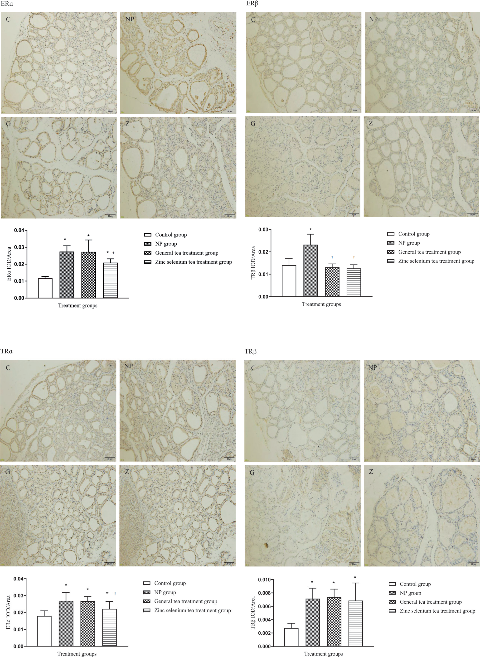
The results of immunohistochemistry showed the protective effect of zinc-selenium tea on thyroid injury caused by NP. Previous studies have identified that EEDs, represented by NP, could interfere with normal thyroid function by disrupting the expression of hormone receptors in the body after being ingested by the body [37] Another study showed that drinking green tea was negatively correlated with the risk of thyroid cancer in postmenopausal women and positively correlated with the risk in premenopausal women [38]. Therefore, some scholars believe that the intake of tea may lead to changes in the activity of estrogen, and estrogen could play a certain role in the occurrence of thyroid tumors, so they believe that tea may prevent the occurrence of thyroid diseases [39-44]. An increase in the dose of NP would lead to an increase in the expression of ERα, ERβ, TRα, and TRβ proteins, and led to changes in the structure and function of thyroid tissues have been confirmed. Interestingly, it was found that the ERα, ERβ, and TRα protein levels in the general green tea group and the zinc-selenium tea group were significantly lower than those of the NP group, and were similar to the control group, although the NP content in the body was not decreased. The changes in the zinc-selenium tea group were more significant than those in the general green tea group.
Based on the above experimental results, it is preliminarily believed that zinc selenium tea could have a stronger protective effect on thyroid diseases caused by NP exposure, and after ingestion, it was likely to protect thyroid function by interfering with hormone receptor expression. In this study, zinc-selenium tea was as an entry point, which could provide some scientific clues for the follow-up of Guizhou zinc-selenium tea-related research and the development of drugs for thyroid disease.
Conclusions
Chronic NP exposure could lead to the pathological damage of thyroid tissue, imbalance of thyroid hormone, and thyroid hyperplasia disease, as well as changes in the expressions of hormone receptor proteins, which might be involved in the mechanism of NP-induced thyroid dysfunction. Zinc selenium tea could decrease the expression levels of ERα, ERβ, TRα, and TRβ proteins in the thyroid tissue of rats exposed to NP, and provide protective effects for the thyroid damage caused by NP.

