

Introduction
Bacteriophages, as biological control tools, have sparked scientific interest as an alternative to controlling harmful bacteria, especially with rising antibiotic resistance. The concept of phage therapy has steadily gained acceptance in Europe and the United States due to its efficacy in treating particular bacterial infections [1-4]. Despite the potential advantages, questions remain about its safety, efficacy, and feasibility in various settings, agricultural and non-agricultural alike.
Notably, phages are gaining attention for their potential in both agricultural and non-agricultural sectors. They have shown promising results in controlling plant pathogens in farming, significantly reducing the reliance on chemical pesticides [5- 7]. Concurrently, in non-agricultural areas such as human health, food safety, and wastewater treatment, phage therapy is also being explored. Bacteriophages’ inherent capability of specific targeting allows for the management of specific pathogens without disrupting other beneficial microbiota, making them an attractive solution in these fields [8, 9].
A common bacterial pathogen that causes food poisoning is Salmonella. Recent years have seen a surge in interest in the application of Salmonella phages as biological controllers. Due to the vast diversity of the Salmonella genus, attempts are required to find lytic Salmonella phages that target various serovars [10]. Multiple research groups have identified several phages that may be useful in the biological control of Salmonella in foodstuffs [11, 12].
Nonetheless, one of the main challenges is the specificity of phages, which can make locating the right phage for a specific therapy difficult. For this reason, significant research is directed toward understanding the host-phage interaction, methods to expand the host range, and its impact on biocontrol [13, 14].
In this research, we are focused on the potential use of phages for the biocontrol of Salmonella Typhimurium (S. Typhimurium), a common pathogen in poultry and dairy products. Phages for this study were isolated from a wastewater treatment facility in Cairo. This investigation is prime for addressing a significant gap in the literature and further understanding the potential of phages in the food safety area.
Materials and Methods
Bacterial Strains and Growth Conditions
16 distinct Salmonella spp. authenticated bacterial isolates from various serovars were gathered from the American Type Culture Collection (ATCC), Central Public Health Laboratory, Ministry of Health, Egypt (CHL), and US Naval Medical Research Unit No. 3 (NAMRU-3) culture collections and used as hosts for phage isolation and application in this study. The frozen glycerol stock was used to streak all bacterial isolates onto tryptic soy agar (TSA) plates. Each strain was then sub-cultured by selecting a single colony from the TSA plates, inoculating it into tryptic soy broth (TSB), and incubating it at 37ºC to get new overnight cultures, as indicated in (Table 1).
Table 1.
Bacterial strains used to isolate bacteriophages and their sources of isolations.
Samples
Various water and soil samples from animal specimens were collected and examined for the presence of phages, as shown in (Table 2).
Phage Sample Enrichment and Isolation
An optimized modification of a published protocol for the isolation of phages was used for the enrichment of phages from water samples, such as sewage, and solid samples, such as soil [15]. 100 ml of sewage water samples were collected into sterile screw-cap cups (in the case of soil, 10 g of soil were mixed with 40 ml of sterile TSB medium or phosphate buffered saline (PBS) in centrifuge tubes and vortexed for 1-2 hours). Samples were centrifuged at 8000×g for 10 minutes to remove solid particles, and the clarified supernatant was filtered through 0.45 μm syringe filters to remove any endogenous bacteria and stored at 4ºC. Next, the filtered samples were added at a 1:4 (v/v) ratio to pre-enrichment sterile Tryptic Soy Broth medium (e.g., 2 ml of wastewater + 8 ml of broth) and inoculated with 100 μl of a fresh overnight mixture of host cultures from all the available Salmonella spp. bacterial isolates. Enrichment cultures were incubated overnight at 37ºC in a shaker incubator. Afterward, a few drops of chloroform were added to the pre-enriched overnight culture. The pre-enriched samples were screened for phage presence by spotting over double layer agar of different Salmonella isolates and then proceeded with positive plaques. After that, 10 ml of the enriched cultures were transferred to sterile 15 ml centrifuge tubes and centrifuged at 8000×g for 10 minutes. The resulting supernatant was sterilized through a 0.22 μm syringe filter and stored at 4ºC.
Screening Assay
Enriched samples were screened for the presence of phages by simply spotting 10 μl drops of each undiluted enrichment on a lawn composed of each bacterial strain as promising phage hosts. Cultures that yielded clear zones for each sample spot were likely to contain phage(s) and were then picked, plated out, or subcultured. Bacterial strains on which the phages grew efficiently were grown to an O.D. 600 of about 0.7 in broth media. 5 ml of top layer of agar in tubes was carefully boiled in the microwave and swirled gently until they were homogeneous, with no unmelted agar. The top layer agar tubes were then heated to 55ºC in a water bath for up to one hour before use. The cooled top layer agar tubes were made ready by adding ~300 µl of a bacterial suspension to the molten TSA top layer, which was vortexed and poured onto 90 mm petri plates on the surface of the TSA plate. The plates were then allowed to dry for 10–20 minutes at room temperature before spotting.
Table 2.
Sample sources and collection locations for various phages isolation.
Plaque Purification
Plaque purification was done by the streak plate method, where 10 μl of the positive pre-enriched samples were added to one end of tryptic soy agar plates and streaked using sterile wooden sticks. Samples were streaked by running wooden sticks through a flame, allowing the sticks to cool briefly, and rubbing the sterile sticks on the agar plates in a gentle motion. This step was repeated using a sterile stick for each sample for three passes. Bacterial lawns were made by adding ~300 µl of 109 CFU/ml to overnight cultures of bacterial strains that gave positive results on prescreening tests to 5 ml of melted top layer agar tubes in the microwave that were left to be heated to 55ºC in a water bath. Bacterial lawns were poured onto the surface of the streaked TSA plates. After 18-24 hours at 37ºC, plates were checked for plaque formation. Desired phage plaques were picked from the plates by touching each plaque with a 1 ml micropipette tip and re-suspending it in 100 μl of SM buffer. The tubes were then stored at 4ºC. The former steps were then repeated for at least three iterations to ensure that a single, pure phage was isolated. The resulting phage stocks were stored at 4ºC for weeks without significant loss of phage viability [16].
Phage Characterization
Characterization was done to determine similarities and differences, and classify isolated phages according to their genome size, nucleic acid restriction pattern, susceptible host range, and morphology [17].
Plaque Assay
Ten-fold serial dilutions of bacteriophage suspensions down to 10-8 dilution factors were made by adding 100 μl of bacteriophage suspension to 900 μl of TSB buffer or sterile SM buffer in sterile Eppendorf tubes. Three replicates of 10 μl volumes from each dilution were spotted on the dried surface of the bacterial lawn. Once the spots dried, the plates were incubated at 37ºC for 24 hours under aerobic conditions before being examined for plaque formation. After the incubation period, visible clear zones (plaques) of lysis of infected bacteria were counted.
Host Range Analysis
10 µl of each phage of the 28 phages was applied to one bacterial lawn of each of the 16 bacterial isolates used to determine the host range or susceptibility of bacteria to isolated phages [18].
Phage Genomic DNA Preparation
For the preparation of phage genomic DNA, 10 µl of a 1010 PFU/ml suspension of phages was prepared using the centrifuge concentration method. The phages were diluted in 40 µl of TE buffer (10 mM Tris, 1 mM EDTA [pH 7.5]). This was mixed with an equal volume of 1.4% molten agarose (pulsed-field gel electrophoresis [PFGE] grade; from BioRad) in TE buffer and dispensed into plug molds. The plugs were allowed to set at room temperature and were then transferred into a 50 ml centrifuge tube containing 5 ml of lysis buffer (100 mM EDTA, 10 mM Tris [pH 7.2], 1% Sarkosyl [wt/vol], and 0.1 mg of proteinase K per ml; from Sigma). The plates were incubated at 55ºC for 18 h with gentle shaking to lyse the phage capsids and digest the protein components. The lysis solution was discarded, and proteinase K was inactivated by the addition of 5 ml of 1 mM phenylmethylsulfonyl fluoride in wash buffer (50 mM EDTA, 20 mM Tris [pH 7.2]) and incubated for 1 hour at room temperature with gentle shaking. The plugs were then washed three times for 20 minutes each with successive changes of wash buffer at room temperature with gentle shaking [19].
Restriction Protocol
The restriction endonuclease digestion was done for phages after genomic DNA preparation to differentiate between phages according to their nucleic acid restriction pattern. A 2-mm slice of each plug was incubated at 37ºC overnight with 10 U of AvrII restriction enzyme (R0174L, from New England BioLabs) in 100 µl of digestion buffer prepared according to the manufacturer’s instructions. AvrII restriction endonuclease is one of the six types of IIP class restriction enzymes that recognize palindromic or interrupted-palindromic DNA sequences and catalyze robust and specific cleavage of both RNA and DNA strands [20]. The plug digests were then transferred to a 1% agarose gel. The gel was run using a Bio-Rad CHEF DRII CHEF cell (Bio-Rad, Richmond, CA, USA) in 0.5X TBE buffer for 18 h at 200 V with a switch time of 2 to 10 s for better resolution of the smaller DNA bands. A high molecular weight ladder (BioRad) was used as a size marker. Gel was stained with GelStarTM non-toxic nucleic acid gel stain, followed by destaining in deionized water. The restriction enzyme nucleic acid cutting pattern analysis was done with BioNumerics based on the BioRad gel documentation system images [21, 22].
Phage Genome Length Determination Protocol
Phages genome length was determined after the preparation of phage DNA by pulsed field gel electrophoresis (PFGE). A 2-mm slice of each plug was then inserted into the wells of a 1% agarose gel. The gel was run using the Bio-Rad CHEF DRII system (Bio-Rad, Richmond, CA, USA) in 0.5X TBE buffer for 18 h at 200 V with a switch time of 30 to 60 s. High molecular weight CHEF DNA Lambda Ladder #1703707 (BioRad) was used. Gel was stained with GelStarTM non-toxic nucleic acid gel stain, followed by destaining in deionized water. The genome size was determined with the BioRad Gel documentation system ImageLab 6.0 software using the ladder lanes as a standard [23].
Transmission Electron Microscopy
Phage morphologies were visualized using a transmission electron microscope with a negative staining method. Concentrated phage stocks were required for electron microscopy imaging. Therefore, high titer fresh phage stocks were prepared by plating 10 plates of each phage stock by the overlay method to get plates to give confluent lysis. Plates were incubated overnight under 37ºC host conditions. After incubation, 5 ml of SM buffer was added to every plate and shaken at room temperature for 4 hours. The liquid was collected into disposable centrifuge tubes and centrifuged at 3000×g for 5 minutes. The supernatant was transferred into a sterile centrifuge tube and centrifuged at a maximum force of 20000×g for 10 minutes, and the liquid portion was removed. The phage pellet was re-suspended with 10 ml of SM buffer by gentle pipetting. The last wash step was repeated, making it a total of three washes. The phage pellet was then re-suspended with 1 ml of SM buffer by gentle pipetting and filtered through a 0.22 µm syringe filter. This mixture was transferred into a sterile microcentrifuge tube, covered, and packed into an ice box. The titer of the stocks was preferably >1011 pfu/ml to allow good visualization. A stock of purified phages was diluted into phosphate-buffered saline (PPS) to avoid the gelatin microstructures and negatively stained with 0.5% phosphotungestic acid. The fixed phages on a carbon coated copper grid were pictured and analyzed by transmission electron microscopy (TEM) (JEOL 2100, Tokyo, Japan) at 60-200 KV [24].
Phage Biocontrol of the Salmonella Protocol
Fresh boneless, skinless chicken breast fillets and pasteurized milk were obtained from a supermarket on the day of each experiment. Chicken meat samples of 1 cm2 were aseptically prepared by washing in 70% ethanol. All the samples were maintained at a temperature of 4ºC. Salmonella Typhimurium ATCC 14028 (B1), CHL 127 (B4), and CHL 123 (B6) were the serotypes used in this study. Cultures of each serotype (109 CFU/ml) were prepared in sterile 10 ml tryptic soy broth by adding a single colony into a tube and incubating it overnight at 37ºC. The cultures were pelleted by centrifugation at 5000×g for 10 minutes. The supernatant was discarded, and the pellets were suspended in 10 ml of fresh sterile SM buffer. A cocktail containing 3 serotypes was prepared by mixing equal volumes of the three cultures. The inoculum containing 106 CFU/ml was prepared by serially diluting (10 folds) the cocktail in sterile SM buffer. The salmonella phage CUMR17 was used in this study. Cultures of phage (109 PFU/ml) were prepared in sterile 10 ml tryptic soy broth by adding 100 µl of stock to a log phase ATCC 14028 (B1) into a conical flask and incubating overnight at 37ºC. The cultures were treated with 3 ml of chloroform and pelleted by centrifugation at 10000×g for 10 minutes. The pellet was discarded, and the supernatant phage titer was determined by serial dilution of 100 µl in tryptic soy broth and plated over double layer agar with an upper layer of Salmonella 14028.
Biocontrol was done based on a published protocol [25] by surface treatment of Salmonella inoculum on chicken meat with phages and stored under aerobic packaging. Chicken meat was obtained from the market, washed with 70% ethanol to decrease indigenous bacteria, and then equilibrated with SM buffer prior to inoculation with bacteria and phages. Chicken meat was aseptically cut into pieces (1 cm2 of surface and 0.4 cm thick), placed in petri dishes, and pre-equilibrated to 4 and 25ºC (room temperature). 20 μl of 2.6 × 10⁸ CFU/ml Salmonella ATCC14028 were pipetted onto the surface of each meat piece and were allowed to attach for 10 minutes at room temperature. Another 20 μl of each bacteriophage was then pipetted onto the meat with 3.7 × 10¹⁰ PFU/ml for Salmonella phage 14. SM buffer was also added to chicken meat instead of phage as a control. At 2, 4, and 24 h, meat pieces were transferred to a sterile bag, 5 ml of SM buffer was added, and samples were smashed for 2 min. A 1 ml portion of the smashed meat fluid was transferred to a sterile tube, and cells were pelleted by centrifugation at 10000×g for 5 minutes. The supernatant was removed, and cells were re-suspended in 1 ml of SM buffer. Finally, a 100 µl sample was removed, and serially diluted in SM buffer, and 10 µl volumes of each dilution were plated on xylose lysine deoxycholate (XLD) agar for viable cell enumeration. The same biocontrol experiment was done on pasteurized milk after some modifications. Three replicates were performed for each treatment. Phage free and Salmonella uninoculated controls were tested to determine the presence of naturally occurring bacteriophages or Salmonella, respectively.
Statistics
The standard deviation of all data and differences between the means of Log10 of bacterial counts between control and treatment of phage CUMR17 were determined for the biocontrol study by analysis of variance (ANOVA). The comparison of means was carried out by Duncan’s multiple range tests. Significance was declared at p<0.05 using the statistical package for social science (SPSS®).
Results
Bacteriophage Isolation
A total of twenty-eight distinct phage specimens were successfully isolated from soil and sewage samples from four main locations, including Tersa: 30º32′21″N 31º11′449″E, Haram: 29º59′19″N 31º09′43″E, Zennin: 30º02′07″N 31º11′24″E, Faisal: 30º01′55″N 31º06′38″E.
Phage Characterization
Host Range Analysis
In the assessment of host range for the isolated phages, it was observed that all 16 authenticated bacterial isolates derived from three different culture collections, including (ATCC) American Type Culture Collection, (CHL) Central Public Health Laboratory Ministry of Health, Egypt, and (NAMRU-3) US Naval Medical Research Unit No. 3 culture – originating from food, animal, and human sources – displayed susceptibility to a subset of the isolated 28 phages. The phage infections followed precise patterns that hold potential for phage typing of unidentified Salmonella bacterial strains. Notably, phage 20 exhibited a specific affinity for all bacterial isolates, while phages 22-28 has a minimal affinity for different unique bacterial isolates, as shown in (Table 3).
Restriction Digestion of Phage DNA
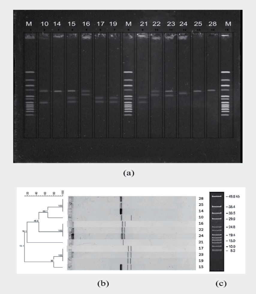
Among the phage genomes, the AvrII restriction enzyme (New England Biolabs, Beverly, Mass, UK) exhibited the capability to digest certain phages while leaving others unaffected. This led to the identification of distinct profiles of restriction digestion patterns for specific phages, while some phages shared similar profiles, substantiating their relatedness or similarity. This investigation further solidified the categorization of these phages as members of the double-stranded DNA virus family. As part of the phage characterization process, the analysis of nucleic acid restriction enzyme digestion was performed for eight of the twelve phages utilizing the AvrII restriction enzyme (phages 10, 16, 22, 24, 17, 23, 19, 15), while the remaining phages (28, 25, 14, 21) were unaffected by digestion. The observed patterns revealed both similarities (phages 17, 13, 19, and 16, 22, 24) and differences (phages 10, 21, 15) in digestion. The outcomes of this experiment were employed in the dendrogram preparation based on endonuclease DNA digestion, and this led to the initial classification of the phages into distinct phylogenetic families, as demonstrated in (Fig. 1). Restriction enzyme digestion patterns show relatedness among phages 17, 13, 19, and 16, 22, 24, while no relation was found among phages 10, 21, 15.
Phage Genome Length Determination
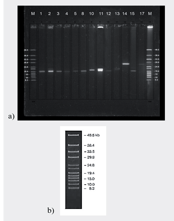
The genome sizes of seventeen out of the 28 phages were successfully determined, spanning a range of approximately 15.7 to 31.5 kilobases. However, eleven phages did not yield clear bands on the gel, which might be attributed to the degradation of their nucleic acids during the extraction process or other experimental variables. Furthermore, within the scope of phage characterization, genome length determination was executed using the PFGE (pulsed field gel electrophoresis) technique for 13 phages. The genome lengths spanned from 19.4 kilobases to 28.2 kilobases, with phage 4 and phage 14 representing the extremes, as detailed in (Table 4 and Fig. 2)
Visualizing Phage Morphology Through Electron Microscopy
The utilization of transmission electron microscopy employing a negative staining method facilitated the visualization of the morphology of three distinct phages (Fig. 3). The results unveiled a diversity of phage virion forms, encompassing hexagonal and icosahedral heads paired with long, flexible, non-contractile tails (Siphoviridae) and icosahedral heads of approximately 82 nanometers in diameter along with stubby, non-contractile tails of 5 nanometers in length (Podoviridae). This insight played a pivotal role in determining the phage family and the nature of their nucleic acid, ultimately categorizing them as Siphoviridae and Podoviridae, respectively, as showcased in (Table 5). After these steps, phages were named, for example, Salmonella CUMR 17, which refers to the host Salmonella, the institution Cairo University, and the primary author Mohamed Raslan phage, number 17. Names were derived from the name of the bacterium, the initials of the institution, the initials of the primary author, and the number of samples.
Salmonella Biocontrol
Selective phages that displayed distinct plaques with varying host ranges were identified. Among these, the Salmonella phage CUMR17 was chosen due to its wide genome size and its effectiveness in controlling the proliferation of the S. Typhimurium ATCC 14028 strain. A statistically significant reduction (p≤0.05) was observed after 2 h of incubation at both 4ºC and 24ºC. This reduction was sustained at 4ºC across different specimens, including chicken meat and milk, for up to 6 h. Notably, the lytic effect exhibited differences in specimens incubated at 24ºC, indicating a prolonged lytic action at 4ºC. Additionally, the phage demonstrated a sustained and potent biocontrol effect even after 24 h at 4ºC, as illustrated in (Fig. 4-7) (Tables 6, 7).
In the experiment involving chicken meat, the CUMR17 phage demonstrated an effective performance in diminishing the number of Salmonella bacteria present, and this impact was temperature dependent. Interestingly, the phage effect over time was more significant at a lower temperature of 4ºC, despite the slower bacteriophage replication under this condition. At this temperature, they noted a reduction of log CFU/ml in the Salmonella cell count in the chicken meat samples. However, when the temperature was increased to 25ºC, promoting more active bacterial growth and subsequent phage replication, there was an initial surge in cell count numbers after 10 h, followed by a steady decrease when the meat was stored at 4ºC, i.e., a non-significant difference between test and control after 24 h at 25ºC. This suggests that the CUMR17 phage can effectively control the growth of Salmonella bacteria in chicken meat, especially when stored at lower temperatures (Fig. 4, 5).
Table 3.
Phages host range analysis.
Fig. 1.
Restriction enzyme gel and phylogenetic analysis dendrogram. a) Restriction digestion of AvrII enzyme pattern as separated by PFGE done on BioRad CHEF DII system (Bio-Rad, Richmond, CA, USA) the following parameters (1% PFGE grade agarose gel, 0.5 X TBE running buffer, running time = 9 hours, V= 6 V/cm, Temperature = 4ºC, Switch Time = 30 to 60 seconds, GelStarTM nontoxic nucleic acid stain) and documentation was done on Gel Doc™ XR+ UV Trans illumination and ImageLab 3.0 software (BioRad); b) Phylogenetic analysis was performed and dendrogram was drawn by using BioNumerics software (Applied Maths NV). Phages were compared according to their genome digestion patterns. Bands were quite different for some phages while being similar for others, which considered to be similar phages due to having the same nucleic acid restriction patterns. c) BioRad CHEF DNA Lambda Ladder #1703707.
Similar tests were done on the CUMR17 phage against S. Typhimurium ATCC 14028 growth in milk samples. With reductions of log CFU/ml at 4ºC, the biocontrol impact was more pronounced in this situation. The number of bacterial cells decreased somewhat when the temperature was raised to 25ºC (by log CFU/ml) and then steadily when it was lowered to 4ºC (Fig. 6, 7). i.e., a non-dramatic variation between the test and control prior to 24 hours at 25ºC. This is potentially linked to sustaining the optimal conditions for bacteriophage replication. These consistently lower cell counts relative to the initial experiment indicate that the CUMR17 phage could potentially be an effective biocontrol agent in the dairy industry as well, inhibiting the growth of harmful bacteria in milk.
Table 4.
Phages genome length determination using Pulsed Field Gel Electrophoresis (PFGE) and restriction analysis.
| Characteristics Phage No. | Phage Genome Size (kb) | Restriction by (Avr II) |
|---|---|---|
| CUMR10 | 21 kb | + |
| CUMR14 | Unknown | - |
| CUMR15 | 19.8 kb | + |
| CUMR17 | 27.6 kb | + |
Fig. 2.
Pulsed Field Gel Electrophoresis (PFGE) Genome Length Determination. a) PFGE for 13 phages out of 28 phages appear as single bands on PFGE which could be used to assess nucleic acid size of phages (1, 2, 3, 4, 8, 10, 11, 12, 13, 14, 15, 17) by comparing it to a ladder. PFGE was done on BioRad CHEF DII system (Bio-Rad, Richmond, CA, USA), the following parameters (1% PFGE grade agarose gel, 0.5 X TBE running buffer, Running Time = 9 hours, V= 6 V/cm, Temp = 4°C, Switch Time = 30 to 60 seconds, GelStarTM nontoxic nucleic acid stain) and documentation was done on Gel Doc™ XR+ UV Trans illumination and ImageLab 6.0 software (BioRad); b) BioRad CHEF DNA Lambda Ladder #1703707.
Discussion
This research sought to isolate and partially characterize particular bacteriophages for Salmonella spp. from natural sources, with the eventual goal of examining phage potential for creating a biocontrol agent. The isolation of phages was successful, proving the applicability of the study’s procedures. It has long been understood that bacteriophages are closely related to their natural hosts. Animals naturally have Salmonella in their gastrointestinal tracts [26-28], so sewage was the best source for phage isolation out of all the used sources (sewage water and soil).
Based on their ability to form plaques on the bacterial lawns of their hosts, all of the isolated phages displayed lytic features. Plaque homogeneity was seen after purification [29, 30]. Several methods have been adopted previously to characterize isolated phages [31, 32]. As part of our study’s methodology, specimens were initially amplified before being characterized using host range analysis, restriction digestion, genome length analysis, and electron microscopy. Based on their host range and genome size, several phages were chosen for comprehensive characterization by restriction digestion of nucleic acids and electron microscopy.
During the investigation of the phage host range, it was found that the majority of the phages had a large host range; however, phages 26, 27, and 28 displayed a relatively restricted host range, which may have been caused by their instability at room temperature. The fact that Phage 20 had the widest host range may have been caused by endolysin or bacteriocin contamination of its stock, which may lyse the bacterial lawns of all Salmonella spp. isolates and produces a falsely large host range while the true host range is less than shown.
The host range of all phages, and 4 out of them, the Salmonella phages CUMR10, CUMR14, CUMR15, and CUMR17 are significant. The most crucial factor for phage taxonomy was ultrastructure, and now it is genome sequence [33-35]. However, they also integrate other important aspects such as nucleic acid type (DNA or RNA), method of replication, host organisms, and the types of diseases they cause. As technology advances, genetic and phylogenetic characteristics are increasingly incorporated into viral taxonomy, providing a more comprehensive understanding of viral relationships and categories [36].
Fig. 3.
Electron micrographs of 4 selected phages (TEM). The TEM micrographs revealed the morphology of 4 lysates (approximately 1010PFU/ml) prepared from phages; a) Salmonella CUMR10, b) CUMR14, c) CUMR15 and, d) CUMR17) that were negatively stained with 1% (w/v) Phosphotungestic Acid on a carbon-coated copper grids (Electron Microscopy Sciences) and examined by transmission electron microscope (JEOL JEM-2100, Tokyo, Japan) at an accelerating voltage of 200 kV. Phages in a) CUMR10, b) CUMR14 and d) CUMR17 were consists of head and long tail while phage c) CUMR15 was consists of short tail and head.
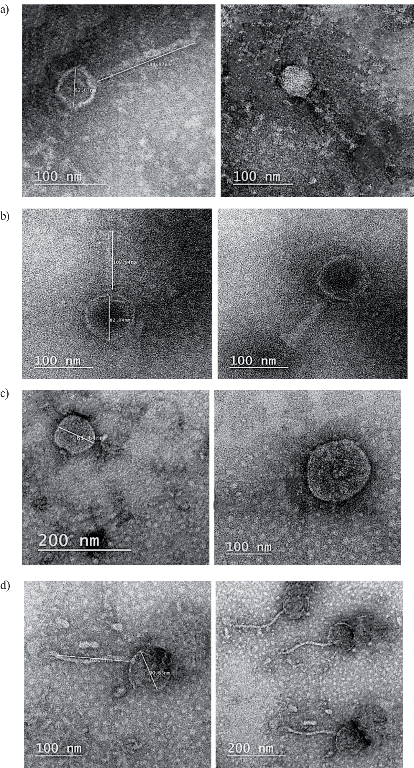
In this study, we used electron microscopy to evaluate the phenotypic structure of the four selected bacteriophages based on variations found in their genome length, restriction pattern, and host range. The phages were recognized using the morphological standards established by the International Committee of Taxonomy of Viruses and the species concept of the published protocol [37-43]. They were then designated as Salmonella CUMR10, CUMR14, CUMR15, and CUMR17 phages using a similar idea to what has been indicated by published protocols [44-50].
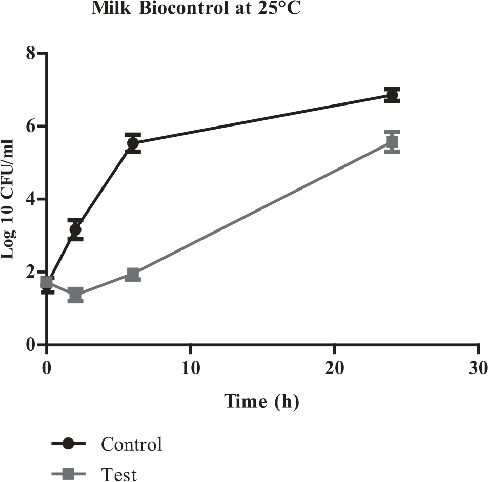
Fig. 4.
Biocontrol of S. Typhimurium ATCC 14028 in Chicken Meat at 4ºC. (Data are represented as means±SD).
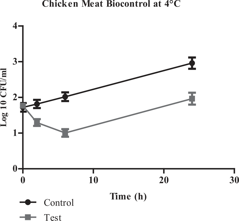
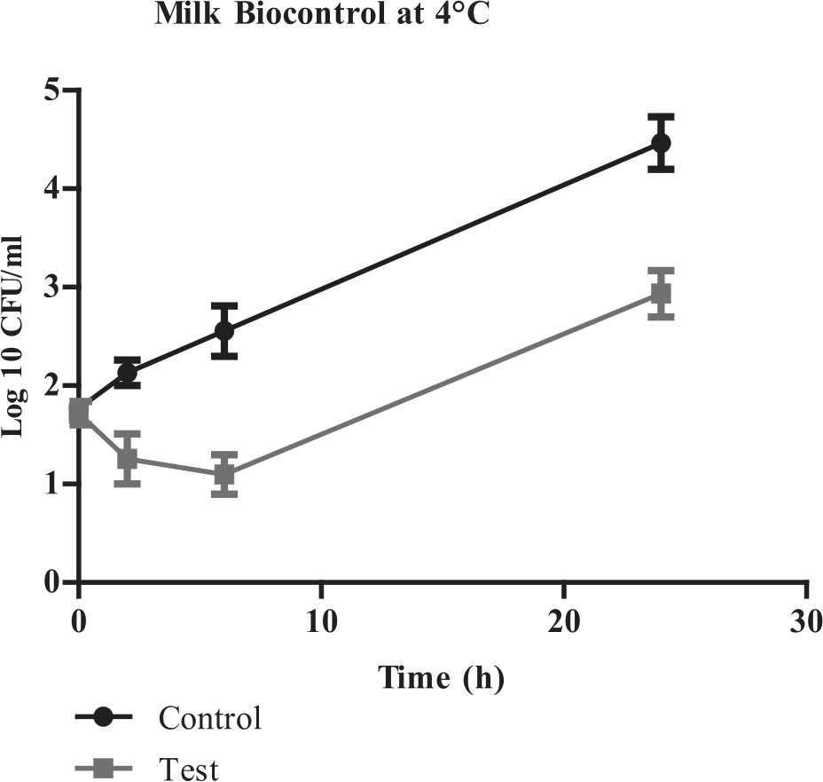
Fig. 5.
Biocontrol of S. Typhimurium ATCC 14028 in Chicken Meat at 25ºC. (Data are represented as means±SD). * At the end of the incubation period results of control likely to be in the same level of control.
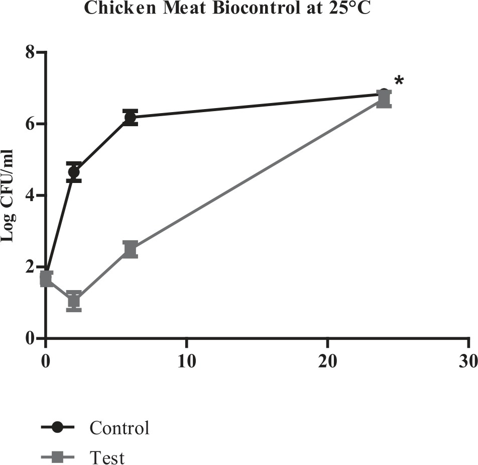
Table 5.
Phage morphological characteristics.
Table 6.1.
Biocontrol of Salmonella in meat using CUMR14 phage at different at 4 and 25 oC. Mean with Standard Deviation for CUMR14 phage.
Table 6.2.
Biocontrol of Salmonella in meat using CUMR14 phage at different at 4 and 25°C. Data significance testing using ANOVA for CUMR14 phage.
Table 7.1.
Biocontrol of Salmonella in milk using CUMR14 phage at different at 4 and 25ºC. Mean with Standard Deviation for CUMR14 phage.
Table 7.2.
Biocontrol of Salmonella in milk using CUMR14 phage at different at 4 and 25ºC. Data significance testing using ANOVA for CUMR14 phage.
The four phages were split between two unique families, Siphoviridae and Podoviridae, which were shown to have different structural, genomic, and functional characteristics. A family of double-stranded DNA viruses known as Siphoviridae is categorized as a bacteriophage, or phage, that infects bacteria. They stand out due to the length of their non-contractile tails. Members of this family have linear genomes and icosahedral, or elongated, heads. They are excellent candidates for phage-based biocontrol because of their general structural capacity to infect many bacterial strains. One well-known member of this family, which infects Salmonella bacteria, is the P22 bacteriophage [51-53].
Pathogens have been responsible for an increase in foodborne illnesses. Controlling the spread and proliferation of infections is therefore crucial. Bacteriophages (phages) have the potential to be a significant player in the biological mitigation, management, and therapeutic management of many foodborne infections because of their advantageous characteristics. The beneficial properties of phages include their selectivity and lack of chemical residues, which not only successfully fight harmful bacteria but also extend the shelf life of food [54-57]. In the present study, a preliminary investigation of isolated phages versus S. Typhimurium ATCC 14028 was reported at different temperatures, where the phage stability is affected by temperature variations as documented in prior studies [56-58]. Further investigations should be conducted to evaluate the phage stability at a temperature of 24ºC. This is critical for assessing the potential use of phages as biocontrol agents in food that is typically stored at ambient temperatures.
Conclusions
There is a phage for every strain of bacteria that exists in the natural environment, which makes bacteriophages more popular and acceptable due to their low cost, animal and human safety, and availability. This research has made initial steps in exploring the potential use of bacteriophages for the biocontrol of S. Typhimurium in food safety applications. Using conventional enrichment techniques, this work isolated and identified bacteriophages unique to Salmonella spp. We were able to develop dependable techniques for bacteriophage isolation from and characterization of sewage and soil. These were carried out using conventional plating, transmission electron microscopy, restriction digestion, PFGE, and host range analysis. Some procedures for the biocontrol of S. Typhimurium in inoculated chicken meat and milk were created. The biocontrol of bacterial growth in milk and chicken meat at 4ºC and in milk just at 25ºC for 24 h was clearly reduced, which was a promising start for this investigation. As a future direction, conducting genomic sequencing of these phages is an essential step, as this would contribute to a more precise understanding of their identity and their structural and functional attributes. Additionally, to ensure the safety and efficacy of phage-based biocontrol, further in vivo studies are recommended. In summary, while this research presents a promising foundation for the application of bacteriophages in food safety, it is important to recognize the need for continued investigation and a comprehensive evaluation of their potential benefits and limitations in real-world food processing scenarios.