

Introduction
Ion beam modification of living cells has been explored as ion beam biotechnology over the past three decades [1, 2]. The technology utilizes ion beams, particularly in a lower energy region, some tens keV, #Ting Wang and Chao Tang contributed equally to this study.
qianwd@sust.edu.cn instead of the MeV higher energy region, to bombard live cells to induce genome-wide mutation or gene transfer into biological cells in a vacuum. Ion beams have unique physical properties, such as diversified radiation parameters, complex track structure and depth-dose distribution, thereby accelerating and shaping natural phenotypic variation of induced mutations [2]. Accumulating evidences have demonstrated that antibiotic resistance can arise both from mutations in the pre-existing genome of a bacterium in the absence of an exogenous DNA and from the uptake of foreign DNA [3]. However, the evolution of bacterial drug-resistance mediated by low energy N+ implantation has not yet been reported.
Staphylococcus aureus (S. aureus) is one of the most frequent and opportunistic pathogen, as a leading cause of morbidity and mortality in humans and animals across the world [4, 5]. In recent decades, the rapid emergence of methicillin-resistant S. aureus (MRSA) is occurring worldwide due to the overuse of β-lactam antibiotics in livestock farming and clinical treatment to treat infectious diseases, thereby posing a serious threat to human health and food safety [6, 7]. The common mechanisms of drug resistance have been well examined, but critical new aspects continue to be discovered. D’Costa et al. report that targeted metagenomic analyses of rigorously authenticated ancient DNA from 30,000-year-old Beringian permafrost sediments and the identification of a highly diverse collection of genes encoding resistance to β-lactam, tetracycline and glycopeptide antibiotics [8]. These results show conclusively that antibiotic resistance might be a natural phenomenon that predates the modern selective pressure of clinical antibiotic use [8]. Mutation is the most source of new genetic variation variety on which natural selection depends in certain environments [9]. However, whether radiation mutation mediated by low energy N+ can accelerate the genetic evolution of bacterial drug resistance has not been explored.
Here, vancomycin-resistant S. aureus by low energy N+ ion implantation mutagenesis was constructed. Then, the vancomycin-resistant S. aureus strains were further examined using drug-resistance phenotypes, and biofilm growth analysis to explore the potential mechanism.
Material and Methods
Chemicals
Mueller-Hinton Broth medium that used for determining minimal inhibitory concentrations (MICs) and sterile filter paper discs were bought from Sigma-Aldrich Chemical Co. (USA). Vancomycin was purchased from local market in China. PrimeScript™ RT Master Mix and TB Green® Premix Ex Taq™ II were bought from purchased from TaKaRa Biotechnology Co. Ltd (Dalian, China). Film Tracer SYPRO Ruby (SYPRO Ruby), WGA conjugated with Oregon Green (WGA), and 4′,6-diamidino-2-phenylindole (DAPI) dyes were bought from Invitrogen (Thermo Fisher Scientific, Waltham, Massachusetts, US).
Strain and Cultural Conditions
The single colony of S. aureus ATCC25923 was cultured in Luria-Bertani broth (LB, Sigma-Aldrich, USA) culture flask at 37ºC with shaking at 150 rpm.
Mutagenesis Method via Low Energy N+ Ion Implantation
Low energy N+ sources were prepared using the ion beam bioengineering equipment developed by the Southwestern Institute of Physics (Chengdu, China). S. aureus ATCC25923 mutation mediated by low energy N+ implantation was conducted as previously described with minor modifications [1]. Briefly, a single colony of S. aureus ATCC25923 was further cultured with shaking at 37ºC in a tube containing 3 mL of LB. After cultured for 12 h, cells were harvested by centrifugation (3000 × g, 5 min) and diluted in 1 mL protection buffer composed of 0.5% glucose to achieve an initial suspension that contains approximately 1 × 10⁸ CFU/mL. Then, 0.1 mL of the original suspension was transferred to a sterile 90-mm plate and plated to form a thin layer manner. Subsequently, the plate was allowed to sterile air dry for 20 min.
For low energy N+ ion implantation, the plate was transferred in a chamber with operating pressure of about 10–3 Pa. Then, the plate was implanted by the N+ ion beam with different dosage (25~150×1014 ions/cm2) under the condition of 15 keV of energy. After implantation, 1 mL LB liquid medium was added immediately to each implantated and control plates, and the cell pellets were resuspended, transferred into new tubes. Finally, the suspensions were cultured for 2 h at 37ºC, and plated on LB medium to screen the S. aureus mutants. The plates were placed in pairs sequentially in the chamber where the cells in the upper plate was implantated via low energy N+ ion beam, whereas those in the lower plate without implantation were utilized as the control group.
Screening and Engineering of Stable Vancomycin-Resistant S. aureus Mutants
After S. aureus ATCC25923 cells exposed to low energy N+ implantation were plated onto LB medium for 24-72 h at 37ºC, the mutants were achieved from the parent strain. Then, the mutant strains were subcultured three generations in LB agr medium with 4 μg/mL of vancomycin at 37ºC to obtain stable genetic vancomycin-resistant S. aureus strains.
Antibiotic Susceptibility Testing of Vancomycin-Resistant S. aureus Mutants
Antibiotic susceptibility profiles of vancomycin-resistant S. aureus mutants were conducted using disc diffusion method [10]. Briefly, the fresh single colony from each mutant was in 3 mL of LB overnight. A 200 μL aliquot of each culture (approximately 105 CFU/mL) were evenly spread on Mueller-Hinton agar (Que-lab, Canada) plates and dried at room temperature. Then, the antibiotic discs (Oxoid, Italy) containing the following antibiotics were applied: vancomycin (30 µg), norfloxacin (10 µg), ciprofloxacin (5 µg), cefoperazone (75 µg), cefotaxime (30 µg), Benzoxacillin (30 µg), clindamycin (2 µg), minocycline (30 µg), doxycycline (30 µg), and gentamicin (10 µg), and placed onto the plates. The diameters of inhibition zones were determined after incubation of 16-18 h at 37ºC, and interpreted following by the ecommendations of the Clinical Laboratory Standards Institute (CLSI) [11]. S. aureus ATCC 25923 was applied as the quality control strain.
Analysis of Drug Resistance Gene Relative Expression
The extraction of total RNAs and quantitative real-time reverse transcription PCR (qRT-PCR) were conducted using the Total RNA Isolation Kit and ChamQ Universal SYBR qPCR Master Mix according to the manufacturer’s instructions (Vazyme Biotech Co. Ltd., Nanjing, China), respectively. All the primers were produced by Sangon Biotech Co. Ltd. (Shanghai, China), and the corresponding oligonucleotide sequences were shown in supplementary Table S1. A relative quantification method (2-∆∆Ct) was applied to determine the changes in the relative expression levels of resistance genes. Nine representative vancomycin-resistant S. aureus mutants were examined using qRT-PCR for the expression of resistance genes (vraSR, fnbA, fnbB, clfA, clfB, mgt, walKR, sle1, altA, and lytM) that confer resistance to antimicrobial agents.
Evaluation of Biofilm Formation of Vancomycin-Resistant S. aureus Strains
The biofilm formation of vancomycin-resistant S. aureus strains was examined by crystal violet, FESEM and CLSM as reported previously [12]. Briefly, vancomycin-resistant S. aureus cultures (1 × 10⁶ CFUs/mL) were prepared in each well of the 24-well plate with a sterilized glass coupon at 37ºC for 48 h. For visualization of biofilm structure and biomass of vancomycin-resistant S. aureus strains, biofilm-coated coupons were washed gently three times with 10 mM PBS solution. Then, washed biofilms formed on the coupons were fixed in 2.5% glutaraldehyde (v/v) immediately at -4ºC for 2 h. The fixed biofilms were then dehydrated using an ascending grades of ethanol (30%, 50%, 70%, 90% and 100%) process. Subsequently, the biofilm samples were evaluated using FESEM.
For characterization of the biofilm matrix of vancomycin-resistant S. aureus, biofilms were formed as described above. Similarly, the biofilms were gently rinsed three times with 10 mM PBS. The resulting biofilm samples were stained individually with the following three dyes: (1) SYPRO Ruby dye, which labels most classes of biofilm-associated proteins;
(2) WGA dye, which stains the polysaccharides component of the biofilm matrix; and (3) DAPI dye, which stains extracellular nucleic acids of the biofilm matrix (eDNA). The fluorescence of three dyes was measured using the following combination of excitation and emission wavelengths: 405 nm/655~755 nm for SYPRO Ruby, 459 nm/505~540 nm for WGA, and 535 nm and 617~635 nm for DAPI, respectively. After 15 min of incubation at 37ºC, the biofilms were gently washed twice with 10 mM PBS and visualized under a CLSM using the 63 × (oil immersion) objective. In addition, fluorescence ratios of the biofilm matrix such as biofilm-associated proteins, polysaccharides, and eDNA were determined using the KS 400 version 3.0 software (Carl Zeiss, Inc, Jena, Germany) and averaged for the images captured in two areas per biofilm from three independent biofilms.
For biofilm examination using optical microscope, the biofilms were washed twice with 10 mM PBS, and then stained with 0.1% (w/v) crystal violet (CV) for 20 min. Finally, the biofilms were examined using the optical microscope at 400 × magnification.
Results and Discussion
Determination of the Optimal Ion Implantation Dose
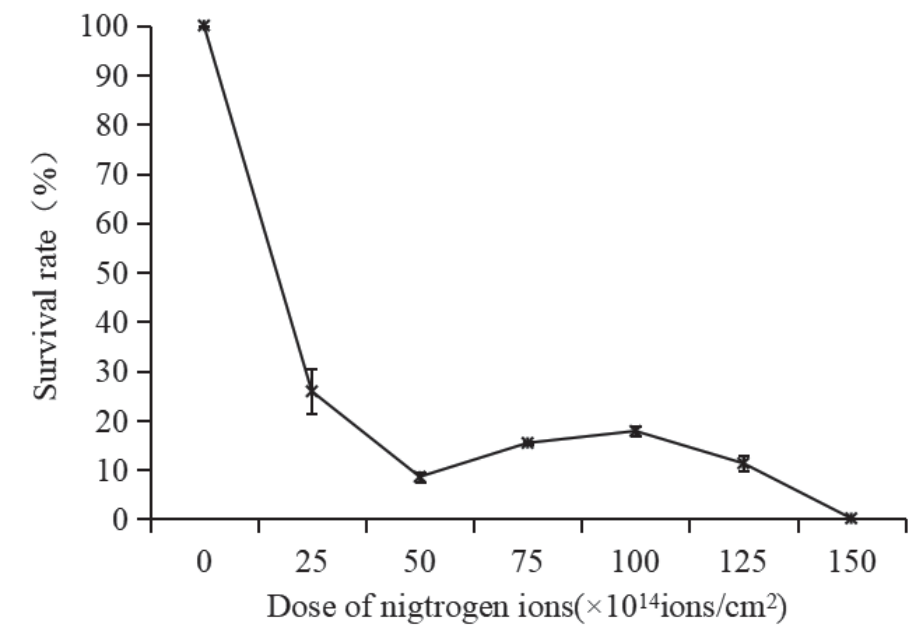
Unique interaction between low-energy ions and biological cells results in a wide mutation spectrum compared to other traditional mutagens. An ion implantation process is mainly determined by two process parameters such as the dose and energy level of low-energy ion implantation. In this study, the dose and energy level of low-energy ion implantation were firstly determined to obtain a particular evolutionary goal. Therefore, the survival rate as a function of ion parameters needs to be exploited. To this end, S. aureus ATCC25923 was implanted by the N+ beam with energy of 15 keV and different doses including 25 × 1014, 50 × 1014, 75 × 1014, 100 × 1014, 125 × 1014 and 150 × 1014 ions/cm2. As shown in Fig. 1, the survival rate was inversely proportional to the ion dosage when the dosage was less than 50 × 1014 ions/cm2. In contrast, the survival rate increased significantly when the dosage between 50 × 1014 and 100 × 1014 ions/cm2 was used. Then, the survival rate decreased even more dramatically when the dose was more than 100 × 1014 ions/cm2. The relationship between the survival rate of S. aureus ATCC25923 and the dose of N+ beam implantation verified the “saddle-shaped” dose-survival effect, which is consistent with results reported in previous studies [13]. Moreover, previous studies indicate that the high mutation rate mainly occurs in survival rates between 10% and 30%. Therefore, N+ beam with 15 keV of energy at the implantation dose of 100 × 1014 ions/cm2 were utilized to mutate S. aureus ATCC25923 strain.
Fig. 1.
The survival rate of S. aureus ATCC25923 treated with various N+ ion implantation dose. Each survival rate value was determined according to three independent experiments using different implantation dose of N+ under the condition of the energy level of 15 keV and indicated as mean standard error (n = 3).
Screening and Engineering of Vancomycin-Resistant S. aureus Strains
To efficiently obtain vancomycin-resistant S. aureus mutants, N+ beam with 15 keV of energy at the dose of 100 × 1014 ions/cm2 was utilized to construct vancomycin-resistant S. aureus mutants by mutation. Followed by a rational screening, 798 vancomycin-resistant S. aureus mutants was obtained using LB broth containing 4 µg/ml vancomycin. After three consecutive generations of cultivation of these vancomycin-resistant S. aureus mutants, 9 genetically stable vancomycin-resistant S. aureus strains were obtained with a mutation rate of 1.1%. The determination process of low energy N+ implantation parameters can be the first prominent step in the engineering of drug-resistant strains. This allows the survival of the sufficient bacterial population until more stable genetically resistant mutants emerge. A recent study shows that that particle pollution should be responsible for 11% of changes in average antibiotic resistance levels across the world, making particle pollution potentially one of the leading contributors of global antibiotic resistance [14]. Volcanoes and lightning can produce certain ions in nature including low-energy ions, which can interacte with bacteria and lead to bacterial drug-resistance. Taken together, our findings might indicate the importance of ions from environments as one of the factors driving global antibiotic resistance.
Antimicrobial Susceptibility of Vancomycin-Resistant S. aureus Strains
The antibiotic susceptibility of 9 vancomycin-resistant S. aureus mutants to 10 antibioticals was examined using disc diffusion method. The antibiotic susceptibility profiles are presented in Table 1, in which each strain was classified as resistant (R), intermediate (I) or sensitive (S), according to the breakpoints proposed by CLSI. All vancomycin-resistant S. aureus strains tested were found to be resistant to both vancomycin and norfloxacin. As exhibited in Table 1, four vancomycin-resistant S. aureus strains (SA-MVR3, SA-MVR5, SA-MVR6, and SA-MVR9) exhibited muti-drug resistant to 4 different antibiotics. Intriguingly, one vancomycin-resistant S. aureus strain (SA-MVR4) was observed to be resistance to 7 different antibiotics including vancomycin, norfloxacin, cefoperazone, cefotaxime, benzoxacillin, clindamycin, and gentamicin. In addition, S. aureus ATCC25923 was susceptible to all antibiotics tested.
Table 1.
The antibiotic profiles of nine vancomycin-resistant S. aureus mutants to 10 tested antibiotical agents.
As shown in Fig. 2, micro broth dilution assay further confirmed that S. aureus ATCC-25923 strain was sensitive to vancomycin with a MIC≤2 μg/mL. In contrast, seven vancomycin-resistant S. aureus strains including SA-MVR1, SA-MVR2, SA-MVR3, SA-MVR4, SA-MVR7, SA-MVR8, and SA-MVR9 were resistant to vancomycin with a 2≤MIC≤4 μg/mL, whereas two vancomycin-resistant S. aureus strains such as SA-MVR5 and SA-MVR6 were more resistant to vancomycin with a 4≤MIC≤8 μg/mL.
Biofilm-Forming Capacity of Representative Vancomycin-Resistant S. aureus Mutants with Muti-Drug Resistance Capacity
In order to reveal the potential mechanism of the drug-resistance formation in vancomycin-resistant S. aureus strains induced by low energy N+ implantation, the biofilm formation ability of vancomycin-resistant S. aureus strains with MDR capacity was investigated. As presented in Fig. 3, 48 h biofilm structures of representative vancomycin-resistant S. aureus mutants with MDR capacity were examined using crystal violet and FESEM. Compared with the original S. aureus ATCC25923, a significant increase in biofilm biomass for vancomycin-resistant S. aureus mutants was shown in Fig. 3. Meanwhile, vancomycin-resistant S. aureus mutants including SA-MVR4, SA-MVR5, and SA-MVR9 formed biofilms containing thick-cell layers though three representative mutants exhibited disparate biofilm-forming abilities, whereas S. aureus ATCC25923 produced thin biofilms with a few, thin scattered cell clusters (Fig. 3). Previous studies show that biofilms are more tolerant to antibiotics than corresponding planktonic cells since the extracellular matrix hinders the penetration of antibiotics, thereby reducing the antibiotic susceptibility of bacterial cells within biofilms [15]. Additionally, the ability of biofilm formation is often positively correlated with the development of multidrug-resistance profile [16]. Futhermore, the close proximity of the bacterial cells entrapped in biofilms appears to provide an excellent platform allowing intense interactions to occur, including horizontal genetic transfer of resistance gene [17]. Therefore, it may be concluded that the increased capacity of biofilm formation by vancomycin-resistant S. aureus mutants results in enhanced tolerance to antimicrobial agents due to the inherent physical and chemical barriers of biofilm matrix.
Fig. 2.
N+ implantation treatment of S. aureus leads to heterogeneous increases in MIC for vancomycin. The MIC of nine vancomycinresistant S. aureus strains for vancomycin was 2≤MIC≤8 μg/mL.
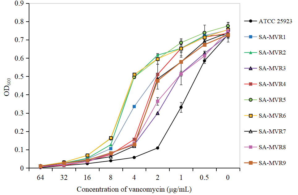
Fig. 3.
Assessment of biofilm formation of representative vancomycin-resistant S. aureus strains using crystal violet and field emission scanning electron microscopy (FESEM). Scale bars represent 10 μm for crystal violet and 10 μm for FESEM.
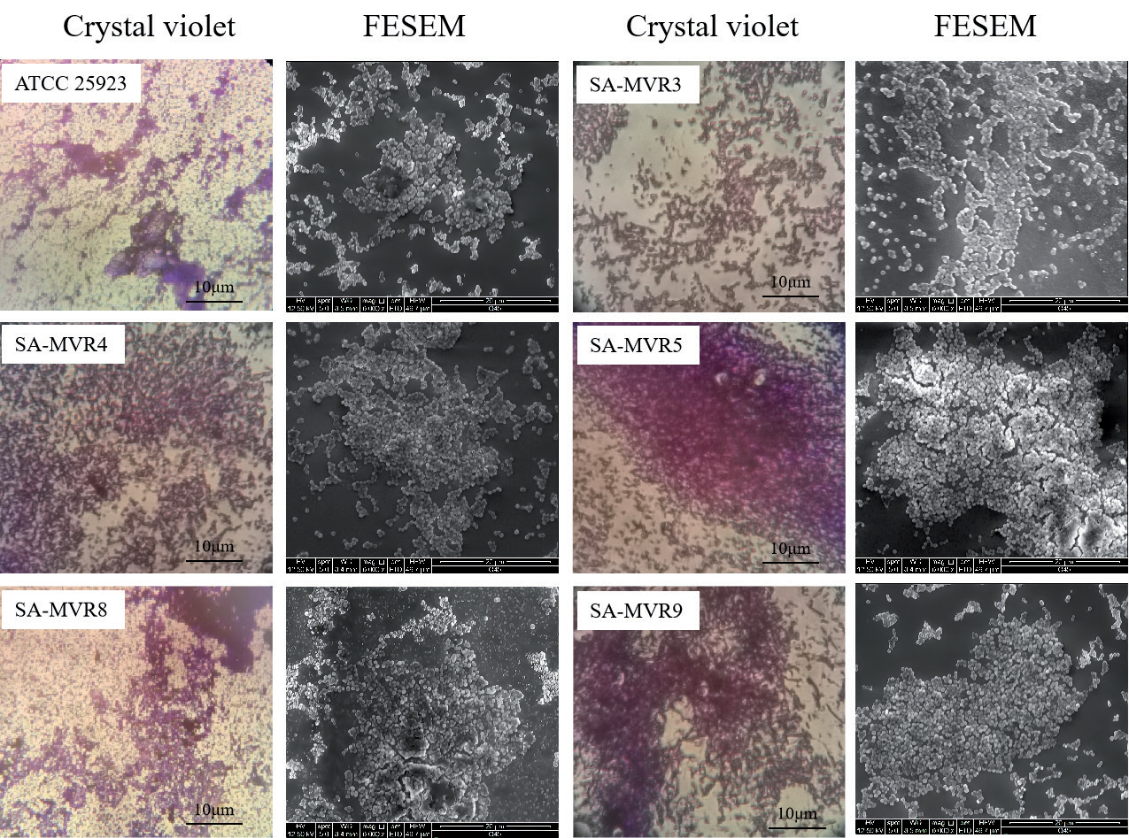
Fig 4.
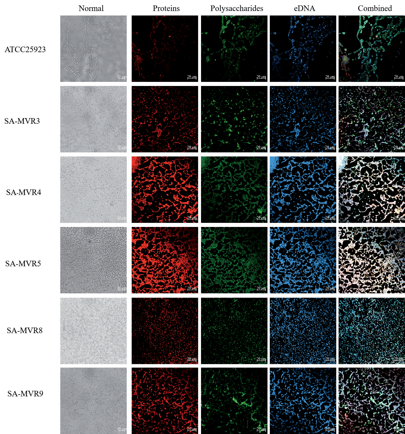
Examination of the biofilm composition levels of representative vancomycin-resistant S. aureus strains with different drug-resistant spectrum using confocal laser scanning microscopy (CLSM). Three fluorescent dyes including Film Tracer SYPRO Ruby, WGA and DAPI were used to stain proteins, polysaccharides and eDNA embedded in the biofilm, respectively. Scale bars represent 20 μm.
Biofilm Composition of Representative Vancomycin-Resistant S. aureus Mutants with Muti-Drug Resistance Capacity
Three different fluorescent dyes were applied to examine the distribution of eDNA, proteins, and polysaccharides embedded in the biofilm produced by representative vancomycin-resistant S. aureus mutants using CLSM. As presented in Fig. 4, eDNA and proteins were at high levels and uniformly distributed in biofilms by three representative vancomycin-resistant S. aureus mutants (SA-MVR4, SA-MVR5 and SA-MVR9), compared with those of S. aureus ATCC25923. In addition, eDNA and proteins were the major components within biofilms by five representative vancomycin-resistant S. aureus mutants (Fig. 4). Similar evidences have displayed that biofilm matrix within the biofilm can prevent drugs from entering the bacterial cell and thus enable bacteria to develop antibiotic resistance, which contributes to the emergence of multi-drug resistant bacteria [18].
Examination of Drug Resistance Genes Relative Expression
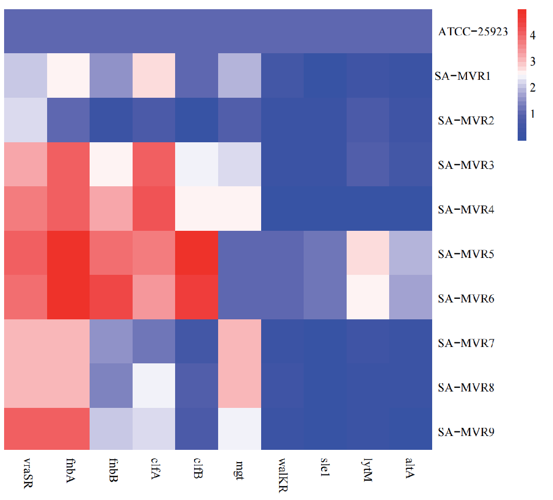
The expression of drug resistance genes in vancomycin-resistant S. aureus strains including vancomycin-resistance-associated response regulator (vraSR), adhesion-related genes (fnbA, fnbB, clfA, clfB), a monofunctional glycosyltransferase (mgt), two component regulator (walKR), cell wall amidase (sle1), and the peptidoglycan hydrolase (lytM) genes were detected with qRT-PCR, where the 16S rRNA gene was used as the internal reference. As shown in Fig. 5, compared with the original strain S. aureus ATCC 25923, four vancomycin-resistant S. aureus strains including SA-MVR3, SA-MVR4, SA-MVR5, and SA-MVR6 showed the significant upregulation in vraSR, fnbA, fnbB, clfA, and clfB gene relative expression, and the significant downregulation in sle1, walKR, altA and lytM gene relative expression in SA-MVR3 and SA-MVR4. In addition, SA-MVR2 strain was found to be the significant upregulation in vraSR gene relative expression, whereas there was significant downregulation in relative expression of other tested genes. The relative expression of sle1, walKR, altA and lytM genes in SA-MVR1, SA-MVR7, SA-MVR8, and SA-MVR9 strains were downregulated, while vraSR, fnbA, clfA, and mgt tested were significantly upregulated. Previous studies showed that vraSR gene can positively regulate cell wall biosynthesis and increase cell wall thickness, thereby promoting vancomycin resistance in the corresponding strain [19]. The fnbA, fnbB, clfA, and clfB genes are mainly involved in bacterial adhesion to the substrate, which might contribute to the biofilm formation, thereby promoting drug resistance [20].
Conclusions
In this study, we engineered vancomycin-resistant S. aureus mutants using the low energy N+ implantation method, and explored the relationship between antibiotic resistance, biofilm formation, and resistance gene of vancomycin-resistant mutants. The current study suggested that the vancomycin-resistant S. aureus mutants have stronger biofilm formation ability compared with the original S. aureus ATCC25923 strain. Furthermore, a positive association between the multiple drug resistance phenotype and biofilm-production for engineered vancomycin-resistant S. aureus mutants was observed. Our study has expanded the potential contributory factors driving antibiotic resistance and promoted the scientific research of environmental exposures that might substantially boost the development of antibiotic resistance.

