

Introduction
Natural product medication development is still greatly influenced by microbes, including bacteria, as they generate a lot of beneficial primary metabolites [1, 2]. Additionally, bacteria create secondary metabolites, which account for a sizeable portion of the medications sold on the market today, to the point that microbial natural products have taken over as the main source of drug lead compounds [3]. Natural products derived from different species of bacteria include a vast collection of different chemical structures, and they will continue to be a source of new therapies, either in their unaltered state or after being improved by synthetic medicinal chemistry. The identification of natural products from bacterial isolates has revived in recent years [4, 5].
In Egypt, the common freshwater snail is called Biomphalaria alexandrina, as reported by Mossalem and Ibrahim [6]. Schistosoma mansoni is a trematode parasite that can infect humans and lead to schistosomiasis [7, 8]. Daphnia magna and Daphnia pulex are crucial bio-monitoring species used to examine heavy metal contamination [9, 10]. D. plux could be present in the aquatic conditions of Egypt [11] and serves as a non-target model to assess the acute and chronic toxicity in freshwater ecosystems [12] because its reproductive mechanism, high fertility rate, and straightforward culture procedure make it suitable for scientific research of water studies [13]. Numerous elements and organic pollutants are also highly toxic to this species [14].
The current research aims to evaluate a variety of bacterial extracts against B. alexandrina snails, identify the most potent bioactive chemicals obtained from bacteria, and determine their effects on B. alexandrina snails’ hematological and biological parameters. The toxicity of the purified molecules has also been tested on D. plux, as a sensitive organism, to ensure their suitability for safe use.
Materials and Methods
Bacterial Cultures and Snails
The Regional Center for Mycology and Biotechnology at Al Azhar University donated 24 distinct bacterial species cultures from their culture collection. Inoculating 250 ml flasks containing 50 ml of Luria-Bertani (LB) broth medium with the proper concentration of 106 cfu/ml of various bacterial cultures. Diverse bacterial inocula were placed in inoculated flasks, which were then kept at 37ºC for 24 hours while being shaken at 140 rpm. The bacterial growth was separated using a 0.45 µm membrane filter [15, 16].
Theodore Billiharz Institute kindly provided B. alexandrina snails (7-12 mm) housed in plastic containers (15 × 20 × 10 cm) containing de-chlorinated water. The snails were fed lettuce and fish food, and the water was changed every week to provide suitable conditions in the laboratory. The containers were kept at room temperature [6].
Preparing Bacterial Extracts
Bacterial isolates were added to flasks that held 100 ml of LB medium apiece, and they were then cultured for 2 days at 37ºC. Following an aseptic filtering procedure in the flasks, the filtrate was concentrated by speed vacuum equipment (Thermo-Fisher, USA). Bacterial secondary metabolites were extracted using methanol/chloroform (1:2, v/v). Each time LB broth from bacterial cultures was concentrated, it was combined with chloroform/methanol. After giving the combination a vigorous shake in a separating funnel, the secondary metabolites settled to the bottom in a dense, watery layer. Every single extract was concentrated using an evaporator and solvent evaporation at a temperature of no more than 45.0±1.0ºC [17-19].
Screening the Molluscicidal Impact
1000 µg/ml of various bacterial extracts was prepared (W/V) using de-chlorinated tap water (pH 7.0-7.5) at 37±2ºC to evaluate the most lethal bacterial species. Three duplicates, each containing 10 snails (6-8 mm)/L of each concentration, were utilized for each test. Each exposure and recovery phase lasted for 24 hours at 37ºC. Extracts were used to create a number of concentrations, including LC50 and LC90 values, and the bacterial extracts were put in the glass containers of snails. The snails were taken out of each concentration and evaluated after exposure. They were carefully rinsed with de-chlorinated tap water and then placed in another container for a 24-hour recovery period. Following the counting of snails, the most poisonous microbial filtrates’ LC50 and LC90 values were calculated. In each experiment, B. alexandrina snails were exposed for 4 hours to sub-lethal doses (LC10 and LC25) of the promising microbial filtrates [20-22].
Detection of Bacterial Effective Molecules versus B. alexandrina
After activation at 80.0±2.0ºC for 30 min for silica gel, 10 ml of various filtrates were put into a column of silica gel (G 100) that was 1.5 cm in diameter and 50 cm long. The column was then exposed to a rinse with chloroform and methanol (90:10) v/v. The gradient volume of filtrates was run through the column in order to balance and stabilize the bed, and the fractions (each containing 1.0 ml) were collected separately (Yoshino et al., 2013; Amal et al., 2019). The most efficient one was then chosen after retesting each of them on the snails. The purified compounds were identified using mass (Shimadzu 2014, Japan) and by using spectral evaluations, analyzing chemicals’ IR spectra: An infinite series FTIR, Perkin-Elmer 1650 SpectrophotometerTM was used to evaluate the infrared absorption spectrum of the isolated effective fraction. 1H-NMR: Nuclear magnetic resonance FT-NMR Bruker Ac 200 spectrometerTM was used to estimate the proton (1H-NMR) spectra [23].
SDS-PAGE Testing
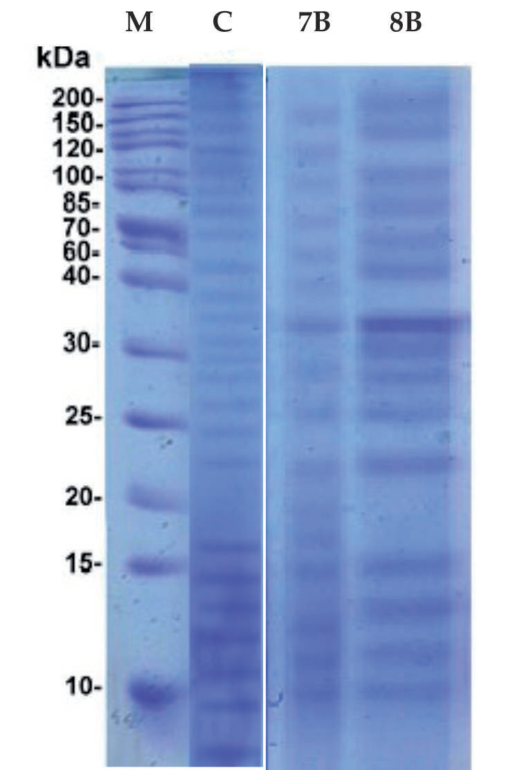
After 24 hours, the hemolymph of the treated snails with purified molecules was collected in a 1.0 ml Eppendorf tube; the hemolymph of 7-10 snails was taken from each group. To pellet hemolymph and other components, hemolymph specimens from various groups were centrifuged at 6000 rpm for 15 min at 5ºC. The pellet was discarded, and a mixture of cell-free hemolymph and sample buffer (4 parts hemolymph to 1 part sample buffer) was made. In a water bath, specimens were boiled for five minutes at 100ºC. To detect hemolymph proteins via electrophoresis, SDS-PAGE (sodium dodecyl sulfate polyacrylamide gel electrophoresis) was used to analyze the protein profiles [24, 25].
Testing Biochemical Variations
The hemolymph of B. alexandrina snails was collected, and various biochemical parameters, including total protein, albumin, glucose, transaminases aspartate (AST) and alanine aminotransferase (ALT), alkaline phosphatases, acid phosphatase, and lipid levels, were detected by Diamond kits from 3bscientific (QCA) Ltd., India. The levels of sex hormones, including progesterone, testosterone, and estradiol, were also detected by gas-liquid chromatography [26, 27].
Testing for Toxicity on D. pulex
The effective chemical was administered to healthy D. pulex (30 per treatment) in petri dishes for 24 hours at sub-lethal doses (LC10 and LC25), with an additional 24 hours serving as a recovery period. Another group was kept in de-chlorinated tap water as a control. Their survival was assessed, and death rates were recorded [28-30].
Results
Impact of Bacterial Metabolites on the Mortality of Snails
As indicated in Table 1, following exposure to various extracts for 24 hours at a concentration of 1000 µg/ml, the effects of twenty-four bacterial metabolites on snails were assessed using a toxicity assay for B. alexandrina snails.
Testing revealed that different bacterial extracts had varying rates of snail death when applied to B. alexandrina. The most lethal metabolites against B. alexandrina snails were discovered to be those from S. aureus ATCC 25923 and E. coli ATCC 25922.
Other bacterial metabolites were shown to have potential properties against B. alexandrina snails, according to the results where Pseudomonas pyocanea ATCC 19429, Enterococcus faecalis ATCC 29212, Eenterobacter aerogenes ATCC 13048, Salmonella typhimurium ATCC 13311, Serratia marscescens ATCC14756, Bacillus subtilis ATCC 6633, Agrobacterium tumefaciens ATCC 4720, Micrococcus luteus ATCC 4698, Streptococcus faecalis ATCC 29212 and Salmonella enterica ATCC 14028 have descending activities.
Table 1.
Snails’ mortality % at a level of 1000 µg/ml of various tested bacterial metabolites after exposure for 24 hours (Data are presented as means±SD).
While Alcaligenes faecalis ATCC 8750, Proteus vulgaris ATCC 8427, Blastococcus aggregatus ATCC 25902, Bacillus licheniformis ATCCC10716, Bacillus cereus ATCC 14579, Pseudomonas aeruginosa ATCC 27853, Saccharococcus thermophiles ATCC43125, Streptococcus pyrogenes ATCC12344, Streptococcus epidermidis ATCC12228, Micrococcus lylae ATCC27569, Enterobacter aerogenes ATCC 13048, and Klebsiella pneumonia ATCC 13883 have no action on snails.
Molluscicidal Impact
Fractionation of S. aureus ATCC 25923 metabolites yielded 33 fractions (Supplement Table 1), where fraction no. 7 gave the highest molluscicidal activity. Meanwhile, fractionation of E. coli ATCC 25922 gave 27 fractions, where fraction 21 had the highest molluscicidal action (Supplement Table 2).
The exposure to various levels of purified molecules of S. aureus for 24 hours showed the best action with values for LC50 = 78.2±1.0 mg/L and LC90 = 98.6±1.1 mg/L compared to the purified compound of E. coli, where values of LC50 = 93.9±1.2 mg/L and LC90 = 110.5±1.1 mg/L, indicating that the purified compound derived from E. coli showed the most promising molluscicidal activity because its LC50 & LC90 were higher than that of S. aureus, which was more potent and had more effect on snails at low concentrations, as illustrated in (Table 2).
Impact of Bacterial Purified Molecules on Biochemical Parameters
Purified molecules from S. aureus and E. coli showed no impact on progesterone, testosterone, and estradiol levels, as well as alkaline phosphatase, acid phosphatase, and glucose levels in snails exposed to LC10 and LC25 concentrations. However, a slight decrease in total protein and albumin was observed in snails subjected to LC10 and LC25 of purified bacterial molecules. On the other hand, purified bacterial compounds significantly increased ALT and ALT (P<0.05) upon using purified bacterial molecules (Table 3).
Table 2.
Molluscicidal activity (µg/ml) of highly active purified bacterial molecules versus adult B. alexandrina (24 hours’ exposure) (Data are presented as means ± SD).
| Species | LC10 | LC25 | LC50 | LC90 |
|---|---|---|---|---|
| S. aureus | 42.9±1.1 | 63.5±0.9 | 78.2±1.0 | 98.6±1.1 |
| E.coli | 55.1±1.0 | 74.6±1.0 | 93.9±1.2 | 110.5±1.1 |
Table 3.
Impact of sub-lethal levels of purified molecules derived from S. aureus and E. coli on some biochemical parameters and steroid hormones of B. alexandrina after 24 hours’ exposure. (Data are presented as means ± SD. *The used kits provided normal reference. # refers to significant difference relative to control P≤0.05).
Table 4.
Impact of sub-lethal concentrations of active compounds from S. aureus and E. coli on mortality percentages of D. pulex. (Data are presented as means ± SD).
| S. aureus | Escherichia coli | |||
|---|---|---|---|---|
| D. pulex | LC10 | LC25 | LC10 | LC10 |
| Number of animals tested | 50 | 50 | 50 | 50 |
| Number of dead animals | 0±0 | 5±1 | 0±0 | 3±1 |
| Percentage of mortality (%) | 0±0 | 10±1 | 0±0 | 6±1 |
Impact of Bacterial Purified Molecules on D. pulex
Following application of the corresponding active substances from S. aureus and E. coli, there were no substantial variations in the mean lifetime of D. pulex (Table 4).
Comparative Protein Profile
With regard to the protein pattern of the snails exposed to S. aureus and E. coli compounds for 24 hours, the results revealed various consequences on protein synthesis, providing a pattern of polypeptides. Many bands were not visible in treated snails but were in controls, and vice versa. Many bands (10, 15, 25, 40, 70, 100, 120, and 140 40 k.Da) could be seen in snail groups exposed to S. aureus and E. coli purified compounds. Numerous more bands were either visible in the control sample or in other samples that had been treated with bacterial-purified molecules, as illustrated in (Fig. 1).
Spectroscopic Analysis
1H – NMR Analysis
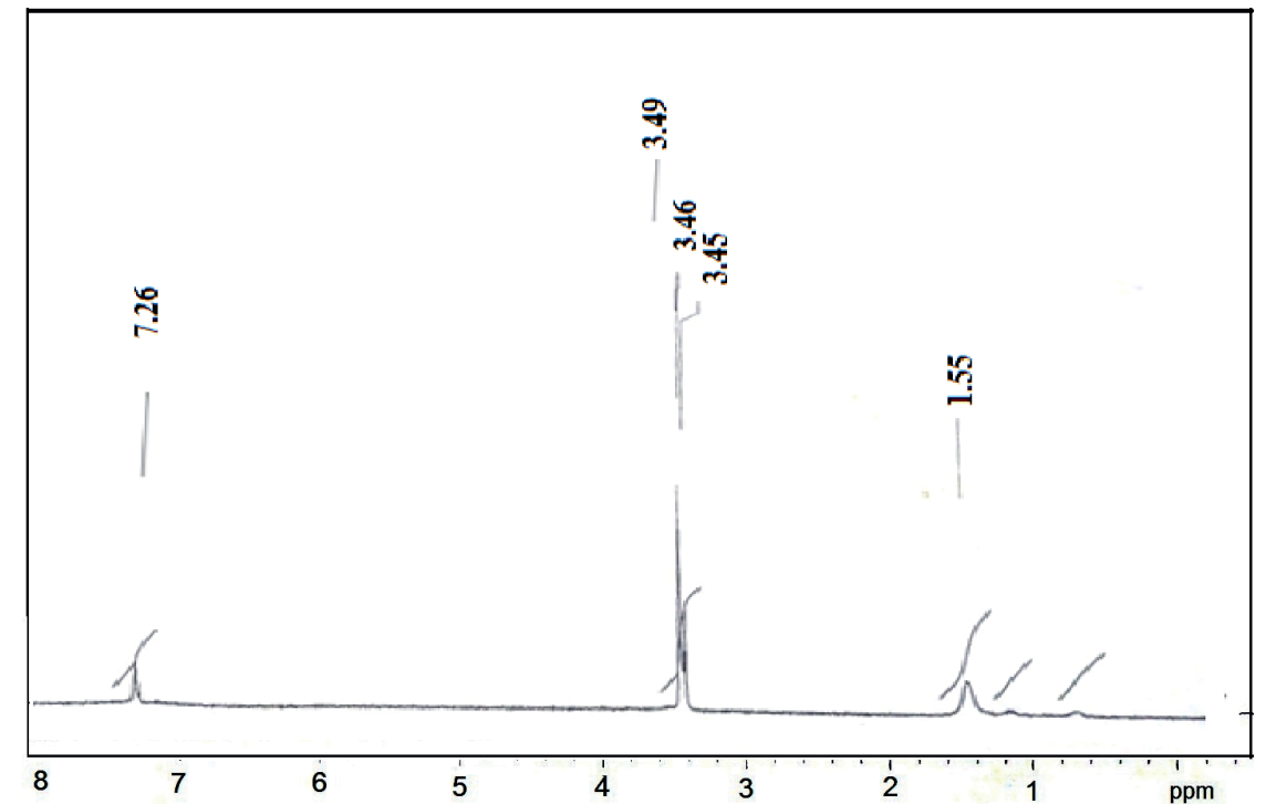
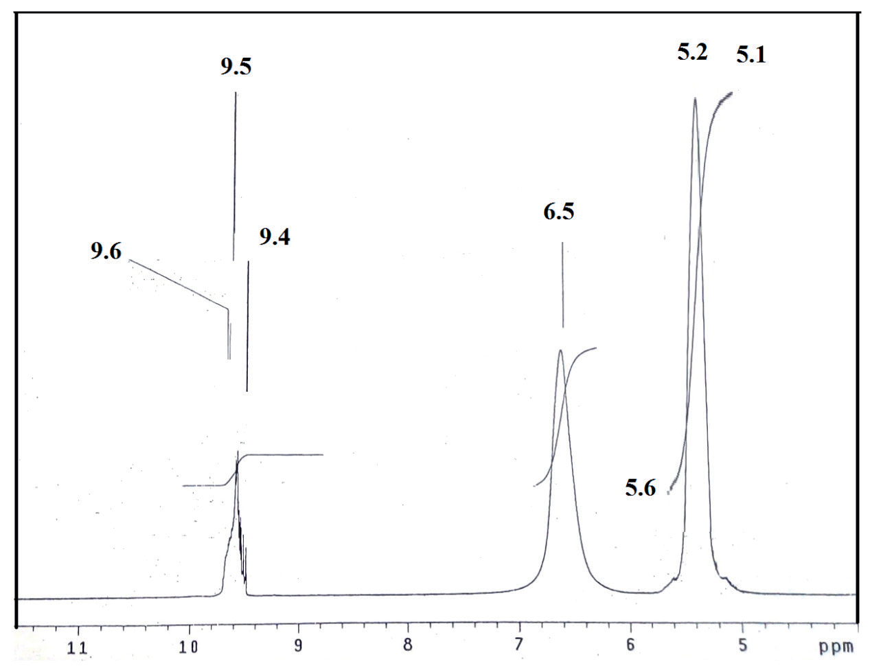
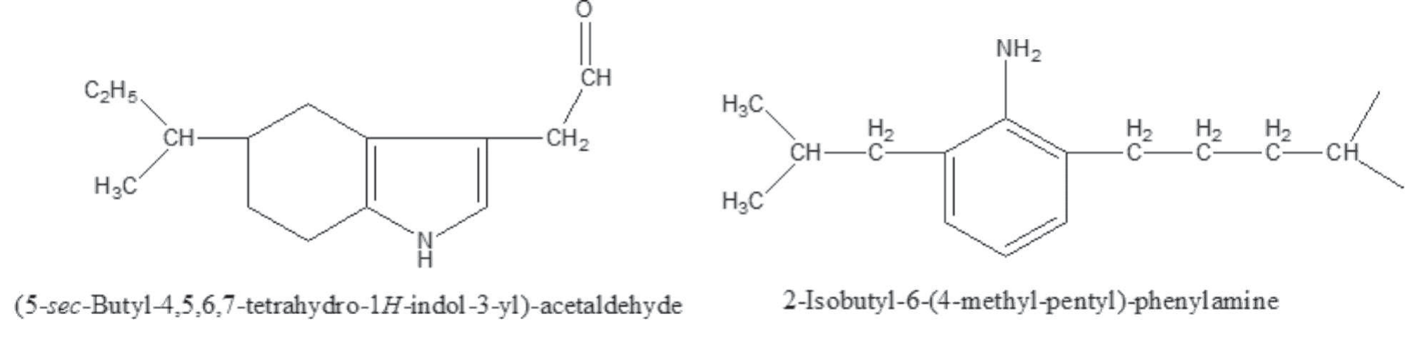
The 1H-NMR spectra of compounds (1) and (2) were measured in DMSO-d6 solvent and show signals consistent with the proposed structures. For compound (1), the peaks at 7.26 and 7.19 ppm are assigned to protons of the benzene ring. The proton signals of the NH2 group are observed at 3.48 and 3.49 ppm. Resonance at 1.55 ppm is due to the methylene protons attached to the benzene ring. Also, the spectrum showed a set of peaks in the 0.81-1.26 ppm ranges, which are assigned to the protons of methyl and methin groups. However, for compound (2), the signals that were observed at 9.4, 9.5, and 9.6 ppm are assigned to the proton of the aldehyde group (-CHO). Resonance appears at 6.5 ppm and is assigned to the proton of the =CH-N group. The peak at 5.6 ppm is due to the proton of the NH group of pyrrol. The peaks at 5.1 and 5.2 ppm are assigned to protons of the -CH2-C=O group. A set of peaks appeared in the 2.5-3.5 ppm range, corresponding to protons of methyl, methylene, and methin groups, as illustrated in (Fig. 2 and 3).
Mass Analysis
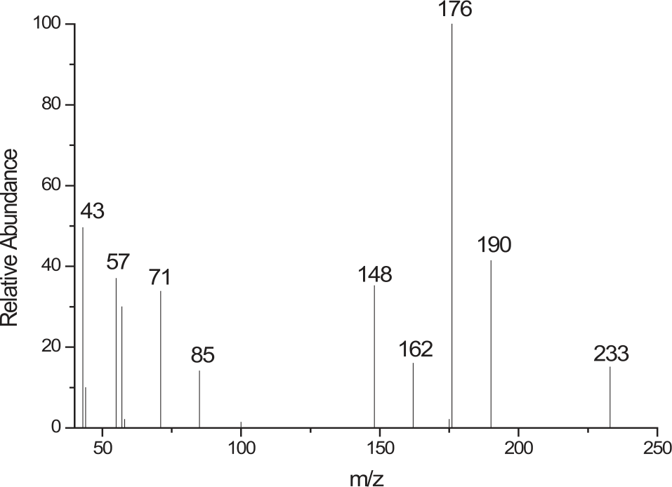
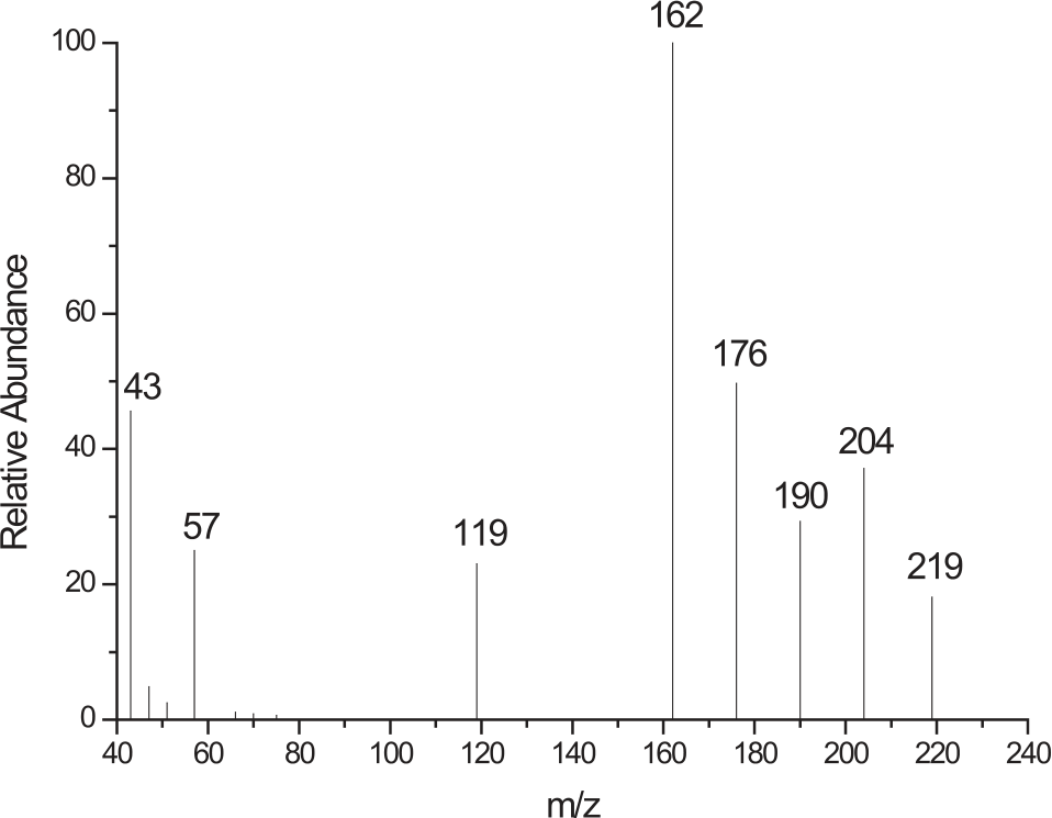
The mass spectra of compounds (1) and (2) confirmed their proposed formulations. The compound (1) shows a peak (m/z) at 233 amu, consistent with the compound’s molecular weight and a base peak at m/z = 176. However, the compound (2) spectrum reveals a molecular ion peak (m/z) at 219 amu, consistent with the compound’s molecular weight, and a base peak at m/z = 162. Furthermore, the fragments observed at m/z = 43, 57, 119, 162, 176, 190, and 204 correspond to C2H3O, C4H9, C8H9N, C10H12NO, C12H18N, C12H16NO, and C13H18NO moieties, respectively, as illustrated in (Fig. 4 and 5).
Additionally, the fragments observed at m/z = 43, 57, 71, 85, 148, 162, 176, and 190 correspond to C3H7, C4H9, C5H11, C6H13, C10H14N, C11H16N, C12H18N, and C13H20N moieties, respectively. The fragments of compounds (1) and (2) are represented in (Table 5 and 6).
FT-IR Analysis
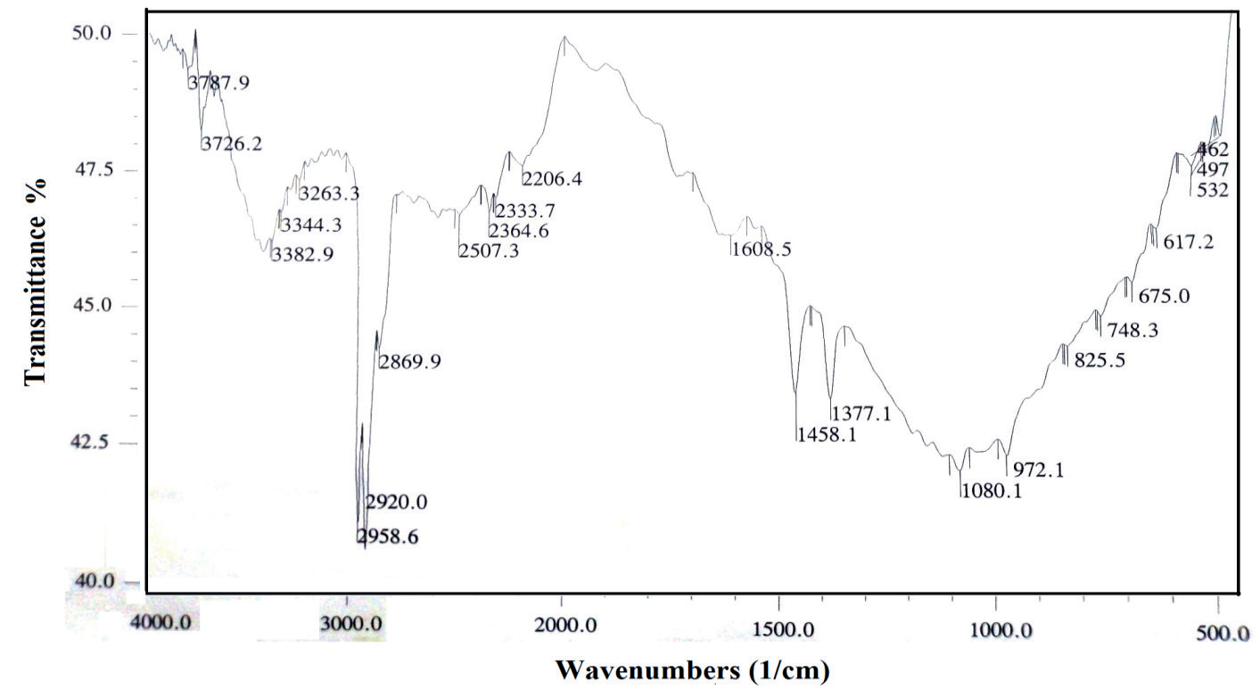
The FT-IR spectra in the region from 4000 to 400 cm−1 were recorded using KBr pellets. The FT-IR spectrum (KBr disk) of compound (1) showed bands at 1608 and 1458 cm-1, which can be attributed to the characteristic stretching vibrations of the benzene ring. The absorption band at 972 cm−1 is assigned to the aromatic ring’s C-H bending (out-of-plane). The absorption band at 3000 cm−1 is assigned to the C-H stretching of the aromatic ring. The medium absorption band at 3445 cm−1 is assigned to the N-H stretching, while the N-H bending appears as medium to strong broad bands at 1550 and 1640 cm−1. The C-N stretching absorption appears as a medium band at 1080 cm−1. The absorption band at 1375 of the methyl group is split into 1370 and 1377 cm−1. These doublet bands are attributed to the characteristic bending vibration of isopropyl groups. The absorption band at 2920 cm−1 is attributed to the C-H stretching vibration of the methyl group as depicted in (Fig. 6).
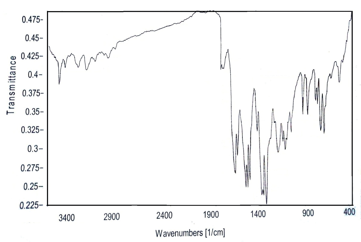
The FT-IR spectrum (KBr disk) of compound (2), as depicted in (Fig. 7), showed bands at 1640, 1597, and 1550 cm−1 attributed to characteristic stretching vibrations of the pyrrol ring. The absorption band at 990 cm−1 is assigned to the C-H bending (out-of-plane) in the pyrrol ring. The absorption band at 3100 cm−1 is assigned to the C-H stretching of the double bond. The medium absorption band at 3450 cm−1 is assigned to the N-H stretching, while the N-H bending appears as a strong, broad band at 1550. The C-N stretching absorption appears as a medium band at 1350 cm−1. The absorption band at 1375 of the methyl group is split into 2 bands of nearly equal intensity at 1370 and 1380 cm−1. These doublet bands are attributed to the characteristic bending vibrations of isopropyl groups. The absorption band at 2923 cm−1 is attributed to the C-H stretching vibrations of the methyl group. The C=O stretching absorption appears as a strong band at 1747 cm−1. The absorption bands at 2750 and 2850 cm−1 are assigned to C-H stretching vibrations, which confirms that an aldehyde functional group is almost certainly indicated. The appearance of the medium absorption band at 1460 cm−1 is due to the bending vibrations of the CH2 group next to the carbonyl group.
Structural features of the extracted compounds were obtained using FT-IR, 1H-NMR, and mass spectroscopy. This combination of techniques was utilized for structure characterization, as illustrated in (Fig. 8).
Discussion
The majority of national and regional schistosomiasis management programs worldwide currently involve the planned massive distribution of anti-schistosomal medication treatment [31]. This strategy has a significant drawback, as persons susceptible to infections, who neglect or refuse therapy, continue to be infected, thereby contributing to the local spread of schistosomal infection [32]. Therefore, some new effective therapies that can halt the parasite transmission mechanism are required [33]. Besides, to manage the intermediate hosts of this parasite, the snails, there is a need for more effective and selective molluscicides [34].
Table 5.
Mass spectra of compound (1).
| m/z | Rel. Int. | Fragment |
|---|---|---|
| 43 | 49.6 | C3H7 |
| 57 | 58.8 | C4H9 |
| 71 | 33.8 | C5H11 |
| 85 | 14.1 | C6H13 |
| 148 | 35.2 | C10H14N |
| 162 | 16.0 | C11H16N |
| 176 | 100 | C12H18N |
| 190 | 41.4 | C13H20N |
| 233 | 15.1 | C16H27N |
Table 6.
Mass spectra of compound (2).
| m/z | Rel. Int. | Fragment |
|---|---|---|
| 43 | 45.6 | C2H3O |
| 57 | 25.0 | C4H9 |
| 119 | 23.0 | C8H9N |
| 162 | 100 | C10H12NO |
| 176 | 49.7 | C12H18N |
| 190 | 29.3 | C12H16NO |
| 204 | 37.1 | C13H18NO |
| 219 | 18.1 | C14H21NO |
This work evaluated twenty-four bacterial extracts in the present investigation against B. alexandrina snails. The results revealed that some bacterial extracts have potential effects on the bacteria, where E. coli and S. aureus had the highest values after 24 hours of exposure time. This is consistent with the previously published molluscicidal action of crude plant extracts, which has been demonstrated for mollusk control in various pesticide test trials conducted worldwide [35, 36].
In the current investigation, purified molecules of bacterial metabolites, including S. aureus and E. coli, illustrated a promising molluscicidal action. Also, Anagallis arvensis extract was tested by Ibrahim and Ghoname [37], who reported that it has a significant molluscicidal potential against B. alexandrina at its LC50 and LC90. Besides, Ibrahim and Bakry [38] demonstrated that chlorophyllin had a molluscicidal impact on B. alexandrina snails. Therefore, compared to other previously examined reports, bacterial compounds in the present findings have the maximum molluscicidal role and should be taken into consideration for applicability.
The present results revealed that purified compounds of E. coli have promising results versus B. alexandrina at LC50 = 93.9±1.2 mg/L and LC90 = 110.5±1.2 mg/L, while bioactive compounds of S. aureus have an efficient impact with LC50 = 78.2±1.0 mg/L and LC90 = 98.6±1.0 mg/L. Exposing B. alexandrina snails for one day to an organophosphorus insecticide showed that LC50 and LC90 values were 39.00 and 73.0 mg/L, respectively [39, 40].
Furthermore, the results of the current investigation showed that the exposure of B. alexandrina snails to sub-lethal doses had a significant impact (P≤0.05) on the functioning of their liver enzymes. According to previous investigations, several substances considerably impact the oxidative enzymes in B. alexandrina quickly after exposure for one day [41].
D. pulex is a kind of aquatic flea that is used to measure toxicity [42]. This work’s potential bacterial metabolites of S. aureus and E. coli demonstrated minimal impact on D. pulex, indicating the suitability of employing these chemicals in some agricultural uses and biological manipulation. According to another study group, various compounds in carwashes have been shown to impact human health and the microbial population in water [43].
The current study analyzes the protein pattern of the hemolymph of B. alexandrina snails exposed to sublethal doses of the purified molecules of E. coli and S. aureus, revealing alterations versus the control group. In accordance with Bakry et al. [44], who applied diazinon, which led to more variation of protein bands of B. alexandrina snails than in the untreated group. The present results suggest that certain bacterial metabolites, especially those purified from S. aureus and E. coli (with lethal and sublethal doses), could potentially be used to combat B. alexandrina with a notable safety level for D. pulex as a non-target, which is present in the same habitat as B. alexandrina.

