

Introduction
The Moraceae family is an important family of flowering plants because of its medicinal and economic value [1]. Members of the Moraceae family are found in tropical and subtropical regions including the equator, but only a few are known from the temperate zone [2]. The study of Zerega et al., [3] shows that the members of Moraceae family are mainly distributed in tropical regions. The family Moraceae includes flowering trees, often referred to as mulberry or fig trees and consists of about 40 genera and more than 1,000 species [2]. However, the study of [4] indicated that the family includes 70 genera and 1,500 species. The study of Clement & Weiblin, [5] stated that the members of the family Moraceae occur in tropical and temperate regions and include 37 genera and 1,100 species. However, the work of Patil & Patil, [6] suggests that the species are restricted to the warm tropical regions of the world, which include about 73 species and 1000 species, but Zhou and Gilbert, [7] mentions that the family has 37-43 genera and 1100-1400 species, which are widely distributed in the tropical and subtropical zone. Recent research studies state that the family was first classified by Linnaeus as it includes 1000 species and was little known in Africa [8]. Reproduction and evolution in Ficus (Moraceae. In Pakistan, the family is represented by 24 species, of which 11 species are native [9]. In addition, the importance of anatomical study of leaves for classification of Ficus species has been emphasized [10-13]. Differentiation of Ficus species has been based on DNA barcoding [14-17]. The study of anatomical features, especially leaf features, is too reliable and comparable in a variety of taxonomic studies [9]. It plays a crucial role in the identification of all non-reproductive organs from sterile stamens, archaeological remains, and fragmentary fossils [18].
The study of anatomical features of leaves alone may not be sufficient to provide important information to delineate problematic taxa, but in combination with anatomical features of leaves it is a good source for taxonomic studies [19]. Epidermal tissue serves a variety of functions; these include defense mechanisms, plant-water relationships, and pollinator attraction. These different functions are due to the presence of many specialized cells, including pavement cells, trichomes, and stomatal cells [20]. The different cells on the epidermis have different degrees of morphological specialization, and it is essential to search for this type of cells on the epidermis to provide us with taxonomic utility. The guard cells of stomata are important because they allow water to enter the plant body and ensure gas exchange during photosynthesis. The distribution and pattern of stomata on the epidermis of each leaf are controlled by genetic material but can change due to variations in environmental conditions, such as CO2 concentration [21, 22]. The stomata are surrounded by three main types of cells, namely pavement cells, guard cells, and subsidiary cells [20]. The characteristics of epidermal cells and the type of stomata present are key tools for plant identification. The pattern and study of the foliar epidermis of Ficus revealed several variations in leaf characteristics, which are also interesting between species and are used as keys for taxa identification [23]. Other leaf epidermis features include trichomes, hydathodes, and epicuticular waxes, which are considered by many taxonomists as taxonomic tools to distinguish plant taxa [24-29]. Hydathodes have also been discussed in genus Ficus by [30]. The hydathodes are evenly distributed on the surface of the leaf lamina in Ficus diversifolia flower. These hydathodes are water pores open to the lower epidermis through circular pores, and 20-30 water pores have been studied on the adaxial surface of the fig family [31]. The aim of the present study is to clarify the qualitative and quantitative characteristics of the leaf epidermis in order to contribute information for species identification and classification of the Moraceae family.
Materials and Methods
Research Site Description
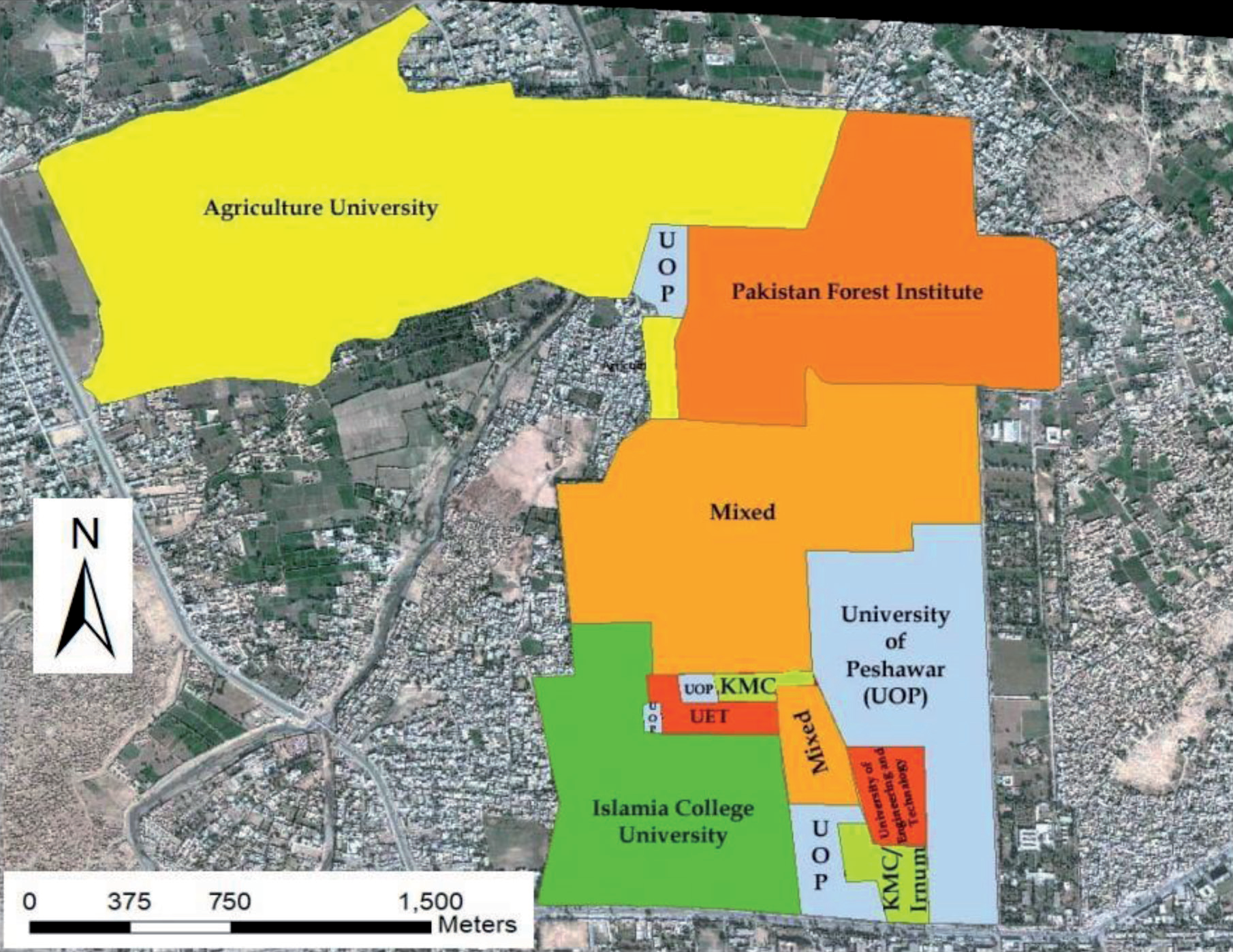
The research Area University of Peshawar campus lies in district Peshawar. The Peshawar district has an area of 1,257 km2, situated between 33°44/ and 34°15/ North latitude and 71°22/ and 71°42/ East longitudes [32] and approximately 1173 feet (358 meter) ALS (Figure1). Surrounded in the west by Mohmand and Khyber Agency, in the north side by Charsadda District, by Nowshera District in the east and the south-east parts are bounded by tribal areas joining Kohat and Peshawar District [33-36]. Peshawar’s climate is subtropical, with mild winters and very hot summers. The average temperature of the coldest month (January) and that of the warmest month (June) as given in Table 1. The majority of the soil is classified as alluvial soil having loamy texture, available phosphorous (40.49 mg/kg), potassium (572 mg/kg), saturation (44%), OM (3.75%), pH (6.91), EC (0.251 dSm-1), bulk density (1.36 gcm-3), and MC (6.401%). Soil properties were analyzed in Chemistry Laboratory, Tobacco Research. Station, Khan Garhi, Mardan. The deposition of silt, sand, and clay carried by rivers, particularly the Indus and its tributaries, creates this type of soil. Alluvial soil is noted for its fertility and is frequently used in agriculture. Soil composition can be influenced by factors such as topography, proximity to rivers, and local geological conditions [37, 38]. Water consumed by humans must be free from pathogenic microorganisms and chemicals that might be harmful to health. The total dissolved salts are (329 ppm), pH (6.175), carbonates ion (283.1 mg/L), bicarbonates ion (365.7 mg/L), copper (2.1 mg/L), manganese (48 mg/L), nickel (2.71 mg/L), lead (0.01 mg/L), chromium (0.04 mg/L), cobalt (0.071 mg/L), conductivity (724.5 μS/cm) were measured in water samples in Chemistry Laboratory, Tobacco Research. Station, Khan Garhi, Mardan.
Plant Collection, Identification and Herbarium Sheets Preparation
A total of 9 species belonging to family Moraceae were collected from University of Peshawar Campus during 2021-22 in flowering season. The collected plants were poisoned, dried, preserved in newspaper, and assigned voucher numbers, and then identified by Herbarium Botanist Dr. Ghulam Jellani and confirmed to be from the native flora (Flora of Pakistan) [39], other published literature [40-43] and The Plant List (TPL) (http://www. theplantlist.org/). Each plant was mounted on herbarium sheets and voucher numbers were assigned to each sheet and then submitted to University of Peshawar Herbarium (PUP) for future record. Herbarium sheets give complete details about each botanical taxon, including the botanical name, family, and locality.
Table 1.
Mean temperature, humidity, and precipitation of research area
Leaf Epidermis Using LM
The anatomical characteristics of leaves of 9 Moraceae species were studied through LM, mostly following the protocol of [44]. The fresh leaves were soaked in water for 2 h to prevent drying and wilting. Subsequently, the leaves were boiled in lactic acid and acetic acid for 10- 15 min following the protocol of Tomaszewski et al. [29]. Both the upper and lower surfaces of the epidermis were removed from the boiled leaves using the peeling technique. Leaves with a thick cuticular layer were treated with formalin, indole acetic acid, and ethanol at concentrations of 1:1:3. The epidermal layer was stained with a glycerol-gel mixture containing with 1% safranin. These stained epidermal walls were examined at LM with different objectives (e.g., 10×and 40×). Various qualitative characteristics of the leaf such as the including shape of the epidermal cells, the pattern of the epidermal wall, and the type of stomata were examined. With the help of SPSS, the quantitative data were tabulated as minimum to maximum value, mean, and standard error. In describing the epidermal cells, we mainly followed the terminology of [28] and [30].
Determination of Stomatal Index
The stomatal index was determined using the following formula proposed by [31].
In this formula, S. I stand for the stomatal index, S for the number of stomata per unit, and E for the number of epidermal cells per unit area.
Foliar Epidermis Using SEM
In the analysis of SEM, we mainly followed the method of [39]. In this method, the waxy layer was removed from the dried leaves by treating the leaves with xylene solution for 24 h. The leaf sections from the abaxial and adaxial surfaces were simply placed on a double-sided tape and then attached to a stub. Specimens were gold-plated in a gold-palladium chamber and then observed at SEM in the Central Resource Laboratory (CRL) of the University of Peshawar. The specimen leaves were photographed using PN 665 Polaroid film.
Results
In this study, the foliar epidermal study of the 9 members of the family Moraceae was examined using light and scanning electron microscopy. Significant variations in the anatomical characters were observed in the studied members, which are shown in Tables 2, 3, and 4.
Foliar Epidermal Morphology
The epidermal cells are differently shaped on both surfaces of the leaf. The cell shapes observed in the studied taxa are irregular, rectangular, pentagonal, and polygonal. The wall of the epidermal cells is thick in most species, except in B. papyrifera, M. alba, and M. laevigata where the epidermal cells are thin on both leaf surfaces. The shape of the epidermal cells is polygonal in most species (Table 2). The current study also focuses on the size of the epidermis and reports that both the length and width of the epidermal cells on the lower and upper epidermis of the leaf vary greatly. The highest epidermal length (42-47.5 µm) on the adaxial surface was recorded in B. papyrifera while the lowest value (9.5-12 µm) of epidermal length on the adaxial surface was recorded in F. benjamina. The width of epidermal length also varied from species to species (Table 3, Figure 2). From Table 3, it can be seen that the species of B. papyrifera have higher value (22.5-25 µm) for epidermis width on the adaxial surface than the other taxa. The size of the epidermal cells on the abaxial surface shows significant differences in both length and width of the cells. Table 3 shows that the epidermis length of M. laevigata has the highest value (32-40 µm) on the abaxial surface, while the lowest value (4-14 µm) was observed in B. papyrifera. The width of the epidermal cells on the abaxial surface ranged from 6 µm to 25 µm according to Table 3. These differences in the size of epidermal cell surface on both leaf surfaces of the leaf were important features of plant morphology.
Table 2.
Qualitative leaf epidermal characters of family Moraceae based on LM
Stomatal Type and Size
The results of the present work show that the stomata and trichomes on the adaxial surface are absent in most species. Both the qualitative and quantitative characteristics of the studied members show that the variations in the shape and size of stomata are important characters that are considered as identification tools for the plant taxa. We found in this study that most species are hypostomatic. Most species have anomocytic stomata on the lower surface of the leaf. The cyclocytic stomata are found only in F. elastic and F. benjamina. The stomatal type, which is rare in the studied species, is cyclocytic, laterocytic, and paracytic. This result shows that the lower surface of the leaf has many epidermal features, and that these epidermal characteristics are of great importance in the identification and classification of plants. The large stomata on the adaxial surface were observed in M. alba with stomata length of (28-31 µm), while the small stomata in F. benjamina with stomata length of (6-12.5 µm) were seen on the upper leaf surface.
On the lower leaf surface, the longest stomata (30-33 µm) were observed in M. alba and the shortest (8-13 µm) in M. laevigata. The width of the stomata on the adaxial surface has different value, ranging from 6 to 25 µm, while the width found on the abaxial surface ranges from 4 to 25 µm. The stomatal pores on the lower surface vary from species to species. We found the stomata on the upper surface stomatal pore in a few species on the upper surface and reported that the highest stomata pore length was found in F. benjamina on the upper surface while the lowest pore length of stomata on the lower surface was observed in F. benghalensis. In addition to stomatal pore length, stomatal pore width on the adaxial leaf surface in some species was also measured that the values were in the range of (3.9-77 µm). On the abaxial leaf surface, the stomatal pore length and width values vary the most, and it can be summarized that the large stomatal pore in F. elastica and the small stomatal pore in M. laevigata are found on the adaxial surface.
Figure 2.
Light micrographs (LM) of the leaf cell shapes, walls pattern, and stomata of Moraceae. (a) Adaxial surface and (b) abaxial surface 1) F. religiosa; 2) F. benghalensis; 3) F. elastica; 4) F. virens; 5) F. benjamina; 6) Ficus racemosa; 7) M. alba; 8) M. laevigata; 9) B. papyrifera
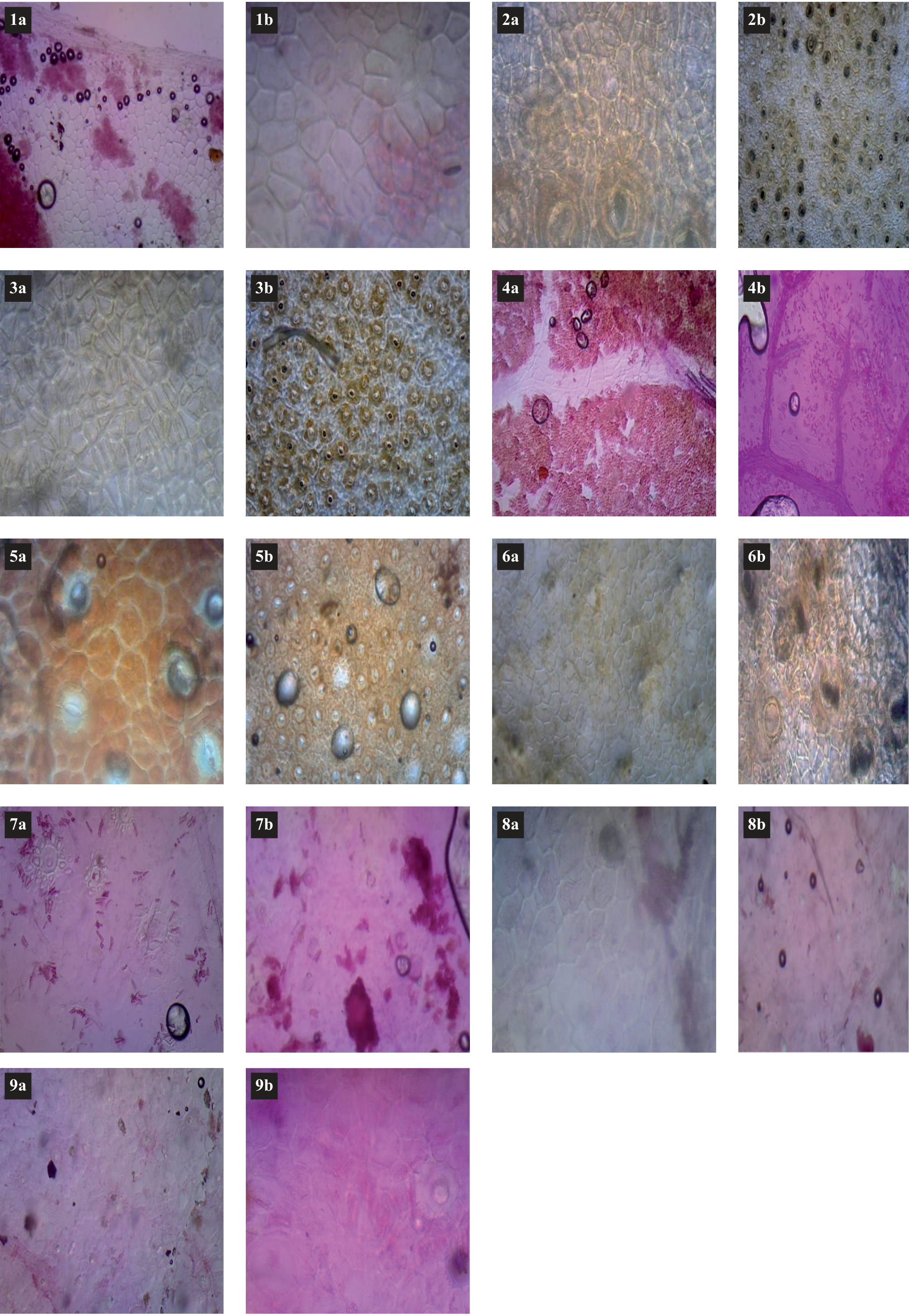
Table 3.
Quantitative leaf epidermal characters of family Moraceae based on light microscopy
Subsidiary Cell Size
The size of the subsidiary cell on both leaf surfaces shows differences in its morphological characteristics. The result shows that the subsidiary cell on the adaxial surface is absent in most species. The highest length (43-47.5 µm) of the subsidiary cell was found on the adaxial surface in F. benghalensis, while the lowest value (14.6-16 µm) was found in F. benjamina. The width of the subsidiary cells is larger in B. papyrifera and smaller on the upper surface in F. benjamina. We also determined the length and width of the subsidiary cell on the lower surface. It was found that the length of subsidiary cells is the largest in M. laevigata (55-61 µm) and smallest in F. benjamina (8-16 µm). It was found that the width of the subsidiary cell on the upper surface ranged from (4-6.1 µm) in B. papyrifera to 35-43 µm in F. racemosa as given in Table 4, Figure 3.
Stomatal Index
The stomatal index found in the studied species shows considerable variation, and this characteristic is useful for distinguishing. Stomatal index is an important characteristic of the leaf; it indicates the transpiration rate of the leaf and can be used as a geographical marker. It was indicated that the stomatal index on the upper surface ranges from 76.7% in B. papyrifera to 2.13% in F. benjamina, while on the lower surface it is highest in F. religiosa (61.8%) and lowest in F. virens (3.06%).
Discussion
The study of leaf epidermis features and epidermal traits of the family Moraceae reveals several important micromorphological characters that are significant for plant identification [45]. The epidermis features have been studied in many angiosperm families and provide valuable data for plant taxonomy [44, 46]. The anatomical characteristics of leaves are widely considered as a tool for species classification and to find morphological similarities among species [13, 47, 48].
Table 4.
Quantitative leaf epidermal characters of family Moraceae based on light microscopy
In this study, the anatomical characteristics of the leaves of the family Moraceae and the variations in leaf characteristics are interesting and interspecific, which serves as a key to taxa identification. A study of the leaf epidermis based on LM and SEM revealed that the cells of the epidermis on the adaxial and abaxial surfaces are highly variable in shape and size; they are rectangular, irregular, polygonal, and pentagonal with thick and thin leaf surfaces [45, 49]. The quantitative and qualitative characteristics of the epidermal cells are the focus of this study. Quantitative analysis shows that leaf epidermis cells vary greatly in both length and width on both leaf surfaces [4], as the smallest epidermis length is found on the adaxial surface in F. benjamina while the highest epidermis length and width are found on the adaxial leaf surface in B. papyrifera and the least epidermal cell width in F. benjamina, on the abaxial surface least epidermal length in B. papyrifera, while the highest epidermal length in M. laevigata and the least epidermis cell width in F. elastica, while the least epidermal width was found in F. benjamina. The anatomical study of leaf epidermis cells [23, 50-52] can be considered as a good taxonomic tool to identify and distinguish taxa.
Figure 3.
Scanning electron micrographs (SEM) of the leaf cell shapes, wall patterns, stomata, and trichome of Moraceae. (a) Adaxial surface and (b) abaxial surface 1) F. religiosa; 2) F. benghalensis; 3) F. elastica; 4) F. virens; 5) F. benjamina; 6) Ficus racemosa; 7) M. alba; 8) M. laevigata; 9) B. papyrifera
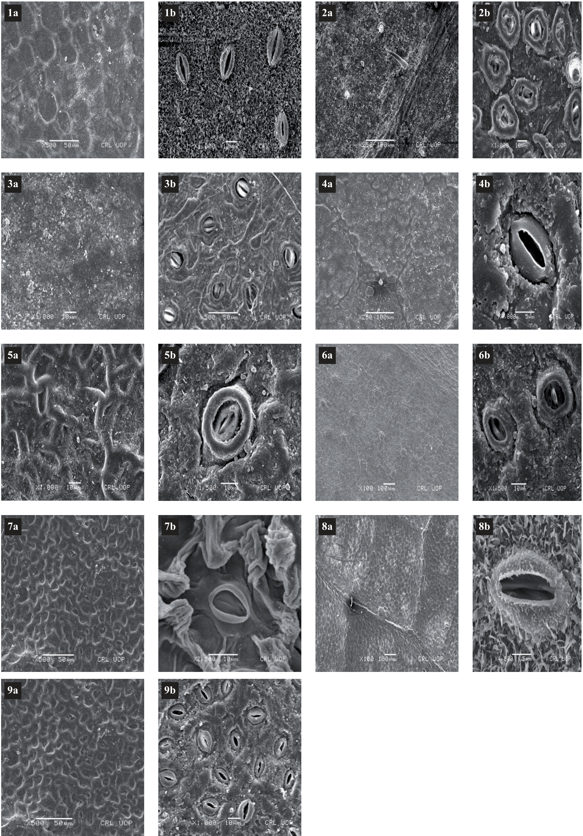
Epidermal characteristics of foliars, including stomata and trichomes, are considered an important tool for the taxonomic delimitation of different plant species [24, 25, 53-55]. It was found that species of the family Moraceae, especially Ficus species, are hypostomatic. This was confirmed by the study of [4, 56-59] on the study of foliar epidermis of Ficus species. The result shows that the anomocytic stomata were evenly distributed on the abaxial leaf surface which is similar to the result of Klimko and Truchan, 2006 [45], but [23, 60] reported that most Ficus species have a paracytic type of stomata. Moreover, all the members studied had stomata on the abaxial surface, but on the adaxial leaf surface, the stomata were only found in F. religiosa, F. elastica, F. virens, F. racemosa, and M. laevigata. In this result, it was found that the cyclocytic type of stomata was found on the lower surface in F. elastica, it was absent on the upper surface in the same species. The most important features of the epidermis are the presence or absence of stomata.
The result shows that the trichome was found in the majority of species and is considered informative in plant systematics [61-64]. However, the trichome on the adaxial surface was absent in F. religiosa, F. elastica, F. racemosa, F. virens, and M. laevigata. In [60], it was reported that the trichome was absent on both leaf surfaces in F. religiosa, Ficus neriifolia Sam., and Ficus virens, supporting current studies. [45] divided the studied taxa into two groups based on the absence or presence of trichomes: a glabrous group, which consist of species without trichomes on both leaf surfaces, and a pubescent group, which includes taxa with one or more trichomes on both leaf surfaces. The present work shows that variation in stomata cell size, epidermal cell size, stomata pore size, subsidiary cell size, and stomata index has been studied, and these large differences have been considered important characters in distinguishing plant taxa. The stomatal index of the studied species shows considerable differences, and this character is useful for distinguishing the taxa.
Stomatal index is an important leaf characteristic that indicates the transpiration rate of the leaf and can also be used as a geographic marker [64]. However, the study of [9] states that stomatal index is considered useful for determining atmospheric CO2 content. Therefore, the level of atmospheric pollution is determined by this characteristic.
Conclusions
LM and SEM-based foliar epidermal study of 9 species of the family Moraceae indicated that leaf epidermis study provides us with valuable and significant information for the systematic position of plant. This information is used for the correct identification of plant taxa. According to the results of the current study, foliar epidermal characteristics have a significant taxonomic value. These systematic characteristics are important for distinguishing between Moraceae species. The study concluded that leaf epidermal characters are important tools used in the authentication of problematic taxa. We concluded from this result that leaf anatomical characters are very helpful in plant identification and classification. The micro morphological study will be crucial for genetically based taxonomy, molecular studies, and chemotaxonomy. It is strongly recommended that further research be conducted to highlight the links between individual members of the Moraceae family based on foliar epidermal characteristics and phylogenetic relationships.

