

Introduction
The marine environment is an ecosystem made unique because of the diversity of the organisms it supports [1]. From estuaries to abyssal trenches to hydrothermal vents, algae demonstrate incredible ecological plasticity. The latter constitutes an enormous reservoir of potentially active natural molecules [2, 3]. Studies have made it possible to isolate and identify a very large number of new molecules of great structural originality, many of which have interesting biological activity. Furthermore, the plant origin of the active ingredients isolated from algae constitutes a real and important advantage at a time when ingredients of animal origin are irrelevant [4]. Faced with the appearance of resistant forms of several bacteria to certain antibiotics, the search for new active molecules has become a necessity [5]. In addition, natural antioxidants and antimicrobial agents may provide solutions to the public’s health issues because they can constitute a safer and more effective alternative to synthetic products whose toxicity is proven [6]. Likewise, these bioactive compounds are of great interest in the medical field, which is in an ongoing search for more powerful antibiotics to combat the antibiotic resistance of pathogens [7, 8].
Algeria is a country with a maritime coastline stretching over 1622 km. It constitutes a rich source of marine algae, which, however, remains little studied and is not yet exploited by pharmaceutical or cosmetic companies, while algae constitute a considerable economic development issue. The existing work on the algal flora of Algeria is essentially of the floristic inventory type. This is why our study aims at another aspect of greater interest and of growing interest which is the composition of polyphenols and flavonoids as well as the study of the antioxidant and antimicrobial potential of marine algae, in particular in Ulva lactuca, Cystoseira amentacea var. stricta, and Corallina elongata.
Materials and Methods
Harvesting and Processing Algae
The harvest of algae was carried out on the Algerian West Coast. The algae were identified by specialists in the field using the standard identification keys. Algal specimens were collected by hand and stored in plastic bins, filled with seawater, until processed the same day in the laboratory. After removing debris, small crustaceans, and other algae, the thalli are rinsed with distilled water several times to clean the epiphytes completely. These algae are dried in the open air and away from light for 7 to 10 days until complete dehydration, then ground into a very fine powder, then sifted and stored in a dark and dry place [9].
Extraction
The extraction method adopted was maceration by mixing two organic solvents. 50 g of each powdered algae is added to 400 ml of two solvents: 200 ml of methanol and 200 ml of toluene. The first is polar, and the second is non-polar. The resulting mixture was stirred continuously for 48 hours in the absence of light at room temperature, then filtered with Wattman No1 paper. The filtrate obtained was evaporated under reduced pressure at a temperature of 40°C using a “Büchi” type rotary evaporator [10].
Each residue was weighed, and the extract recovered with a few drops of methanol was stored in sterile pill bottles at 4°C until its use for the different dosages [11].
Determination of Yield
The determination of the yield of the dry extract is performed by applying the following formula: R (%) = (P1 – P2/PE) × 100 knowing that: P1: Weight of the balloon after evaporation. P2: Weight of the empty flask before evaporation. PE: Weight of dry algal material.
Dosage of Flavonoids
A volume of 1 ml of the extract is added to 1 ml of 2% aluminum trichloride. The mixture was shaken and incubated for 30 min at room temperature in the dark, then the absorbance was measured at 430 nm on a spectrophotometer, with quercetin used as a reference standard. The results are expressed in micrograms of quercetin equivalent per milligram of dry extract (μg EQ/mg extract) [12].
Dosage of Polyphenols
Add 200 µl of the extract in a tube to the mixture (1 ml of Folin-Ciocalteu reagent diluted 10 times and 0.8 ml of 7.5% sodium carbonate). Shake the tube and store at room temperature for 30 minutes. Use a spectrophotometer to measure the absorbance at 765 nm against a blank. Use gallic acid as a positive control and run the calibration curve in parallel under the same operating conditions. Results are expressed in milligrams (mg) of gallic acid equivalent per gram of dry algae (mg EAG/g DM).
Dosage of Chlorophyll a, b, and Total Carotenoids
The determination of chlorophyll and carotenoids was carried out according to the spectral method [13, 14]. It uses characteristic wavelengths which correspond to the maximum absorbances of chlorophylls and carotenoids. 3 g of crushed algae were added to 30 ml of methanol. After adding 300 mg of magnesium sulfate, the mixture was incubated for 24 hours at 4°C. The mixture obtained was filtered and then centrifuged at 5000 rpm for 10 minutes. The supernatant was collected and the absorbances were read at 666, 653, and 470 nm for chlorophylls a, b, and total carotenoids, respectively. The chlorophyll and carotenoid contents were calculated in μg/ml according to the following formulas:
Ca (μg/ml) = 15.65 A666 – 7.340 A653
Cb (μg/ml) = 27.05 A653 – 11.21 A666
Cc (μg/ml) = 1000 A470 – 2.860 Ca – 129.2 Cb/245
Evaluation of Antioxidant Activity
Trapping of the Free Radical DPPH (2,2-Diphenyl-1-Picrylhydrazyl)
This method is based on measuring the ability of antioxidants to trap the DPPH radical [13]. A volume of 50 μl of different concentrations of each extract is added to 1950 μl of the freshly prepared methanolic solution of DPPH (0.025 g/l). Concerning the negative control, the latter is prepared in parallel by mixing 50 μl of methanol with 1950 μl of a methanolic solution of DPPH at the same concentration used. The positive control is represented by a methanolic solution of a standard antioxidant (Ascorbic acid). After incubation in the dark for 30 min and at room temperature, the absorbance reading is carried out at 515 nm using a spectrophotometer against the white, according to the formula: DPPH (%) = (Ac – At) /Ac) × bra100 DPPH (%): Percentage reduction in DPPH. Ac: absorbance of the negative control At: absorbance of the extract
The IC50 value is the extract concentration, which ensures the 50% reduction in DPPH, determined graphically by linear regression, for each extract from the curve of the percentage reduction as a function of concentration [15].
Evaluation of Antibacterial Activity
The bacterial activity was evaluated on seven bacteria (Pseudomonas sp, Escherichia coli, Bacillus subtilus, Bacillus cereus, Methicillin-resistant Staphylococcus aureus or SARM, Klebsiella sp, Staphylococcus aureus). In order to evaluate the antimicrobial activity of the extracts of the three algae, we used the diffusion method on the Muller Hinton agar medium [14]. Sterile Wattman filter paper discs measuring 6 millimeters in diameter are impregnated with 40 μl of the different extracts. Using sterile forceps, the discs are placed on the surface of a medium seeded with a microbial suspension with an optical density of 0.5 McFarland (108CFU/ml). After diffusion, the plates are incubated for 18 to 24 hours at 37°C. Discs impregnated with different solvents will serve as controls.
All analyses were performed in triplicates and the experimental data were expressed as means±standard deviation using Microsoft Office Excel 2007.
Results and Discussion
Extraction Yields
Yields of crude extracts were determined relative to 50 g of plant material, and the results are shown in Fig. 1.
The method of extracting bioactive materials from plants plays an important role in providing consumers with a high-quality herbal product, and modern “advanced” extraction methods are superior to conventional methods. Extraction is considered one of the most crucial procedures in the manufacture of herbal products, which affects the active ingredients in the sample both qualitatively and quantitatively [16].
The best yield extraction obtained is that of Cystoseira amentacea var. stricta with a value of 6.4%, followed by that of Ulva (4.50%), and the lowest is recorded by the extract of Corallina elongata, with a rate of 1.2%. Results that are close to ours put forward a yield value of 2.53% for the methanolic extraction of Corallina elongata [17].
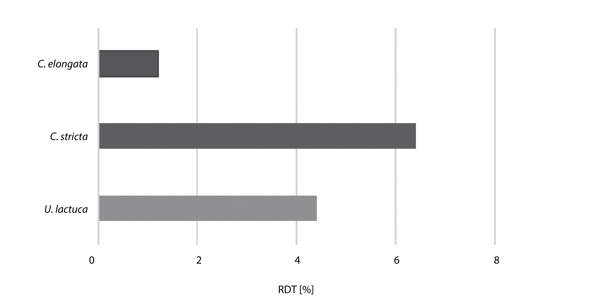
Table 1.
Total phenol and flavonoid contents of crude extracts of Ulva lactuca, Cystoseira amentacea var. stricta and Corallina elongata.
| Crude extrats | Polyohenols (mg EAG/g extrait) | Flavonoid (mg EQ/g extrait) |
|---|---|---|
| Ulva lactuca | 79.77±0.72 | 65.17±4.19 |
| Cystoseira amentacea | 73.92±0.53 | 36.2±3.33 |
| Corallina elongata | 219.43±5.79 | 61.6±1.37 |
Calculations of extraction yields are based on several factors, namely the extraction temperature, the initial plant material, and humidity [18]. This difference between species can also be attributed to the polarity of the compounds present in these algae, in addition to the nature of the solvent used and the chemical properties of the molecules to be extracted [19]. Likewise, the extraction method (mace ration, decoction, infusion) also plays an important role in determining the yield and the chemical composition of the prepared extract [20].
Dosage of Phenolic Compounds
The contents of polyphenols and total flavonoids in our samples are reported in Table 1. A single dissolvent cannot extract all classes of phenolic compounds simultaneously and at a maximum concentration [21]. Therefore, we have used a mixture of two solvents.
From the results of a dosage of polyphenols we find that the methanol-toluene extract of Corallina elongata is rich in total polyphenols with a value of 219.43 mg/g, followed by the green algae extract Ulva lactuca with 79.7743 mg/g, while the extract of the brown algae Cystoseira amentacea is lower than the other two with a content of 73.92 mg/g. These results are not in agreement with other studies [22], which indicate that brown algae have higher polyphenolic contents compared to green and red algae. On the other hand, the values found are close to those [23] which put forward more or less comparable concentrations in Cystoseires: Cystoseira crinita (56.5 mg/g), C. sedoides (50.3 mg/g), C. compressa (61 mg/g), and which [24] recorded values of 37.5 mg/g of polyphenols in Corallina elongata.
According to the literature, methanolic extracts are the richest in phenolic compounds, therefore methanol remains the best solvent for extracting these compounds. This is due to the ability of methanol to inhibit the action of polyphenol oxidase, which causes the oxidation of polyphenols in plant tissues [25, 26].
A wide range of algae species, including Ulva fasciata, were screened for antimicrobial activity. Researchers stated that drying reduced the bioactive compounds in the chosen algal species, and they found that methanol: toluene was the best solvent for extracting the antimicrobial compounds from fresh algae, and that the antimicrobial activity from chosen algae was due to a lipophilic compound that was stable over a broad range of temperatures (30–60°C) [27].
Many works suggest that maceration time affects the extraction rate [28–30] and the increasing temperature leads to an increasing extraction rate [31].
For flavonoids, we see that the Ulva extract has a higher content of flavonoids with a value of 65.17 mg/g, coming just after the Corallina red algae extract with 61.6 mg/g, the highest content. The lowest was recorded in Cystoseira with a value of 36.2 mg/g.
There exists little work on the flavonoid content in marine algae [32–34] in contrast to the numerous studies on flavonoids from terrestrial plants. A study on the distribution of flavonoids in 27 species of marine algae (6 Chlorophyta, 11 Phaeophyta, and 10 Rhodophyta) showed that they had a completely different flavonoid composition from fruits and vegetables [35].
The dosage of flavonoids is very important because they are a class of secondary plant phenols possessing potent antioxidant activity, thereby scavenging a wide range of reactive oxygen species and inhibiting lipoperoxidation, making them a potential therapeutic agent against a wide variety of diseases [36].
Dosage of Chlorophyll a, b, and Total Carotenoids
The results of the dosage of chlorophyll obtained are summarized in Table 2 and indicate that the brown alga Cystoseira amentacea var. stricta is richer in chlorophyll a and b (20.49 and 15.10 µg/ml) respectively than the green alga Ulva lactuca (7.96 and 14.12 µg/ml) and the red alga Corallina elongata (7.54 and 11.61 µg/ml). On the other hand, the highest carotenoid level is recorded by U. lactuca. These variations are probably linked to the species, geographic distribution, and age of the algae [17]. The effect of the solvent is not spared, although methanol is among the best solvents extractable from chlorophylls.
The main function of polar solvents is to improve and facilitate the permeability of the cell wall of the chemical substances, which allows better contact between the solvent and the solute, thus increasing the percentage yield of the extraction [37].
Table 2.
Contents of chlorophyll a, b and total carotenoids in the extracts.
Table 3.
Percentages of DPPH inhibition by algae extracts.
Evaluation of Antioxidant Activity
According to the results obtained for anti-radical activity presented in Table 3, the increase in anti-radical activity is proportional to the increase in the concentration of the extract. At the lowest concentration of 50 (μg/ml), the reduction rate of Ulva was the lowest with a value of 23.46% followed by Corallina elongata extract with 30.54%, and finally the extract from Cystoseira with 35.8%. At moderately high concentrations of 350 (µg/ml), the extracts show a progressive increase in DPPH reduction varying between 66.55%, 54.32%, and 58.9%, for Ulva lactuca, Cystoseira amentacea, and Corallina elongata, respectively. At the highest concentrations (750 µg/ml) we recorded values of 68.06%, 72.43%, and 66.02% for Ulva lactuca, Corallina elongata, and Cystoseira amentacea, respectively.
According to our results, the IC50 has a value of 275.7 μg/ml for Corallina elongata, 329.38 μg/ml for Ulva lactuca, and 352.91 μg/ml for Cystoseira amentacea var stricta, and these values remain significantly higher than that of ascorbic acid (63.24 μg/ml), which is in agreement with the low percentages of inhibitions recorded by the different extracts.
The most effective antioxidant power was recorded with the red algae Corallina. Our results show very high IC50s. The latter was also reported by others [17] who recorded IC50s at 1787.7µg/ml, and 3450 µg/ml for methanolic extracts of Corallina elongata and Cystoseira stricta, respectively. We know that the IC50 is inversely linked to the antioxidant capacity of a compound. However, an extract with low antioxidant activity cannot be considered a poor source of antioxidants, because an extract is composed of chemicals with different functional groups and polarities, allowing it to behave differently depending on the reaction mixture [38].
Because of different protocols and reaction conditions, IC50 values for the same extracts vary a lot. Thus, it turns out to be more difficult to compare the results of different laboratories [39]. Light, oxygen, and pH of the reaction mixture also affect the absorbance of the DPPH solution [40].
Antimicrobial Activity
A study of 82 different marine macroalgae from the Chlorophyceae, Phaeophyceae, and Rhodophyceae classes were screened for antimicrobial activity [41]. Among the selected taxa, Phaeophyceae were reported to be the most active, with 84% of the algae from this taxa showing antimicrobial activity.
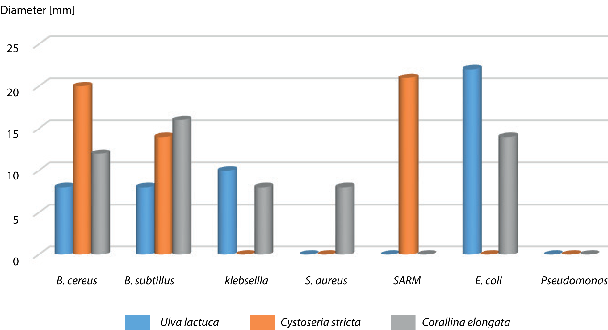
The results obtained for the antibacterial activity, reported in the histogram in Fig. 2, and expressed in terms of the diameter of the inhibition zone, showed that all bacteria are sensitive to one or other of the extracts except Pseudomonas which revealed its resistance for all three algae. The crude extracts of Ulva act on B. cereus, B. subtilus, and SARM with diameters of (8 mm), (10 mm), and (22 mm), respectively. This is consistent with the work [42] that noted the activity of methanolic extracts of U. lactuca on SARM with inhibition zones greater than 10 mm. Unlike those [43], who showed that antimicrobial activity is absent in Ulva algae extracts. A previous study [44] reported that the methanolic and aqueous extracts of 19 marine algal species collected along the western coast of Libya against 4 Gram-positive and 4 Gram-negative bacteria revealed that the observed inhibition zones values with methanolic extract of U. lactuca ranged between 11 mm for S. aureus and Bacillus subtilis and 14 mm for P. aeruginosa and Klebsiella spp. They also reported that Cystoseira crinite (Phaeophyceae) of the 19 examined algae, was the most potent against tested isolates.
The same results are noted for Cystoseira extract. Indeed, the latter acts on B. cereus, B. subtilus, and SARM with inhibition zones of (20 mm), (14 mm), and (21 mm), respectively. Researchers [42] who worked on two brown algae Cystoseira humilis and C. bifurcata showed that they had good activity against SARM with an inhibition zone diameter greater than 15 mm. Cystoseira crude extract has no effect on Klebsiella, Staphylococcus, E. coli, and Pseudomonas, contrary to other researchers [45], who reported that different Cystoseira species exhibited potent antibacterial activity against Gram-negative bacteria: Salmonella typhi, E. coli, P. aeruginosa, and Klebsiella sp.
The extracts of the two algae Ulva and Cystoseira have no effect on E. coli. These results are consistent with others [42, 46] which noted that only a few species of algae presented weak inhibitory activity against E. coli. However, a study [47] reported that the antibacterial activity of methanolic extracts of U. lactuca and Enteromorpha compressa (Chlorophyta), against 4 Gram-negative and 2 Gram-positive isolates showed that U. lactuca extract exhibited the most inhibitory effect for K. pneumonia, S. aureus, P. vulgaris, B. subtilis, and P. aeruginosa, followed by E. compressa. Nonetheless, the methanolic extract of U. lactuca was inactive against E. coli. Reports [48] have been made about the antibacterial activity of methanol/methylene chloride of Ulva lactuca, Codiumto mentosum, and Hypnea musciformis collected from the Suez Canal, Egypt. The latest study showed that U. lactuca has inhibitory activity only against Salmonella typhimurium, K. pneumonia, E. coli, Shigella boydii and S. aureus with inhibitory zones ranging between 6–9 mm.
As for the Corallina extract, its effectiveness is noted against most strains except SARM and Pseudomonas. The inhibition zones vary from (16 mm) against B. subtilus, to 8mm for Kleibseila and Staphylococcus, passing through an inhibition zone of (12 mm) for E. coli.
Our results agree with those [49] which reported that high activity was shown on E. coli (32.00±1.73 mm) and E. faecalis (21.66±0.57 mm) with a methanolic extract of the red alga Corallina officinalis. Works [50] about the antimicrobial activities of some marine algae from the Mediterranean Sea (Algeria) show that the chloroform extracts of Ulva lactuca and Corallina elongata had the highest activity against E. coli and Salmonella sp.
Conclusions
The marine environment is a rich source of biological and chemical diversity. This diversity is at the origin of new chemical compounds likely to be used in industrial applications such as pharmaceutical products, cosmetics, and nutritional supplements. Finding new sources of purely natural bioactive substances is the main goal of most researchers today. This work is aimed at exploring the natural potential of marine algae from the west coast of Algeria. The various activity tests performed on the three species of algae show that the extracts studied contain a mixture of bioactive substances with a broad spectrum of activities which must be carefully studied and deepened in order to be able to use them for therapeutic purposes (antioxidants, antibiotics).

