

Introduction
There are a lot of fungi in the environment, and fungal pathogens are becoming more and more common causes of infection [1]. As a result, opportunistic infections, such as fungal infections, have also increased in frequency. As a result, antifungal therapy is becoming more prevalent in medical care, and natural sources for novel antifungals are now being screened more frequently [2-4]. Fungal diseases are still frequently controlled with synthetic chemical fungicides. However, strict regulatory policies have been imposed on their use as a result of increased awareness of food safety and human health [5]. Additionally, the emergence of new physiological pathogen races renders many of these synthetic chemicals ineffective, requiring the discovery of novel natural antifungal compounds.
Bacteria, which are microscopic organisms, have a significant impact on their surroundings. While most bacteria are beneficial and harmless germs, some pose significant threats to public health. Over the past few decades, pathogens have developed resistance to antibiotics, which is causing antibiotics to gradually lose their effectiveness against bacteria. Antibiotic resistance has evolved, resulting in ineffective treatment, infection persistence, and spread [6]. Therefore, new biological targets and antibiotics are required to combat antibacterial resistance. Recently, scientists have become more aware of the health risks posed by chemical additives and synthetic drugs [7]. As a result, the use of natural treatments over synthetic ones, like drugs derived from plants, is the new trend [8] or animals [9]. It has been demonstrated that the most efficient method for obtaining bioactive peptides from protein sources is enzymatic hydrolysis [10].
Plant extracts from numerous higher plants have been shown to possess antibacterial, antifungal, and insecticidal properties during laboratory trials. Among these plants, Alstonia scholaris and Millettia pinnata [11]. On other hand, plant protection plans that aim to lessen biotic and abiotic stresses often use plant cell responses to pathogenic and non-pathogenic fungus species [12, 13].
Various diseases, including hypertension, diabetes, cancer, and aging, have been linked to the production of reactive oxygen species by the body’s natural metabolism. These species can cause oxidative damage to all important cellular components [14]. Human health might benefit from antioxidants. Therefore, novel natural antioxidants are absolutely necessary [15]. Antimicrobial peptides are one of the proposed therapeutic antifungal, antibacterial, and antioxidant strategies [16]. Additionally, antioxidant, antibacterial, and antibiofilm hydrolysates and peptides have been reported to be produced by enzymatic hydrolysis of whey proteins, which has the potential to improve food quality and human health [17]. Minerals, vitamins, and fats abound in camel milk, which is primarily produced in Southeast Asian, Middle Eastern, and North African nations. It is known that camel milk has a different physicochemical composition than milk from other domesticated dairy animals [18]. Overall, camel milk is known to have more water, vitamins, minerals, antimicrobial factors, and carbohydrates than other milk. Casein and whey protein are the two main components of camel milk proteins. About 25% of all proteins are made up of whey protein [19]. Camel milk contains a lot of proteins that have the potential to kill bacteria [20]. Additionally, camel whey proteins have demonstrated superior antimicrobial activity to that of other milk-derived whey proteins [21]. Alcalase® is an extract of several proteinases from Bacillus licheniformis with varying specificities. Alcalase has been extensively used to make soy protein-soluble hydrolysates [22]. The antibacterial and antifungal properties of camel whey protein hydrolysates have only been the subject of a few studies [23]. Alcalase-produced camel whey hydrolysate’s antifungal activity against plant pathogenic fungi has not previously been investigated to our knowledge. In the current study, camel whey protein was hydrolyzed with alcalase and evaluated as antioxidant, antibacterial against pathogenic bacteria, and antifungal against plant pathogenic fungi.
Materials and Methods
Materials and Chemicals
The local market provided camel milk. The camel milk was skimmed by centrifugation at 2,326 xg for 20 minutes at 10ºC. The skimmed milk was then acid precipitated with 1 N HCl at pH 4.6, and centrifuged at 2,326 g for 10 minutes at 4ºC to separate the caseins from the whey, as described in [24]. After that, whey samples were lyophilized for subsequent experiments. Alcalase (FG; EC 3.4.21.6) was obtained from B. licheniformis from Sigma (St. Louis, MO, USA).
Preparation and Characterization of Camel Whey Protein Hydrolysates (CWPH)
CWPH Preparation
After being dissolved in distilled water (100 g/L) and hydrolyzed batch-by-batch using alcalase (E/S ratio of 1:200 “w/w”) at 55ºC and pH 7.8, lyophilized camel whey (CW) was used. After allowing the hydrolysis to continue for 12 hours, 1 M NaOH was added to keep the pH at 7.8. During hydrolysis, the degree of hydrolysis (DH) was measured every two hours [25].
CWPH Characterization
The percent of trichloroacetic acid (TCA) ratio method was used to determine the degree of hydrolysis, as described in [26]. To produce 10% TCA soluble material, 20 ml of protein hydrolysate was added to 20 ml of 20% (w/v) TCA following hydrolysis. After the mixtures were centrifuged at 8000 x g for 10 minutes to allow precipitation, they were left to stand for 30 minutes. The Kjeldahl method was used to look for protein in the supernatant. The protein content of the hydrolysate sample was also examined. The formula that follows was used to determine the degree of hydrolysis (DH)
Antioxidants Activity Estimation
CWPH was measured as an antioxidant after 0,2 4, 6, 8, 10 and 12 h at 200 μg/mL to pick the highest activity of hydrolysates by using DPPH assay. The highest-active CWPH (after 8 h) was evaluated for antioxidant activity using the following protocols.
DPPH Radical Scavenging Activity Assay
The antioxidant activity of CWPH (after 8 h) at different concentrations (0-800 μg/mL) was estimated by their ability to scavenge DPPH (2,2-Diphenyl-1- picrylhydrazyl) radicals according to the method of [27] as described in [28]. The absorbance of WPH samples (1000 µL) plus 3000 µL (15 mM DPPH solution in ethanol 95%) was recorded at 520 nm after incubation for 30 min using a spectrophotometer (JENWAY, 6405 UV/Vis, U.K.).
The following equation calculated the radical scavenging activity:
Linear regression of percentage scavenging curves with sample concentration was used to determine the sample concentration that scavenges 50% of the DPPH radicals (SC50).
ABTS Radical Scavenging Assay
The antioxidant activity of CWPH (after 8 h) at different concentrations (0-800 μg / ml) was estimated by their ability to scavenge ABTS (2,2’-azino-bis(3-ethylbenzothiazoline-6-sulfonic acid) radicals according to the method of [29]. The ABTS reagent was prepared by the reaction of 7 mM of ABTS solution in 2.45 mM potassium persulfate. The blend was preserved in the dark at 25ºC for 16 h before utilization. To achieve an absorbance of 0.7 at 734 nm, the solution was diluted with distilled water and equilibrated at room temperature. The absorbance of each sample (500 µL) plus 2500 µL (ABTS solution) was recorded at 734 nm after incubation for 20 min using a spectrophotometer (JENWAY, 6405 UV/Vis, U.K.).
The radical scavenging activity was calculated by the following equation:
Where; Abst and Abs0 = absorbance of the sample at t = 20 min and t = 0, respectively. Abct and Abc0 = absorbance of the negative control (solvent) at t = 20 min and t = 0, respectively
Linear regression of percentage scavenging curves with sample concentration was used to determine the sample concentration that scavenges 50% of the DPPH radicals (SC50).
Antibacterial Activity Estimation
CWPH was measured as an antibacterial against G- (Escherichia coli, and Salmonella typhi) and G+ (Staphylococcus aureus, and Listeria monocytogenes) after 0,2 4, 6, 8, 10, and 12 h at 1000 μg/mL to pick the highest activity of hydrolysates by using Kirby-Bauer disc diffusion assay [30]. The highest-active CWPH (after 8 h) was evaluated for antibacterial activity using the following protocols.
Minimum Inhibitory Concentration (MIC)
The lowest concentration at which bacterial growth on culture plates cannot be observed is the minimum inhibitory concentration. After incubation, readings on the culture plates were used to determine this. The tube dilution method and agar dilution procedures are the most often used methods. Bacterial growth media is used to serially dilute the products. After that, the test organisms are added to the dilutions of the product, incubated, and their growth is graded. This method is a common antibacterial assay [31]. The MIC of CWPH was performed against pathogenic bacteria G- (Escherichia coli, and Salmonella typhi) and G+ (Staphylococcus aureus, and Listeria monocytogenes) by a serial agar dilution technique as described by Bauer [30]. The bacterial suspensions were applied to the nutrient agar plates’ surfaces. Then, 6-mm diameter sterilized paper discs soaked in protein solutions at a serial dilution concentration from 10.000 µg ml-1 to 15 µg ml-1 were put on the surface of nutritional agar media with adequate spacing between them. The protein discs (6 mm) were subtracted from the overall zone diameters after the nutrient agar plates were incubated at 37ºC for 24 hours. The lowest concentrations without visible growth were defined as MICs.
Transmission Electron Microscopy (TEM)
The ultrastructure of S. aureus (Gram positive bacteria) and S. typhi (Gram negative bacteria) was evaluated using transmission electron microscopy (TEM), as stated by [32]. Before performing transmission electron microscopy (TEM) imaging, the bacterial strains were cultivated in nutritional broth. The broth was either supplemented with 1 MIC of CWPH or without CWPH. The cultures were then incubated at their respective optimal temperatures for 24 h.
Antifungal Activity Estimation
In vitro Antifungal Activity
The impact of CWPH at several concentrations (0, 250, 500, and 1000 µg/ml) was tested on the linear growth of Botrytis cinerea using a potato dextrose agar (PDA) medium. In an incubator, the plates were incubated at 25ºC. Colony diameters were measured daily until the fungal growth covered the control Petri plates. The following equation was used to calculate linear growth reduction (LGR).
LGR: linear growth reduction; CG: control growth; TG: treatment growth
Scanning Electron Microscopy (SEM)
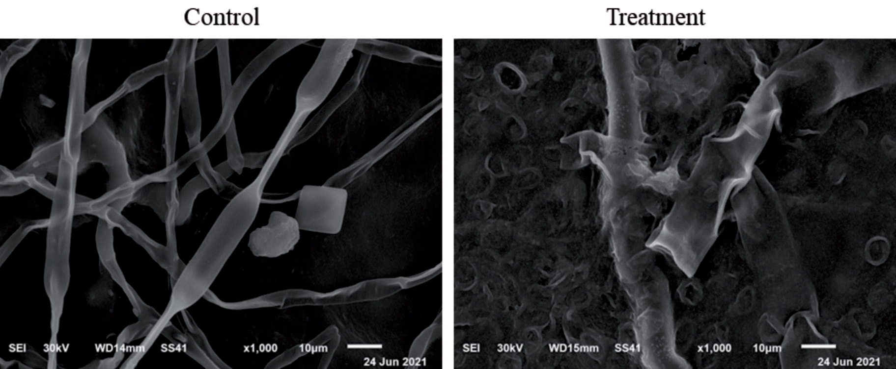
Scanning electron microscopy (SEM) of Botrytis cinerea, treated with CWPH at 250 µg/mL for four h at room temperature, was estimated according to Sitohy et al., 2013 [32] as compared to control (without treatment).
Results and Discussion
Whey Protein Hydrolysate Characterization and Antioxidant Activity Evaluation
The effect of the degree of hydrolysis of CWPH with alcalase at different intervals (0, 2, 4, 6, 8, 10, and 12 h) on antioxidants activity of CWPH at the same intervals is presented in Fig. 1. The highest DH was 47%, which was obtained after 12 h in comparison with those obtained after 2, 4, 6, 8, and 10 h; 20, 26 32, 39, and 45 %, respectively). Our results indicate that the link between hydrolysis time, DH and antioxidant activity is direct. After 8 h proteolysis presented the highest antioxidants activity against DPPH free radical (70% at a concentration of 200 µg/mL.
Fig. 2 depicts the DPPH and ABTS radical scavenging activity of whey protein hydrolysates (WPH) produced with alcalase at various concentrations (0-800 g/mL) (E/S ratio of 1:200, w/w). The DPPH radical scavenging activity assay is one of the in vitro methods for evaluating an antioxidant’s capacity to neutralize free radicals. The sample’s antioxidant activity is correlated with the degree of color changes. Antioxidants quickly reduce the relatively stable radical ABTS. The ability of peptide fractions to act as donors of electrons or hydrogen in free radical reactions is demonstrated by their scavenging behavior against the cationic radical ABTS [33].
The hydrolysates’ antioxidant activity is expressed as SC50 (µg mL-1). Antioxidant activity is high when SC50 values are low [34]. Hydrolysis of CWP by alcalase increased the antioxidant activity with increased concentration (Fig. 1). When compared to DPPH and ABTS, the respective SC50 values of the CWPH were determined to be 140 and 160 µg/mL, respectively.
Previous studies have reported the generation of biologically active peptides containing amino acids that possess antioxidative properties at their C-terminal residues through the action of the chymotrypsin enzyme. This enzyme is known for its specificity towards carboxylic groups of aromatic or other hydrophobic amino acid residues [35].
Antibacterial Activity Evaluation
The antibacterial activity of CWP and CWPH against four harmful microorganisms was evaluated using the disc-diffusion method. The antibacterial properties of CWP, including its lysozyme, lactoferrin, and immunoglobulins, may be the cause of this [36]. CWP’s antibacterial activity was enhanced by alcalase enzymatic hydrolysis. Alcalase’s cleavage of antimicrobial peptides may be responsible for this outcome. Similar results were presented by [25]. CWPH lower MIC values compared to those of CWP confirmed its potent antibacterial activity. The bacteria S. typhimurium and E. coli were most affected, with MIC values of 78 and 625 µg mL-1, respectively (Table 1). The antibacterial activities of whey proteins and derived peptides depend on how they interact with bacterial cell walls and membranes [37]. The peptide’s molecular structure (acid sequence and composition), size, hydrophobicity, and charge distribution all play a role in this interaction [38]. The targets of the electrostatic binding of positive-charged peptides are lipoteichoic acids on the surfaces of G+ bacteria or lipopolysaccharides on the outer membrane of G-bacteria [39].
Fig. 1.
Alcalase hydrolyzed camel whey protein hydrolysates (CWPH) at various times (0, 2, 4, 6, 8, 10, and 12 h) and DPPH radical scavenging activity (DPPH-RSA) for WPH (200 g/mL) were used to determine the degree of hydrolysis (DH).
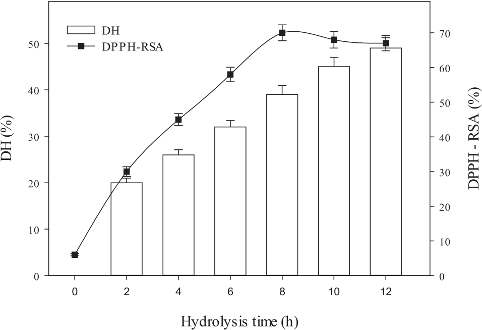
Fig. 2.
Whey protein hydrolysates (WPH) at various concentrations (0-800 g/mL) produced with alcalase (E/S ratio of 1:200, w/w) at 37ºC and pH 8 for 8 hours demonstrated their DPPH and ABTS radical scavenging activity.
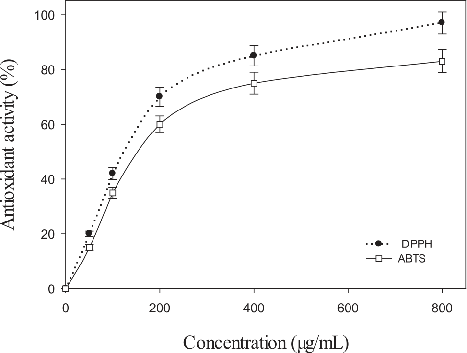
Table 1.
Minimal inhibitory concentration (MIC; µg/mL) of camel whey protein (CWP) and their hydrolysates (CWPH) against pathogenic bacteria G- (Escherichia coli, and Salmonella typhi) and G+ (Staphylococcus aureus, and Listeria monocytogenes).
| Microorganisms | MIC (µg/mL) | |
|---|---|---|
| CWP | CWPH | |
| Gram (+) | ||
| S. aureus | 10000 | 1250 |
| L. monocytogenes | 10000 | 2500 |
| Gram (-) | ||
| E. coli | 5000 | 78 |
| S. typhimurium | 5000 | 625 |
The mode of action of CWPH as an antibacterial agent against untreated and treated S. aureus and E. coli cells were examined using TEM. The normal morphological and structural characteristics of the cells in S. aureus and E. coli that had not been treated were confirmed by TEM images (Fig. 3). According to TEM images, the S. aureus and E. coli antibacterial effects of CWPH ranged from cell distortion to cell lysis. Adsorption of the antimicrobial CWPH to the cell’s surface could be thought to be the first step in the interaction between the CWPH’s peptides and the bacterial cell. Phosphate groups in the outer membrane of Gram-negative bacteria or lipoteichoic acids on the surface of Gram-positive bacteria are examples of negatively charged components in the bacterial cell wall that interact with the cationic peptide. A definitive feature of the antibacterial mode of action of cationic antibacterial peptides is their ability to associate with membranes [40]. Additionally, biologically active peptides from camel and bovine whey proteins released after enzymatic hydrolysis using Trypsin and Chymotrypsin have been previously investigated by Salami et al. [41].
Antifungal Activity Evaluation
Antifungal activity of CWPH at several concentrations (0, 250, 500, and 1000 µg/mL) on mycelial growth are presented in Fig. 4a) and 4b). Evidently, CWPH inhibited B. cinerea mycelial growth in a concentration-dependent manner. Fungal growth of B. cinerea was reduced by 44%, 56%, and 78%, respectively, when CWPH was administered at 250, 500, and 1000 µg/mL. SEM images of Botrytis cinerea after being exposed to CWPH (250 µg/mL) for four hours at room temperature are shown in Fig. 5. The untreated normal fungal had typical hyphae. Both fungal hyphae’s anatomical features have been significantly altered by CWPH treatment, which completely destabilized and distorted their shape at 250 µg/mL. SEM revealed that the fungal hyphae had deformed and appeared shriveled up after being exposed to the CWPH in PDA. B. cinerea treated with CWPH exhibited this trait, which suggests a connection to the protein-protein interaction that influences membrane permeability [23, 25].
Fig. 3.
Transmission electron microscopy (TEM) was used to examine the effects of one MIC of camel whey protein hydrolysates (CWPH) on control and treated gram-positive bacteria (S. aureus) and gram-negative bacteria (E. coli).
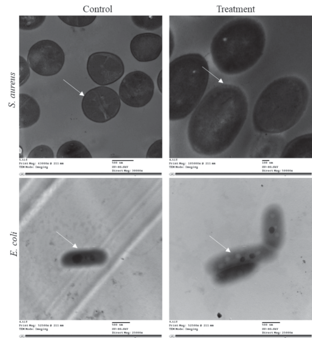
Fig. 4.
Effect of different concentrations (250, 500, and 1000 µg/mL) of camel whey protein hydrolysates (CWPH) compared to control against inhibition of B. cinerea mycelial growth.
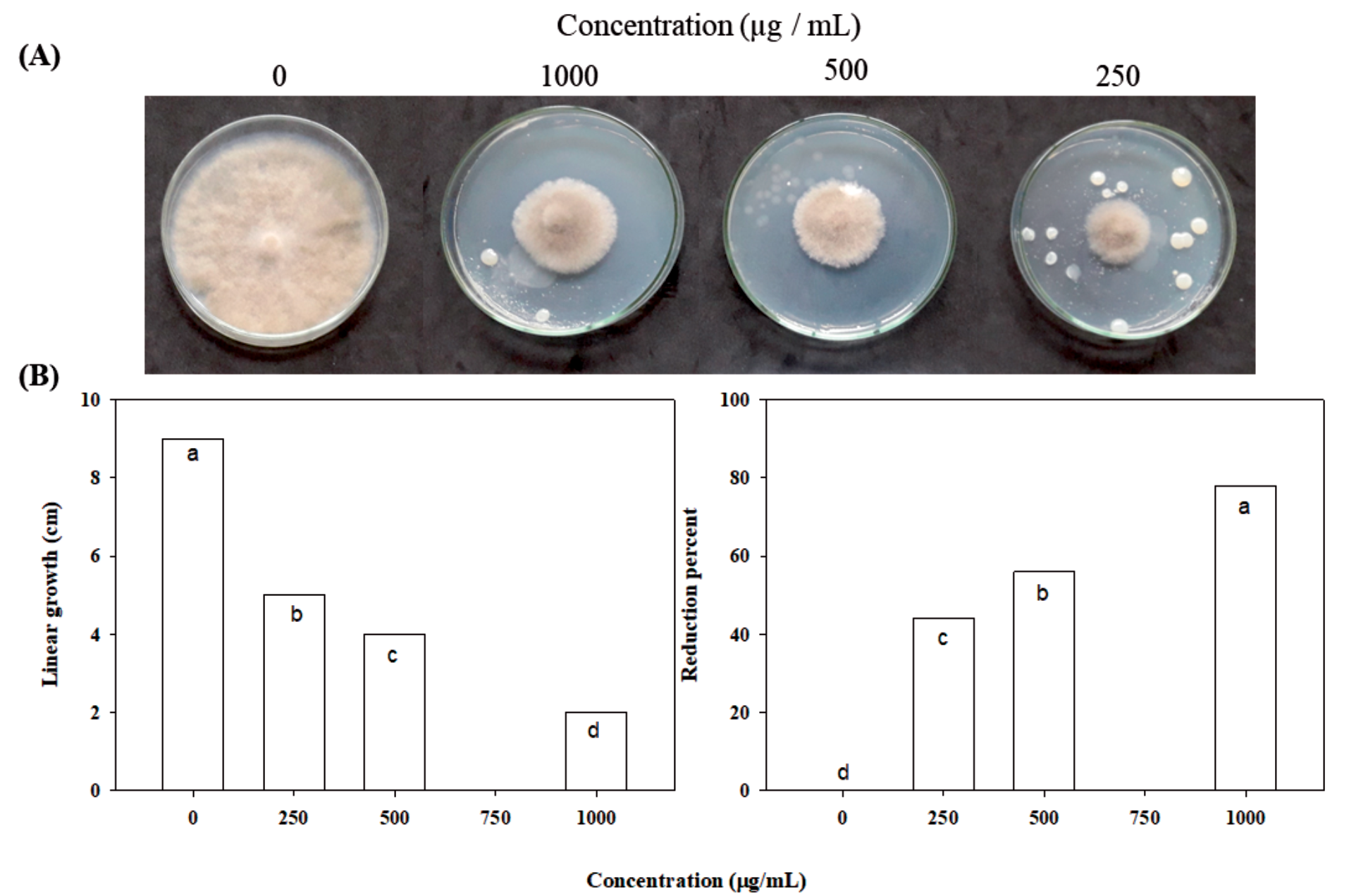
Fig. 5.
Scanning electron microscopy (SEM) was used to examine the effects of 250 µg/mL of camel whey protein hydrolysates (CWPH) on control and treated B. cinerea.
The camel lactoferrin peptides in the P-T hydrolysate, such as lactoferampin, lactoferricin, and lactoferrin chimaera, are very good at killing fungi. These peptides may lead to damage of fungal cell membranes and alter their permeability [42].
Conclusion
In this research, alcalase was used to break down camel whey protein (CWP) for 12 hours at a ratio of 1/200 (w/w) enzyme to substrate. After different amounts of time (2, 4, 6, 8, 10, and 12 hours), the hydrolysates’ antioxidant, antibacterial, and antifungal effects were tested. It can be concluded that camel whey protein hydrolysates have antibacterial, antioxidant, and antifungal activity against pathogenic bacteria and fungi.

