
Introduction
Nanoscale materials are applied across a diverse range of fields including batteries [1, 2], wastewater treatment [3], electronics, magnetics, optoelectronics, biomedicine, pharmaceuticals, cosmetics, energy, environmental science, catalysis, materials science [4], and drug delivery [5]. Manganese oxides e.g., MnO, Mn5O8, Mn2O3, MnO2, and Mn3O4, have attracted unique attention among the 3d transition metal-oxides due to their diverse structural and chemical variations and find applications in batteries, catalysts, and electrochromic and magnetic materials. Among these, Mn3O4 stands out as an effective and affordable catalyst for reducing NOx and CO emissions, offering a potent method for combating air pollution. Additionally, Mn3O4 is being studied as a catalyst for reducing nitrobenzene and oxidizing methane. It has also been found to be a corrosion-inhibiting pigment in epoxy–polyamide and epoxy ester-based primers and topcoats, further highlighting its potential in various industries [6, 7]. Mn3O4 (hausmannite) NPs show a broad range of applications in various fields, including catalytic, energy storage devices, wastewater remediation, biosensors, medical, etc. Their bactericidal and photocatalytic efficiency is significantly affected by their distinct crystalline forms, surface morphologies, synthetic methods, reaction conditions, etc. The fascinating physicochemical properties and distinct structural features of Mn3O4 NPs enable them to be used in wastewater treatment and photocatalytic degradation of dyes under UV and visible light radiations. Their ability to act as antimicrobial agent for the control of drug-resistant microbial populations can be owed to their high reactivity and larger surface-to-volume ratio as compared to bulk materials. The role of Mn3O4 NPs in developing epidermal ointments (used for eczema) and for controlling drug-resistant pathogens is well recognized [8]. The fabricated hybrid nanoclusters of OCFC-MnO2-Mn3O4 on oxidized carbon fiber in aqueous 1.0 M Na2SO4 electrolyte demonstrated remarkable specific capacitance of 1709 F g-1 at 1 A g-1 current density [9].
In the past, both chemical and physical routes have been utilized to fabricate uniform-sized NPs with long-term stabilities. However, these methods often involve the use of toxic chemicals, posing negative impacts on human health, so it is important to explore alternative methods to minimize the hazardous effects associated with traditional physical and chemical approaches [10]. Green synthesis of NPs is gaining popularity as a non-toxic, eco-friendly, clean, less expensive, and novel technique [11, 12]. The environmentally friendly synthesis of NPs with regular form and size, using genetic engineering techniques, molecular cloning, plant extracts, and other biological methods, represents a significant advancement in nanobiotechnology [13, 14]. Plants, fungi, bacteria, yeasts, algae, and actinomycetes may all be used to produce biogenic-metallic NPs, which alters the metal’s characteristics significantly [15]. The growing interest in efficient green chemistry has led to a focus on plant-mediated synthesis of nanoparticles. Plant extracts can reduce and stabilize metal nanoparticles in a single-step synthesis, leveraging their natural characteristics. A variety of natural organic compounds in plant extracts, such as alkaloids, flavonoids, saponins, steroids, terpenoids, and tannins, serve as both reducing and stabilizing agents [16]. Plants are also rich in phenolics and flavonoids, which act as powerful antioxidants [17, 18].
The Coriandrum Sativum (an annual herb) belongs to Apiaceae (Umbelliferae) family and is also known by the names of Chinese parsley, coriander, dhania, cilantro, etc. [19]. It is commonly grown in India, Italy, the Netherlands, Central and Eastern Europe, China, and Bangladesh. Different portions of C. sativum have the ability to yield essential oil, flavonoids, fatty acids, and sterols [20]. Green coriander consists of 84% water; essential oils and fatty oil are its active ingredients. The essential oil content of ripened and dried coriander ranges from 0.03 to 2.6 percent, with fatty oil content ranging from 9.9 to 27.7%; the major component of essential oil is S-(+)-linalool (60–70%) with the existence of monoterpene hydrocarbons as minor active ingredients [21].
Recent studies are focused on the plant-mediated synthesis of Mn3O4 NPs using Coriandrum sativum leaf extract. The biosynthesized NPs were characterized by XRD, FT-IR, SEM, and UV/VIS spectroscopy and also evaluated for their electrochemical and antimicrobial potential.
Materials and Methods
Coriandum Sativum powder, de-ionized water, manganese (II) nitrate hexahydrate (analytical grade), methanol (Analytical grade), and 0.1 N solution of NaOH were used for the green synthesis.
The synthesized NPs’ phase nature and crystallinity were analyzed using an X-Ray Diffractometer. FT-IR spectroscopy was performed in the range of 500–4000 cm-1 using a Carry 630 model. SEM analysis was performed by the JEOL JSM-6480LV model. UV-Visible spectroscopy was performed by a UV-3002 spectrometer in the range of 200 to 900 nm. The electrochemical behavior of the synthesized NPs was investigated using a Galvanostat/Potentiostat of the CS300 model. Cyclic voltammetry was performed by using a reported procedure [22].
Antibacterial activities were performed at the Department of Biochemistry, University of Agriculture, Faisalabad, Pakistan by using the disc diffusion method. The samples were tested against Staphylococcus aureus (Gram-positive) and Escherichia coli ATCC 25922 (Gram-negative). The Institute of Microbiology at the University of Agriculture, Faisalabad, Pakistan, confirmed the purity and identity of the product. Nutrient agar was used as the growth medium. The inhibition zones were recorded in millimeters by a zone reader [23].
Collection and Powdering of Plant Material
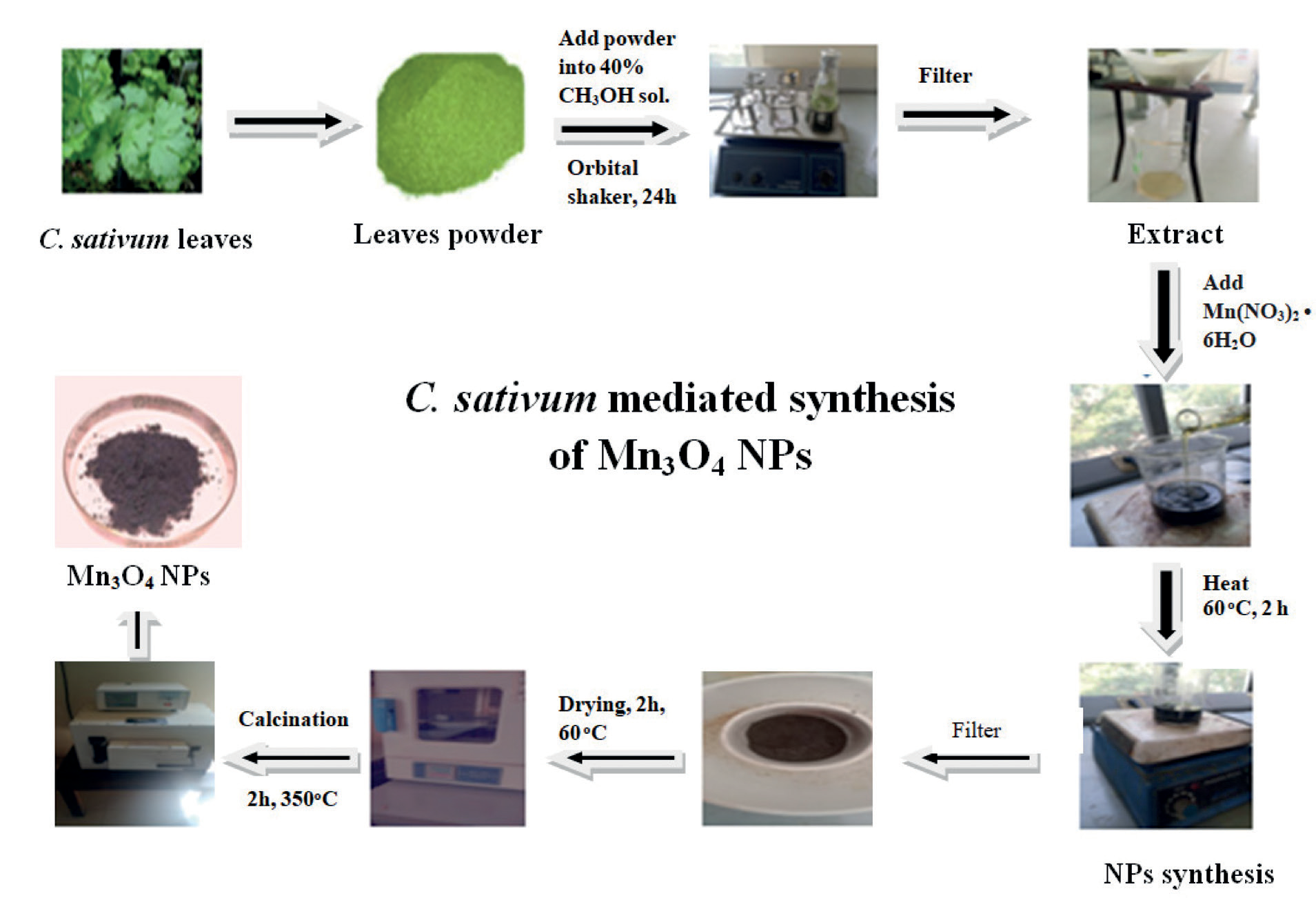
The leaves of Coriandrum sativum (Fig. 1) plant were purchased from Ichra Market of Lahore (Punjab), Pakistan in August 2021. The plant species was verified by the Department of Biology, Lahore Garrison University Lahore, Pakistan.
The C. Sativum leaves were cleaned with fresh water and then with distilled water to remove dirt and contaminants. They were dried at room temperature for 24 hours to eliminate the moisture. The dried leaves were ground in a steel grinder to obtain a homogenized powder, which was then sieved to a mesh size of 100 μm.
Preparation of Extract
30 g of C. sativum leaves powder was soaked in 100 mL of 40% methanol. The solution was stirred for 24 hours at ambient temperature. It was then filtered, and the filtrate was centrifuged to remove any residual impurities, and refrigerated at 4°C for usage within a week.
Synthesis of Manganese NPs
30 mL of 5% manganese (II) nitrate hexahydrate solution in water was taken in a 250 mL beaker. 30 mL of C. sativum leaves extract was added to it, and the pH of the solution was adjusted to 12 by adding NaOH solution (0.1 N) dropwise. The solution was stirred for 2 hours at 60ºC, then cooled and centrifuged at 2000 rpm for 15 min at ambient temperature. The color of the solution changed from dark green to light green, confirming the bio-synthesis of Mn3O4 NPs. The solution was finally filtered to separate the precipitated Mn3O4 NPs (1.75 g). After drying, the NPs were ground into powder and added to a China dish which was finally placed in a muffle furnace at 350ºC for 2 hours for calcination to leave behind the dried green crystals of Mn3O4 NPs (0.65 g).
Results and Discussion
C. sativum leaves powder (30 g) was soaked in 40% methanol (100 mL), stirred for 24 hours, filtered, and centrifuged to leave behind the clear extract of the plant. Then, manganese (II) nitrate hexahydrate solution (5%) was mixed with C. sativum extract (30 mL) and pH was adjusted to 12 by adding NaOH followed by stirring at 60°C for 2 hours, cooling and centrifugation to separate out the green-colored precipitates of Mn3O4 NPs which were calcined at 350ºC to leave behind the final product. The applied synthetic method uses the leaf extract of C. sativum as a reducing and stabilizing agent for the synthesis of Mn3O4 NPs. The synthesized NPs were subjected to XRD, FTIR, SEM, and UV-visible analyses. They were also tested for electrochemical potential by cyclic voltammetry and their antibacterial activity was also evaluated by the disc diffusion method.
XRD Analysis
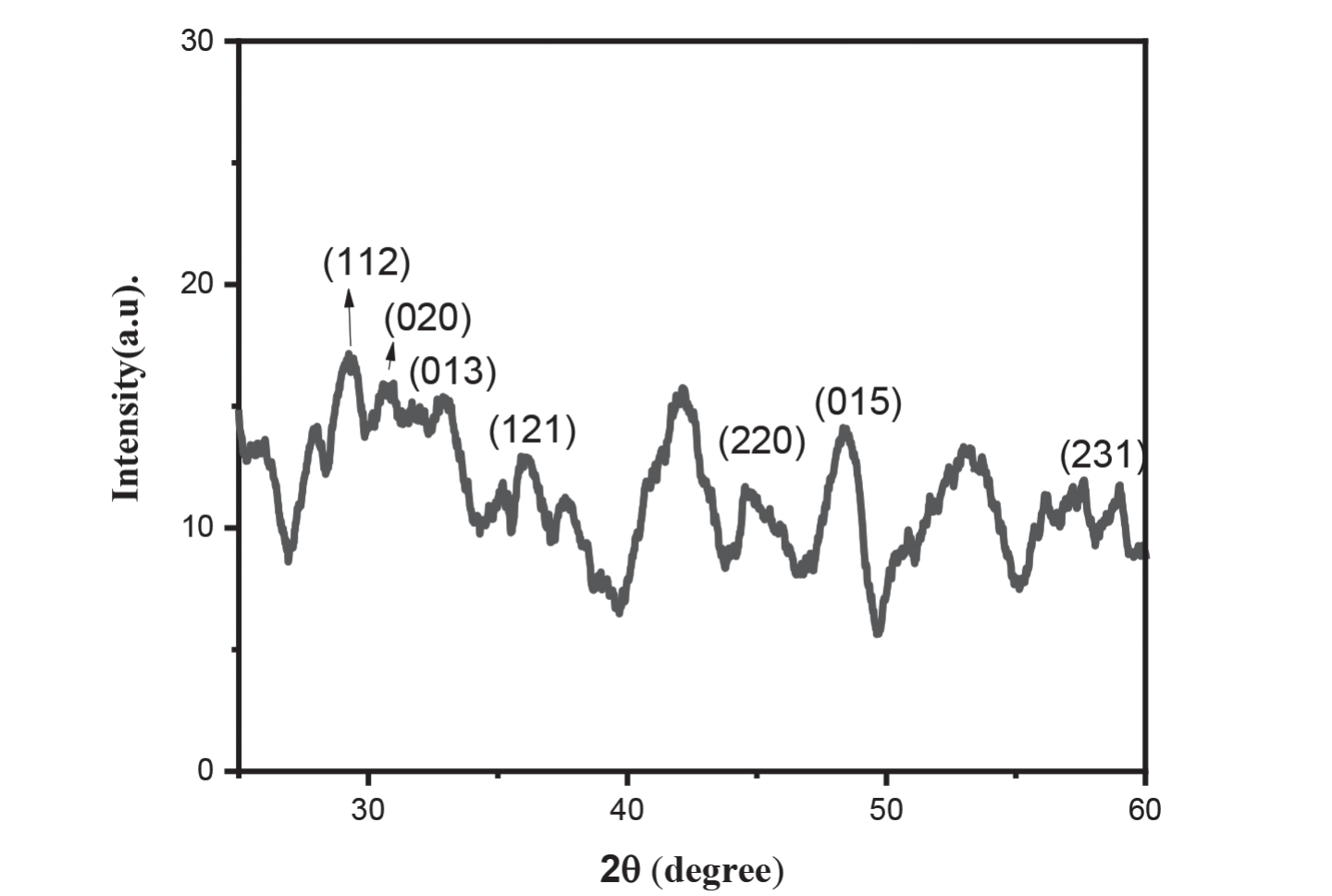
The XRD pattern (Fig. 2) exhibited prominent diffraction peaks at 2θ values of 29.38°, 30.91°, 33.01°, 36.13°, 38.19°, 44.80°, 49.6°, and 58.99°, corresponding to the Miller indices 112, 020, 013, 121, 022, 220, 015, and 231, respectively. These peaks are consistent with the JCPDS CARD number 03-065-2776, indicating the tetragonal crystal structure characteristic of Mn3O4 [24]. The crystallite sizes of Mn3O4 NPs were calculated by applying the Debye–Scherer Equation 1 [25].
Where K = 0.94, which depends on the shape of crystallites, β represents the peak’s highest width at half maximum, λ is the wavelength (0.154), D is particle size, and θ is diffraction angle. Using the Debye-Scherer equation, the average grain size of Mn3O4 NPs was found to be 23.62 nm.
FT-IR Spectroscopy
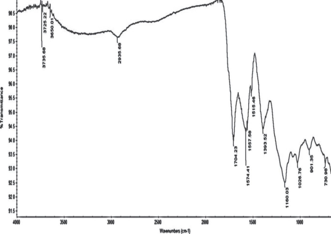
FT-IR spectrum of synthesized NPs was recorded in the range of 4000 to 500 cm-1 and is shown in Fig. S1 (Supplementary Material). A peak at 3650 cm-1 indicates the presence of the -OH group of H2O [26]. The broad band at 2935 cm-1 shows the presence of the CH2 stretching vibrations of the coriander leaves [27]. A shift in the peak of the C=O group from 1624 cm-1 to 1708 cm-1 is attributed to the formation of Mn3O4 NPs. Three peaks ranging from 1574 to 1515 cm-1 represent the C=C group. The broad band at 1160 cm-1 shows the presence of the C–N vibrations whereas the band at 1026 cm-1 is associated with the stretching vibration of the C–O–C or C–O groups that, together with C=C groups, are related to phytochemicals which are present in the plant as flavones, alkaloids, phenols, and anthracenes that can help to generate metallic nanoparticles according to the literature [28]. A peak at 1557 cm-1 shows the C–H bending vibration [29]. The bending of the adsorbed OH vibration combined with Mn NPs occurs at 1393 cm-1 [30]. The 730 cm-1 peak indicates the MnO stretching in tetrahedral and octahedral sites [29].
SEM Analysis
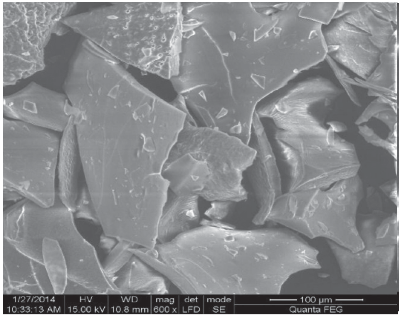
The surface structure and size of the Mn3O4 NPs were examined using Scanning Electron Microscopy (SEM); the obtained image is shown in Fig. 3. The surface morphology of the Mn3O4 NPs appeared agglomerated. The aggregated form of Mn3O4 NPs also exhibited a flake-like structure [31]. The particle size of Mn3O4 NPs was 34.17 nm.
UV-Visible Spectroscopy
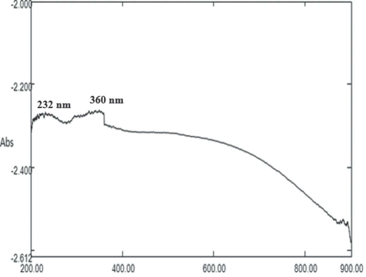
The absorption peaks were observed at 232 nm and 360 nm in UV-Visible absorption spectrum of Mn3O4 (Fig. 4) and correspond to the permissible charge transfer transitions of O2-→Mn2+ and O2-→Mn3+, respectively [32].
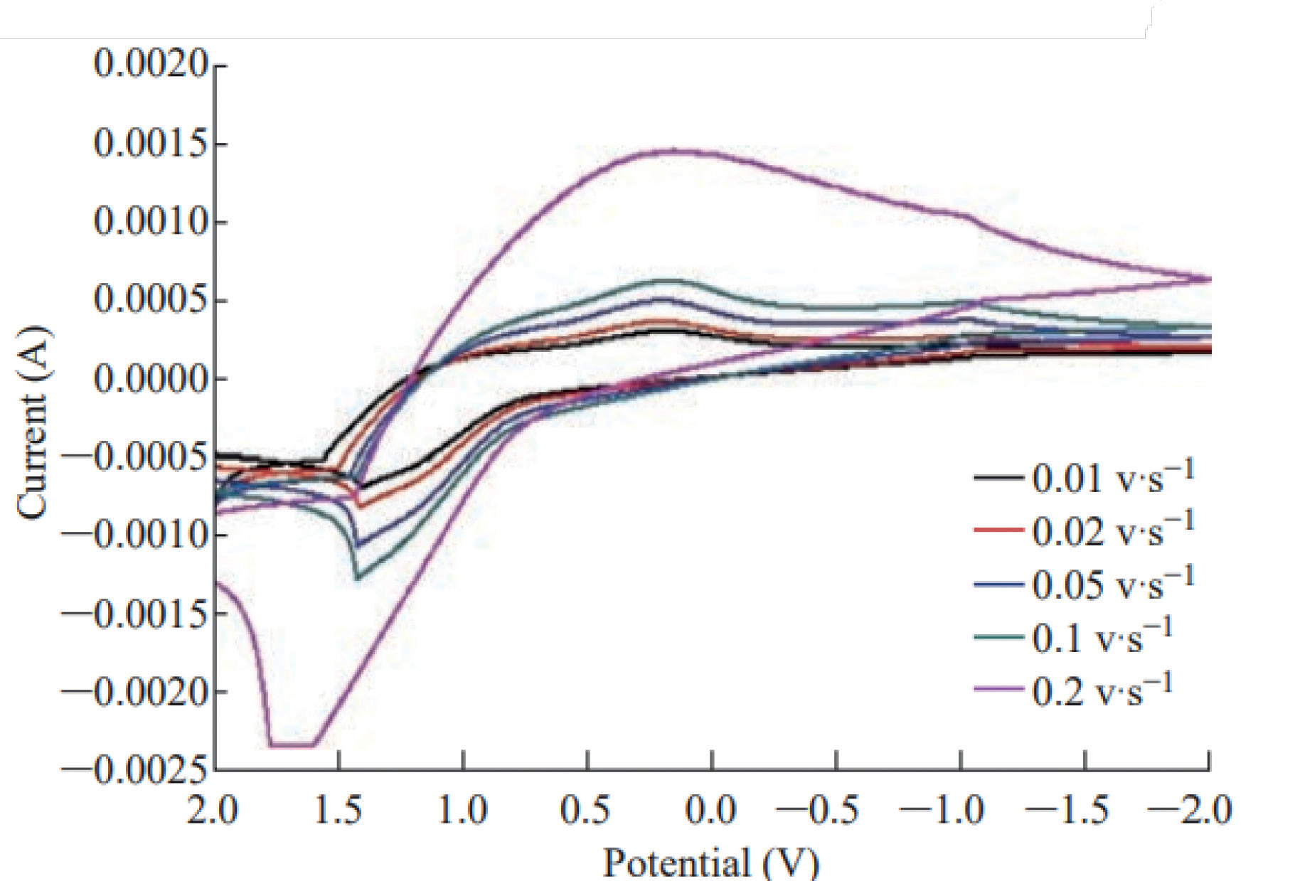
Table 1.
Calculation of redox current and redox potential of Mn3O4 NPs.
Cyclic Voltammetry (CV)
The CV curves of Mn3O4 NPs at various scan speeds are shown in Fig. 5, together with data on the transfer of electrons, kinematics, and transportation characteristics of the electrolytic reaction. The current flow was measured relative to the supplied linear voltage. The processes that take place at the surface of the electrode can be better understood by examining the current fluctuation that results from the different electrode potentials.
Cyclic voltammetry (CV) was employed to assess the electrochemical behavior of Mn3O4 NPs in redox reactions at an electrode in distilled water, using scanning speeds ranging from 0.01 to 0.2 V/s and a voltage range of 2 to -2 V. At a scanning rate (υ) of 0.1 V/s, two oxidation peaks were observed for Mn2+ to Mn3+ and Mn3+ to Mn4+ transitions. The oxidation peaks were detected at Epox1 = -0.735 V and Epox2 = -1.050 V, with corresponding anodic peak currents Ipox1 = 1.6×10⁻⁴ and Ipox2 = 4.868×10⁻⁴, respectively. A single reduction peak was observed at Epred = -0.526 V, corresponding to an anodic current maximum Ipred = -1.05×10⁻⁴ A. The voltammetry results of an electrode containing Mn3O4 NPs in the presence of 1 ppm MnSO4 solution at different scanning rates (0.01, 0.02, and 0.05 V/s) are presented in Table 1.
Antibacterial Activity
NPs were evaluated for their antibacterial potential against Escherichia coli (G-negative) and Staphylococcus aureus (G-positive) by the disc diffusion method. Ciprofloxacin was used as a positive control, while discs containing only water were employed as a negative control. A zone reader was used to measure zones of inhibition in mm. The zones of inhibition (ZOI) of Mn3O4 NPs were found to be 12 mm and 3 mm against E. coli and S. aureus, respectively; the observed activities were significantly smaller as compared to 37 mm and 40 mm zones of ciprofloxacin against E. coli and S. aureus, respectively.
Conclusions
A simple, environmentally sound, cheap, and novel green approach was employed for the synthesis of Mn3O4 NPs from Coriandrum sativum extract. The characterization of the NPs was performed by XRD, FTIR SEM, and UV-Visible analyses. X-ray diffraction (XRD) studies revealed the tetragonal configuration with a crystallite size of 23.62 nm. FTIR vibrations at 730 cm-1 were attributed to the Mn-O stretching mode. There was involvement of alcohol and ethers in the reduction of metal ions and capping of formed NPs. SEM analysis confirmed the aggregated and flake-like structures of Mn3O4 NPs with a particle size of 34.17 nm. UV-Visible spectroscopy has shown the absorption peaks at 232 and 360 nm, which correspond to correspond to the permissible charge transfer transitions of O2-→Mn2+ and O2-→Mn3+, respectively. Cyclic voltammetry has verified the redox nature and reversible behavior of synthesized material. The synthesized NPs were tested for their antibacterial activities by disc diffusion method using the standard drug ciprofloxacin and have shown some antimicrobial potential against E. coli and S. aureus.