

Introduction
Methotrexate (MTX) is a medication used in cancer treatment and as an immunosuppressant. It acts by competitively blocking the enzyme dihydrofolate reductase, disrupting the synthesis of nucleic acids by interfering with folic acid [1, 2]. MTX is frequently prescribed to treat various types of cancer, as well as conditions such as multiple sclerosis, dermatomyositis, sarcoidosis, psoriasis, and rheumatoid arthritis, among other inflammatory diseases [3]. However, its clinical use leads to several side effects, including hepatic toxicity, ranging from mild hepatitis and cholestasis to fibrosis, cirrhosis, and renal toxicity. Several studies have confirmed that MTX administration leads to an increase in lipid peroxidation and oxidative stress, amplifying the production of reactive oxygen species (ROS), leading to cellular degradation of proteins, lipids, and DNA [4]. Consequently, several natural and potentially antioxidant products have been used to mitigate oxidative cell damage and to improve the antioxidant defense system in MTX-exposed animals [5, 6]. In this regard, astaxanthin (3,3'-dihydroxy-β, β'-carotene-4,4'-dione, AX) is a natural carotenoid extracted from the microalgae Haematococcus pluvialis, possessing antioxidant and anti-inflammatory properties. It is recognized as one of the most effective antioxidants present in nature, reducing oxidative stress by neutralizing oxygen free radicals and inhibiting the propagation of chain reactions triggered by these free radicals [7]. Chlorella, spirulina, and astaxanthin are three such natural compounds that have been reported to possess antioxidant, anti-inflammatory, and cytoprotective properties, making them promising candidates for the prevention of MTX-induced toxicity [8, 9]. Chlorella, a green algae, has been shown to have antioxidant and anti-inflammatory effects, which may help protect against MTX-induced oxidative stress and inflammation [10, 11]. Spirulina, a cyanobacterium, is rich in antioxidants and has been reported to have hepatoprotective and nephroprotective effects [10, 12]. Astaxanthin, a carotenoid pigment, has potent antioxidant and anti-inflammatory properties, which may help mitigate MTX-induced oxidative stress and inflammation [13, 14]. Furthermore, as chlorella, spirulina, and astaxanthin have potential health benefits, long-term exposure may also cause potential side effects in some people, including digestive issues, sun sensitivity, worsened autoimmune conditions, increased bowel movements, and red stool color [15, 16]. Therefore, the present study aimed to evaluate the efficacy of astaxanthin, spirulina, and chlorella in preventing oxidative stress and nephrotoxic and hepatotoxic effects of methotrexate in mice.
Materials and Methods
Biological Materials
Fifty-four Swiss male mice, weighing between 34 and 36 g, were provided by the Pasteur Institute of Algiers and acclimated in the animal house of our Institution maintained with a temperature of 25°C and a photoperiod (12 hours of light/12 hours of darkness). The mice received an energetically balanced concentrate food (ONEP of El-Taref, Algeria, (Rodent food production industry)) and water ad libitum. The mice were kept in these conditions for an adaptation period of 10 days before the start of the experiment.
Chemical Materials
Methotrexate was obtained from Fisher Scientific SAS - Boulevard Sébastien Brant - F67403 Illkirch Cedex - France, astaxanthin from Nanjing NutriHerb BioTech Co., Ltd, China, Chlorella from Teramer, Mistral 34400 St Just France, and spirulina from Teramer, Mistral 34400 St Just France
Methods
After 10-days of acclimation period, mice were divided into eight main groups:
Group 1: Control mice vehicle receiving an oral administration of soybean oil 0.3 ml followed by an intraperitoneal injection of physiological water 0.5 ml (on the 5th day).
Group 2 [AX n=6]: Mice receiving an oral administration of astaxanthin of 8 mg/kg dissolved in soybean oil.
Group 3 [MTX n=6]: Mice receiving an intraperitoneal injection of methotrexate at 3.4 mg/ kg body weight (bwt) (on the 5th day).
Group 4 [AX/MTX n=6]: Mice receiving an oral administration of astaxanthin of 8 mg/kg bwt followed by an intraperitoneal injection of methotrexate (3.4 mg/kg/bwt) on the 5th day.
Group 5 [CH n=6]: Mice receiving an oral administration of chlorella extract at 0.5 g/kg bwt.
Group 6 [CH / MTX n=6]: Mice receiving an oral administration of chlorella extract of 0.5 g/ kg bwt followed by an intraperitoneal injection of methotrexate 3.4 mg/kg bwt diluted in physiological water.
Group 7 [SP n=6]: Mice receiving an oral administration of spirulina extract of 0.5 g/kg bwt.
Group 8 [SP/MTX n=6]: Mice receiving an oral administration of Spirulina at 0.5 g/kg followed by an intraperitoneal injection of methotrexate at a rate of 3.4 mg/kg/bwt.
After 8 days of treatment, the animals were sacrificed by decapitation using sterile scalpel blades. The blood was immediately collected by polyethylene tubes labeled EDTA and then directly centrifuged at 5000 rpm for 25 min. The plasma obtained was separated into several fractions in Eppendorf tubes and stored at -18°C. Blood samples were directly transferred to the clinical laboratory for the measurement of biochemical parameters. The kidney and liver of each animal were removed, rinsed in a 0.9% sodium chloride (NaCl) solution, weighed, and then stored in a 40% formaldehyde solution until the histological evaluations.
Biochemical Evaluations
Serum levels of urea, creatinine, GOT, and GPT were colorimetrically determined by an autoanalyzer (Siemens Healthcare Diagnostics, Marburg, Germany) using a commercially available kit (Bayer. Diagnostics, U.K.).
Table 1.
Changes in some physiological parameters in control and mice treated with astaxanthin (“AX”, per os, 8 mg/kg bwt), AX + MTX, chlorella extract (“Ch E”, per os, 0.5 mg/kg bwt), Ch E + MTX, spirulina extract (“SP”, per os, 0.5 mg/kg bwt) for 8 days, and methotrexate (“MTX”, i:p, 3.4 mg/kg bwt) given on the 5th day.
Evaluation of Oxidative Stress
The kidney and liver sections were homogenized using an ultrasonic disruption machine (Ningbo. Xin-zhi Biotechnology Co. Ltd., China), centrifuged at 3000 rpm for 15 min, and the supernatant was collected for determination of oxidative stress makers. The renal and hepatic tissue level of glutathione (GSH) and the enzymatic activity of glutathione -S-transferase (GST) and catalase were measured according to previously reported methods [17], [18], and [19], respectively.
Histopathological Examination
The fixed kidney and liver samples from each group were cut into 2 μm thick slices using a Cryostat Microtome (CM1850, Leica Microsystems Nussloch GmbH, Germany), deparaffinized, rinsed, and stained with hematoxylin and eosin (HE) for histopathological examination [20].
Results and Discussion
Effect on Physiological Parameters
As indicated in Table 1, body weight, food, and water intake significantly decreased in mice treated with MTX (p<0.001), AX+MTX, Ch E + MTX, and SP+MTX (p<0.05) since the relative kidney and liver weights were significantly increased in MTX (p<0.001), AX + MTX, Ch E + MTX and SP+MTX (p<0.05) compared with those of control mice. However, AX, Ch E, and SP treatments showed slight but non-significant changes in these parameters compared to the control group. As a result, the combined treatments showed a significant improvement (p<0.01) in the mentioned physiological parameters as compared to the MTX group. The obtained results proved the effective beneficial effects of chlorella, spirulina extracts, and astaxanthin in alleviating the effect of MTX-induced hepato-renal toxicity in mice. The present study revealed that the intraperitoneal injection of MTX at 3.4 mg/kg body weight significantly decreased body weight and food and water intake. These results concord with those previously reported [21, 22]. MTX likely causes suppression of food appetite, resulting in reduced food consumption, increasing the metabolism rate, leading consequently to body weight loss [23]. In addition, methotrexate can damage the gut lining, causing ulcers and inflammation [24]. This discomfort can discourage eating and hinder nutrient absorption, even if the rat eats normally. Also, the decreased water intake in methotrexate-treated animals may be due to the reason that malnutrition from decreased food intake can lead to weakness and lower activity levels, resulting in less need for water, in addition to diarrhea induction associated with dehydration and decreased water intake [25]. Additionally, MTX treatment caused a marked increase in relative liver and kidney weights, and this is almost similar to that previously reported [26]. MTX-induced liver and kidney weight increase is likely explained by the cellular damage, leading to cell death and inflammation, in addition to interfering of MTX with folate metabolism, essential for cell growth and repair, which can hinder normal cell turnover, leading to a relative increase in the proportion of non-dividing cells in the organ, potentially contributing to weight changes [27]. On the other hand, the altered physiological parameters were improved in MTX supplemented with AX, Ch E, and SP extracts, and this can be explained by the potential antioxidant and anti-inflammatory properties of these bioactive molecules [28]. Similar works have also reported the beneficial effects of Ch and SP [29, 30] and [31] against other toxicant-induced physiological changes.
Fig. 1.
Concentrations of serum urea and creatinine in control and mice treated with astaxanthin (“AX”, per os, 8 mg/kg bwt), AX + MTX, chlorella extract (“Ch E”, per os, 0.5 mg/kg bwt), Ch E + MTX, spirulina extract (“SP”, per os, 0.5 mg/kg bwt) for 8 days, and methotrexate (“MTX”, i:p, 3.4 mg/kg bwt) given on the 5th day. Error bars represent the error of the means.” Values with superscripts are statistically different p value ns (not significant), *p< 0.05, ***p< 0.01, and **p< 0.01 versus the control group. ap< 0.05, bp< 0.01, and cp< 0.001 versus methotrexate (MTX) group.
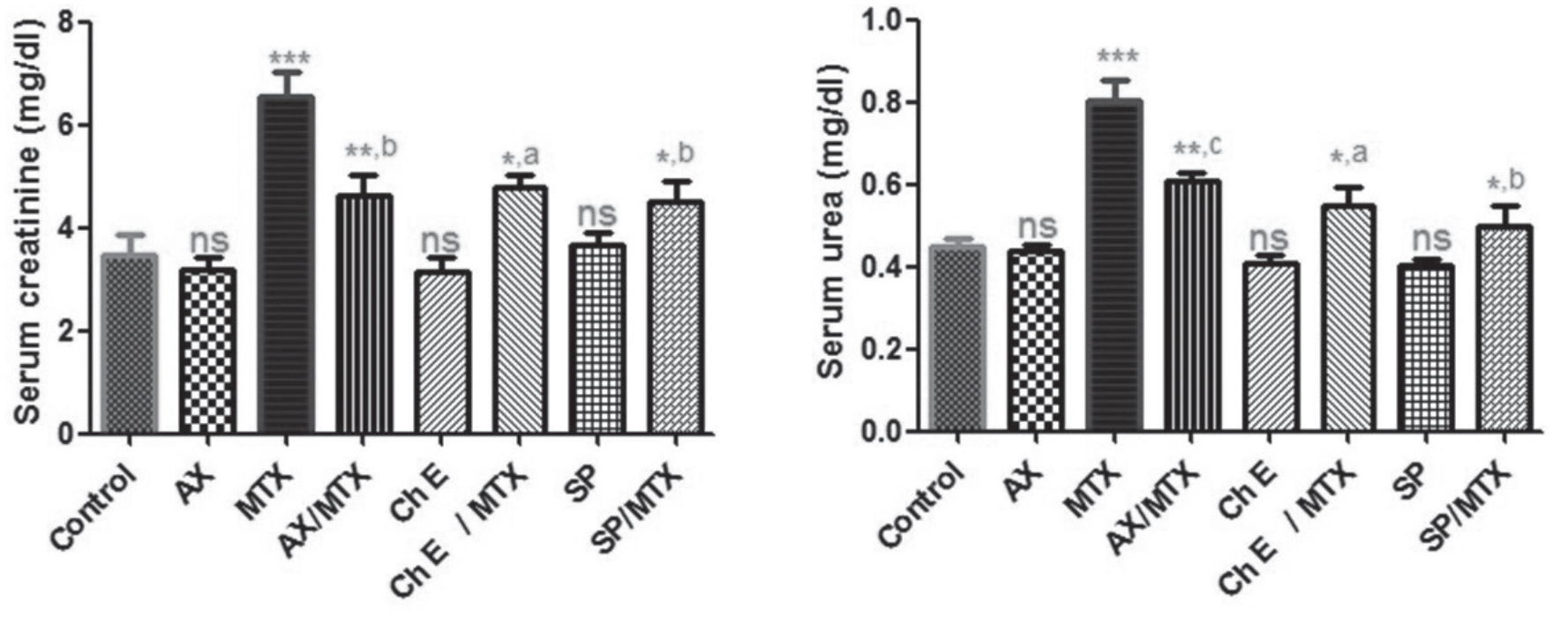
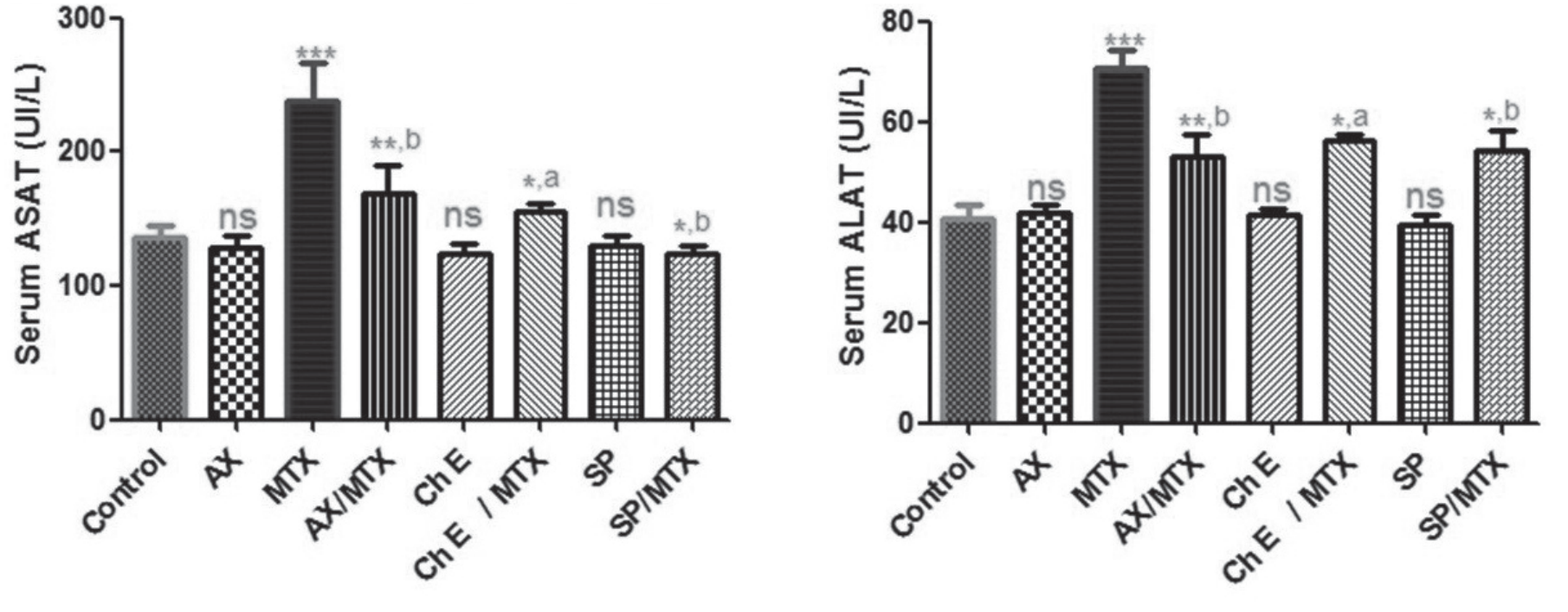
Fig. 2.
Enzymatic activity of serum aspartate aminotransferase (ASAT) and alanine aminotransferase (ALAT) in control and mice treated with astaxanthin (“AX”, per os, 8 mg/kg bwt), AX + MTX, chlorella extract (“Ch E”, per os, 0.5 mg/kg bwt), Ch E + MTX, spirulina extract (“SP”, per os, 0.5 mg/kg bwt) for 8 days, and methotrexate (“MTX”, i:p, 3.4 mg/kg bwt) given on the 5th day. Error bars represent error of the means.” Values with superscripts are statistically different p value ns (not significant), *p< 0.05, ***p< 0.01 and **p< 0.01 versus control group. ap< 0.05, bp< 0.01 and cp< 0.001 versus methotrexate (MTX) group.
Effect on Biochemical and Stress Parameters
As shown in Figs. 1 and 2, methotrexate (MTX) induces significant nephrotoxicity and hepatotoxicity, as evidenced by increased serum urea, creatinine, ASAT, and ALAT levels (p<0.001) as compared with the control group. Also, combination treatments (AX + MTX, Ch E + MTX, SP + MTX demonstrate a significant decrease in the mentioned parameters in AX + MTX (p<0.01), Ch E + MTX, and SP + MTX (p<0.05) as compared with the control group. No significant changes in these biochemical parameters have been noticed in AX, Ch E, and SP-treated mice compared with controls, and consequently, the studied parameters decreased significantly in AX/MTX, SP/MTX (p<0.01), and Ch E/MTX (p<0.05) as compared with MTX treated mice. The serious hepatotoxic and nephrotoxic effects of MTX are evidenced by a significant increase in the serum levels of aspartate aminotransferase (ASAT), alanine aminotransferase (ALAT), creatinine, and urea. This result has been reported in previous studies [32, 33]. Interestingly, these adverse effects of methotrexate are somehow related to the induction of liver and kidney injuries. Methotrexate-induced liver injury, leading to elevated levels of liver enzymes, specifically AST (aspartate aminotransferase) and ALT (alanine aminotransferase), explains the leakage of these enzymes from damaged liver cells into the bloodstream, indicating potential liver problems [34]. Additionally, the nephrotoxicity associated with increased serum levels of creatinine and urea in methotrexate-exposed experimental animals is elucidated mainly by the severe damage of tubular renal cells, which are responsible for reabsorbing essential substances and filtering waste products. This damage can disrupt these vital functions, leading to kidney impairment as evidenced by elevated levels of waste products like creatinine and urea in the bloodstream [35, 36]. On the other hand, the supplementation with chlorella, spirulina extracts, and astaxanthin has reversed the altered biochemical parameters of liver and kidney functions. Similarly, some previous works have proved the protective effects of chlorella, spirulina extracts, and astaxanthin [37] against chemical-induced health conditions and organ dysfunctions. Furthermore, Fig. 3 revealed a significant decrease (p<0.001) in catalase and GST activity in the MTX group, AX/MTX, Ch E/MTX (p<0.01), and SP/MTX (p<0.05) compared with the control group. However, these antioxidant parameters showed a significant increase in AX/MTX, SP/MTX (p<0.01), and Ch E/MTX (p<0.05) as compared with those of the treated group. This observation has been noticed in other previous studies [38] investigating the effect of apigenin on MTX-induced liver and kidney oxidative damage. Methotrexate was reported to induce oxidative stress-mediated generation of reactive oxygen species (ROS), and the subsequent weakening of the antioxidant defense system poses a potential threat to kidney and liver health and overall cellular function [39]. However, the improved level in the major antioxidant parameters following supplementation of astaxanthin, chlorella, and spirulina extracts has been previously reported [40, 41]. Noteworthy, the effective antioxidant activity and the beneficial effects are owed to their powerful ability to scavenge free radicals and subsequently reduce oxidative damage. More precisely, astaxanthin, a carotenoid compound, acts as a chain-breaking antioxidant by forming an ortho-dihydroxyconjugate polyene system that scavenges reactive oxygen species (ROS) [42], and similarly, Chlorella and Spirulina contain antioxidant compounds like phenolics, tocopherols, and carotenoids that contribute to their antioxidant properties [43].
Fig. 3.
Enzymatic activity of catalase and glutathione S transferase (GST) in liver and kidney homogenates of control and mice treated with astaxanthin (“AX”, per os, 8 mg/kg bwt), AX + MTX, chlorella extract (“Ch E”, per os, 0.5 mg/kg bwt), Ch E + MTX, spirulina extract (“SP”, per os, 0.5 mg/kg bwt) for 8 days, and methotrexate (“MTX”, i:p, 3.4 mg/kg bwt) given on the 5th day. Error bars represent error of the means.” Values with superscripts are statistically different p value ns (not significant), *p< 0.05,***p< 0.01 and **p< 0.01 versus control group. ap< 0.05, bp< 0.01 and cp< 0.001 versus methotrexate (MTX) group
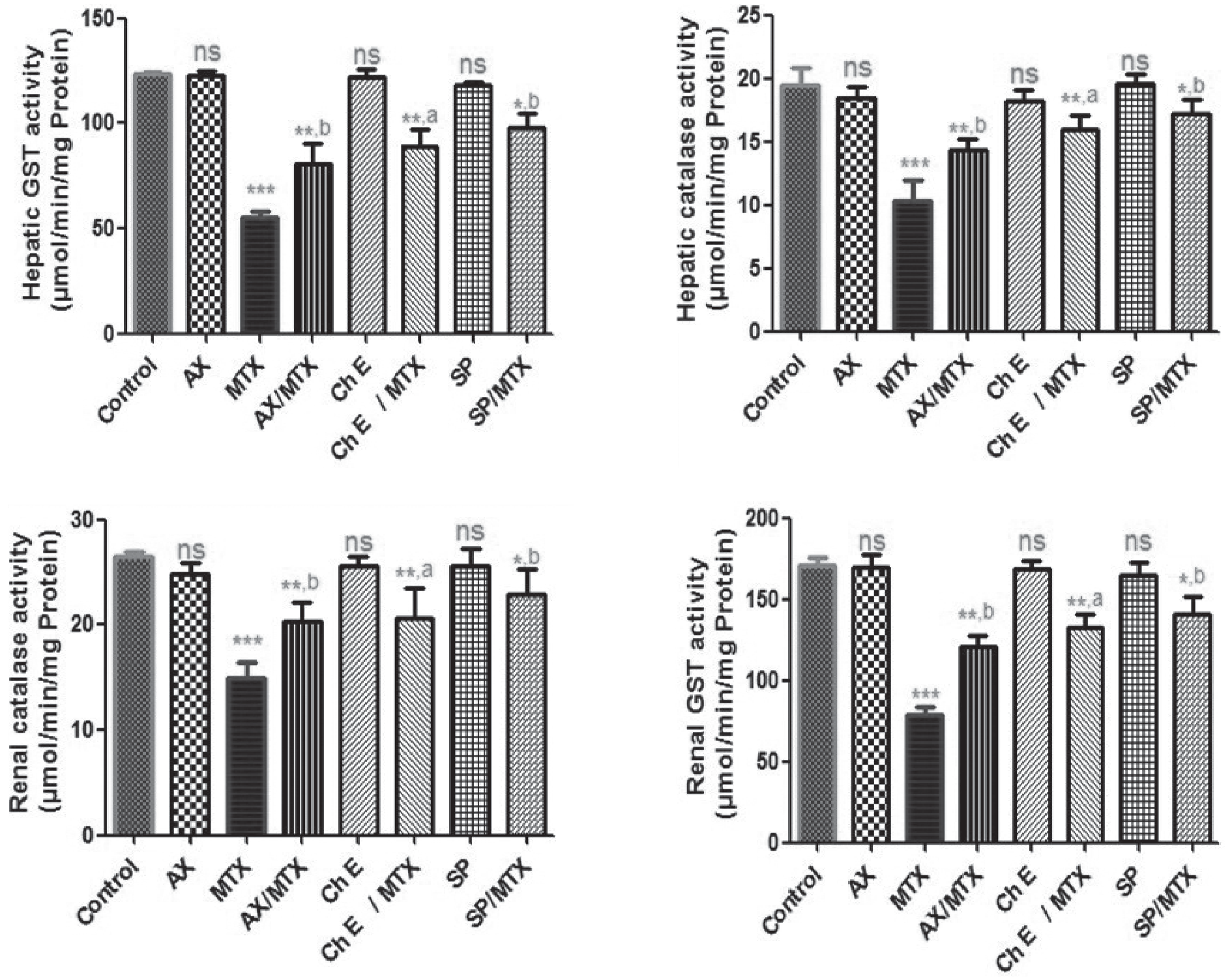
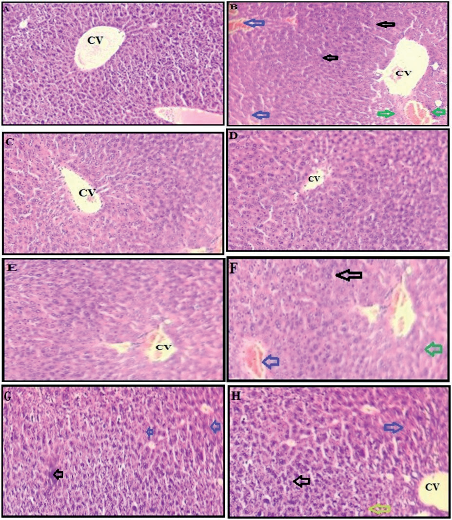
Fig. 4.
Histological liver sections of control (A) and mice treated with methotrexate (“MTX”, i:p, 3.4 mg/kg bwt) given on the 5th day during 8 days (B), astaxanthin (“AX”, per os, 8 mg/kg bwt) (C), chlorella extract (“Ch E”, per os, 0.5 mg/kg bwt) (D), spirulina extract (“SP”, per os, 0.5 mg/kg bwt) for 8 days (E), AX + MTX (F), Ch E + MTX (G), SP + MTX (H). 400X (scale bar=100µm) magnification. CV: central vein, blue arrows indicate hemorrhage, black arrows indicate mononuclear cell infiltration, and green arrows indicate microvesicular steatosis.
Liver and Kidney Histopathological Results
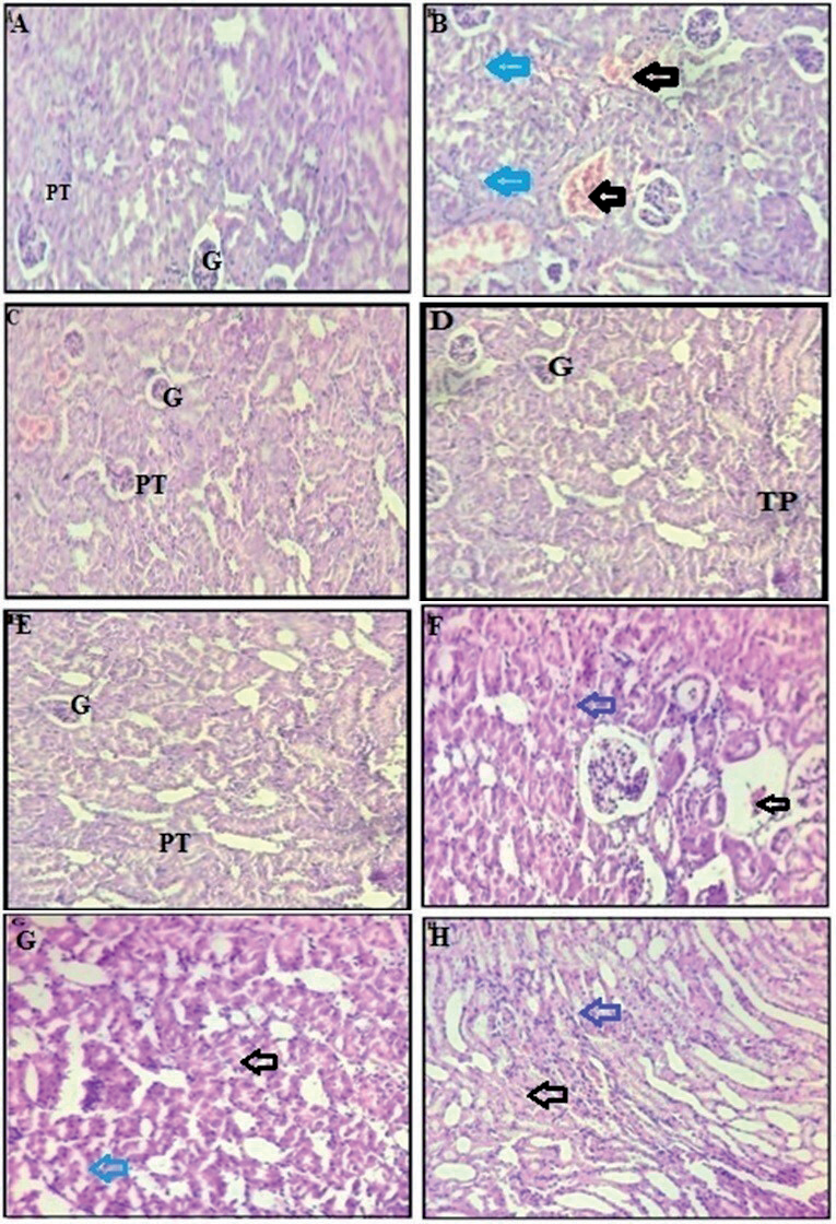
The tissue sections of the control group liver (Fig. 4A) showed normal hepatocytes arranged around central veins with no inflammation or congestion. However, liver tissue samples from the MTX-treated mice (Fig. 4B) revealed severe histological changes, including vacuolar degenerations, necrosis around the central veins, sinusoidal dilatation and hemorrhage, mononuclear cell infiltration congestion, and microvesicular steatosis. The treatments with AX (Fig. 4C), Ch E (Fig. 4D), and SP (Fig. 4E) separately did not cause any marked histological alterations, and thus, they appeared to have close liver histological agriculture to the control group. Accordingly, the combined treatments AX/MTX (Fig. 4F), Ch E/ MTX (Fig. 4G), and SP/MTX (Fig. 4H) revealed fewer liver histological alterations as compared with MTX-treated mice. Further, kidneys from control mice (Fig. 5A) revealed normal kidney histology indicative of healthy tissue, showing a normal structure of glomerulus and proximal tubule cells. However, the kidneys of mice treated with MTX (Fig. 5B) showed significant histological damages evidenced mainly by tubular necrosis, congestion of renal blood vessels, and glomerular degeneration. On the other hand, the kidneys from AX (Fig. 5C), Ch E (Fig. 5D), and SP (Fig. 5E) treated mice showed no marked histological alterations, and thus their kidney structures look almost close to those of controls. Thus, the supplementation of AX, Ch E, and SP to MTX-treated mice (Figs. 5F - H) has effectively attenuated the kidney histological damages caused by MTX. These histopathological alterations in the kidney and liver of MTX-exposed mice have been well documented [44, 45]. Importantly, the effective protective effect of chlorella, spirulina extracts, and astaxanthin against the oxidative nephrotoxic and hepatotoxic effects in association with histopathological alterations of methotrexate in mice is somehow owed to the typical biological properties of these bioactive substances. In this regard, the toxic effects of some toxicants induced health conditions have been previously reported attenuated by chlorella vulgaris and/ or spirulina platensis extracts, and astaxanthin. Also, some other studies suggested that these substances might also support the body's natural detoxification processes and possess anti-inflammatory properties, which could be beneficial in mitigating the triggered inflammatory response [46]. As our study is the first study investigating the beneficial effect of chlorella vulgaris, spirulina platensis extracts, and astaxanthin against methotrexate-induced liver and kidney oxidative damages, we refer that the reduced toxic effects of methotrexate by these substances are likely owed to their effective antioxidant and anti-inflammatory properties.
Fig. 5.
Histological kidney sections of control (A) and treated mice with methotrexate (“MTX”, i:p, 3.4 mg/kg bwt) given on the 5th day during 8 days (B), astaxanthin (“AX”, per os, 8 mg/kg bwt) (C), chlorella extract (“Ch E”, per os, 0.5 mg/kg bwt) (D), spirulina extract (“SP”, per os, 0.5 mg/kg bwt) for 8 days (E), AX + MTX (F), Ch E + MTX (G), SP + MTX (H). 400X (scale bar=100µm) magnification. G: normal glomerulus, PT: proximal tubules; black arrows indicate hemorrhage and congestion of renal blood vessels; blue arrows indicate renal tubular necrosis and glomerular degenerations in the cortex.
Conclusions
The study demonstrates that chlorella, spirulina extracts, and astaxanthin effectively alleviate the hepato-renal toxicity induced by methotrexate (MTX) in mice. These findings suggest a protective role for these substances against MTX-induced organ damage. Also, the study highlights the potential of these natural products in reducing the side effects of MTX treatment. However, it is important to note that this is the first study investigating this specific combination against MTX-induced toxicity, and further research is needed to confirm these findings.

