

Introduction
Currently, the green synthesis of nanoparticles (NPs) has attracted a special attention of the researchers due to its eco-friendly, cost-effective, sustainable, simple and simple protocol [1, 2]. This process involves the suitable selection of the solvent medium, reducing agent and non-toxic substances during the production of nanoparticles (NPs) [3] and is advantageous in terms of energy efficiency, product selectivity and safety for human health and the environment [4]. The green way for the synthesis of NPs involves various biological moieties such as microbes and plants as reducing agents [5, 6]. Especially, the use of plants extracts for NPs production [7] has attracted considerable attention due to involvement of quick, easier and economically affordable procedures. The plants contain biological components (such as carbohydrates, proteins, and coenzyme) that have exceptional potential to reduce the metallic salts into metal/metal oxide NPs [8] and also regulate morphology of NPs [9]. The metal/metal oxide NPs find numerous applications e.g., in purification of air and water, medical technology, energy storage, biosensors [10], photocatalytic, anti-termite [11], biomedical [12], antibacterial, magnetic, electrical, and catalytic activity. The properties of NPs are determined by their sizes, shapes, composition, morphology and crystalline phase [3]. The biologically synthesized metal/metal oxide NPs are used in the treatment of wastewater at industrial and level due to their valuable properties such as synthetic dyes quenching, metal sensing, antimicrobial effectiveness and catalysis [9].
Titanium(IV) oxide (titania) NPs are one of the most fascinating materials which have attracted attention due to their distinctive features [13] and numerous benefits associated with their high specific and large surface area, appropriate electrical band structure, high quantum efficiency, chemical innerness and durability [14]. They are involved in the formation of numerous high value-added products due to their low price, biocompatibility, high availability, non-toxicity and exceptional photo catalytic activity. They can impart UV-protective, cleaning and antibacterial characteristics to the textile products [15]. They find wide applications such as in dental treatment, bones implantation [1], in diagnosis and treatment of diseases, production of surgical instruments, tissue engineering, imaging, sensing, in the field of electronics, making of batteries, energy production [16], in pigments, cosmetics, catalysts, and photocatalyst [13]. Their photo catalytic efficiency for the purification of contaminated water and removal of contaminants from the environment is widely investigated [16]. Composites of TiO2-activated carbon are used in the photo catalytic destruction of naphthol from wastewater [14]. TiO2 functions as a nanocatalyst in the breakdown of methyl orange dyes [17]. The antibiotic levofloxacin can be removed from water below its detection limit by TiO2 photo-catalysis [18]. There are also reports for 97% removal efficiently of levofloxacin by using graphene/TiO2 composites utilizing 300 W of UV power and 45 minutes of exposure time [19]. TiO2NPs are also used in the treatment of osteosarcoma and chonrosarcoma [20], nondermatologic illnesses such as hyper-pigmented skin [21] and in the reproduction of silkworms [22]. They have the ability to improve plant immunity and photosynthetic rate, which may causes an increase of agricultural yield up to 30% [23]. Zea mays sprayed with nano-TiO2NPs during the crop’s reproductive phases had shown greater pigmentation and increase in the crop output [24]. For dye-sensitized solar cells, TiO2 nanoparticles are used as a semiconductor because they are non-toxic, readily available, and inexpensive [25]. They may serve as a tracer for other nanoparticles with a comparable size and aggregation to TiO2 [26]. In the aerospace industry, titanium alloys are used in numerous applications such as aircraft, engines, helicopters, and spacecraft [27]. TiO2 finds uses in the pharmaceutical and food processing industries because it is non-toxic and compatible with human skin. It is utilized as a filler and pigment (being a white) while making medications and tablets [28].
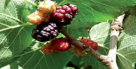
Plants are famous for their useful phytochemical and antioxidant contents [29, 30]. The black mulberry, Morus nigra L. (Fig. 1) is a plant of South west Asia, Europe and the Mediterranean. Because of its painkiller and anti-inflammatory properties, M. nigra has been utilized as a herbal medication for both the human beings and animals and also has high tolerance for pollutants in the air [31]. M. nigra is rich in phenolics and flavonoids with 276 milli gram quercetin equivalents and 1422 milli grams Gallic acid equivalents in 100 grams of its fresh material. Linoleic acid (53%), palmitic acid (19%) and oleic acid (7%) were the three primary fatty acids reported in mulberry fruits [32]. M. nigra L. has 1.50 percent acidity and a pH of 3.52. The mineral compositions of N (0.84 % N), P (234 mg/100 g), K (1142 mg/100 g), Ca (139.1 mg/100 g), Mg (109.4 mg/100 g), Na (59.8 mg/100 g), Fe (4.2 mg/100 g), Cu (0.5 mg/100 g), Mn (5.0 mg/100 g) and Zn (4.1 mg/100 g) has been reported in the mulberry species [33].
Keeping in view the importance of TiO2 nanoparticles and common availability of Morus nigra L., current studies were performed on the green synthesis of TiO2 NPs by utilizing the leaves of Morus nigra L., their structural characterization and biological activities.
Materials and Methods
The research was performed at the Department of Chemistry in the Lahore Garrison University Lahore, Pakistan. The analytical grade chemicals (titanium tetraisopropoxide and ethyl alcohol) were used for synthesis of NPs. Muffle furnace (S.X-2.5-10), Electric balance (BSM-520.3), Hot plate with magnetic stirring (85-2), oven (WHL-25A) and Pyrex origin glassware were used during the research. Fourier Transform Infrared Spectroscopy (FTIR) was performed in the range of 4000-600 cm-1 by Perkin Elmer L1600301 Spectrum Two FT-IR Germany Sr No. 114337, Lantrisant, USA. Raman spectra were recorded by Reinshaw in Vis Reflex. The X-Ray diffraction (XRD) studies performed by Bruker AXS, D8 Advance. The NPs were subjected to SEM (Scanning electron Microscopy) analysis by Hitachi S4800. SEM analysis.
The antibacterial potential of the TiO2 NPs was evaluated by disc diffusion method [35] against two bacterial strains (Proteus Mirabilis and Staphylococcus aureus) using ciprofloxacin as a standard drug. Their toxic hemolytic effects were evaluated on red blood cells by a reported procedure with respect to Triton X-100 (0.1percentv/v) as a positive control and phosphate buffer solution as a negative control [36].
Collection of Morus nigra Leaves and Sampling
The Morus nigra leaves were collected from the Lidher village of Lahore, Punjab (Pakistan) in October 2021. The plant species was identified and confirmed by Biology Department, Lahore Garrison University, Lahore, Pakistan. The plant leaves were washed with distilled water for the removal of dust particles, dried under shade and ground into fine powder with grinder and passed through a sieve of 80 mesh size. The fine powder was was stored in polythene bag at room temperature for further use [37].
Preparation of Aqueous and Ethanolic Leaves Extract of Morus nigra
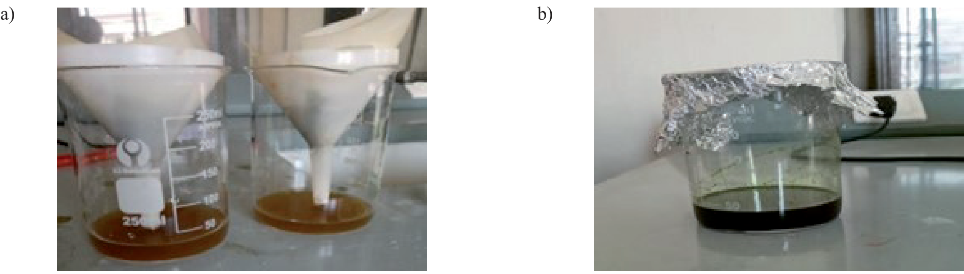
10 g dried leaves powder of Morus nigra was added in 100 ml distilled water and the mixture was stirred for 24 an hour in a magnetic stirrer. The resulting mixture was heated for 1 hour at 50oC on a hot plate until its volume was reduced to 2/3rd. It was subsequently cooled and filtered to obtain the aqueous extract which was finally preserved at 4oC for further use (Fig. 2a). Same procedure was employed to prepare the ethanolic extract (Fig. 2b) of leaves by using ethanol solvent in place of distilled water.
Synthesis of TiO2(Aq) and TiO2(Et) NPs
80 ml solution of titanium tetra-isopropoxide (TTIP) was prepared by dissolving 10 ml of TTIP in 70 ml of distilled water. This solution (80 ml) was added to 30 ml aqueous extract of Morus nigra leaves and the resulting mixture was stirred by a magnetic stirrer for 3 hours at 50oC till the color was changed from light brown to dark brown indicating the formation of nanoparticles. The prepared NPs were washed repeatedly with plenty of water in the reaction flask followed by decantation of liquid each time. The mixture was filtered to separate out the prepared TiO2 NPS which were dried in oven at 100ºC for 24 hours and then calcinated in a muffle furnace for 3 hours at 400oC to leave behind the final product of TiO2(Aq) NPs.
The same procedure was repeated to prepare the TiO2 NPs with ethanolic extract of Morus nigra leaves. Here, 30 ml ethanolic extract of Morus nigra leaves was stirred with 80 ml solution of TTIP (10 ml of TTIP in 70 ml of distilled water) for 2 hrs at 50oC till the color was changed from dark green to light green. At this stage, the reaction mixture was repeatedly washed (with distilled water) followed by filtration, drying and calcination as described above to produce the final product of TiO2(Et) NPs.
Results and Discussion
The current study was carried out to synthesize two kinds of TiO2 nanoparticles (TiO2(Aq) and TiO2(Et)) by treating aqueous/ethanolic extract of Morus nigra leaves with the solution of titanium tetraisopropoxide (TTIP) in distilled water. The synthesized NPs were characterized by FTIR, X-ray diffraction (XRD) analysis, scanning electron microscopy (SEM), and Raman spectroscopy. They were also tested for their antibacterial potential against Staphylococcus aureus and Proteus Mirabilis by disc diffusion method and their toxic hemolytic effects were also evaluated.
Structural Properties
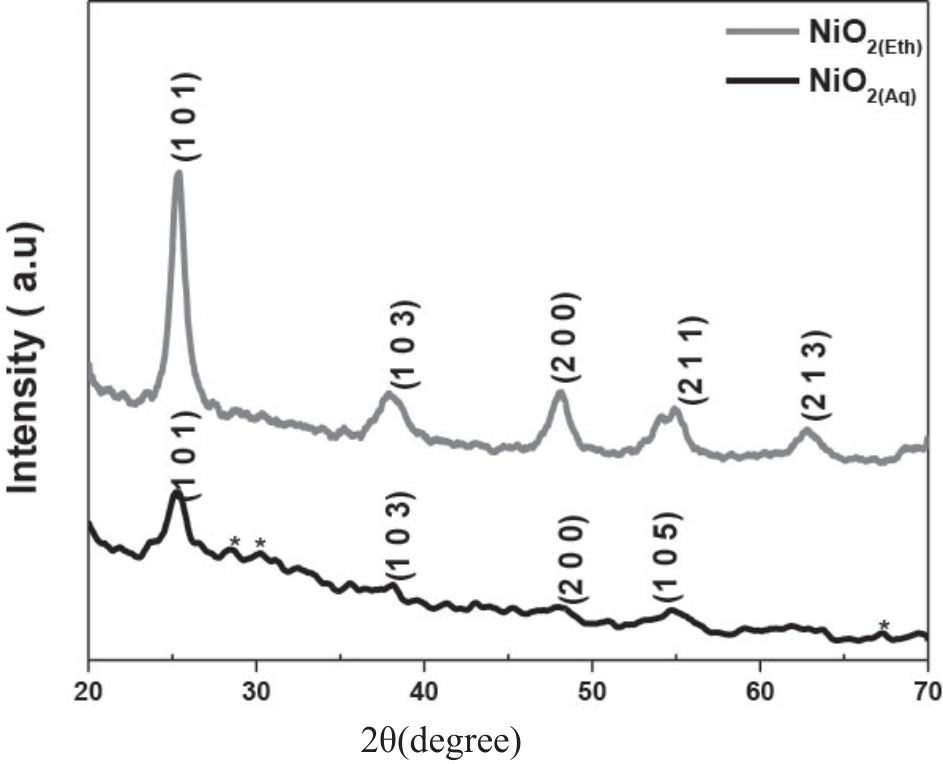
The crystalline structure and crystallite sizes of the NPs can be determined by using XRD analysis [38]. The XRD patterns of the synthesized TiO2(Aq) and TiO2(Et) NPs are shown in Fig. 3 and match well with the standard JCPDS card number 21-1272.
XRD is consider as an important technique which can be used to determine phase composition, crystal structure and grain size of sample. The size of nanoparticles can be determined by Scherer equation (Eq. 1) given below:
Where D denotes the crystal size, K represents the Scherrer constant, β is full width half maximum (FWHM), λ indicates X-Ray wavelength (1.54 Å), and θ represents the angle of diffraction.
In TiO2(Aq), the peaks at 2θ = 25.28º, 38.57º, 48.04º, 53.89º correspond to (1 0 1), (1 0 3), (2 0 0) and (1 0 5) crystal planes, respectively whereas the peaks at 2θ = 25.28º, 36.94º, 48.40º, 55.06º and 62.68º were associated to (1 0 1), (1 0 3), (2 0 0), (2 1 1) and (2 1 3) crystal planes, respectively in TiO2(Et). The crystallite sizes of TiO2(Aq) and TiO2(Et) NPs were found to be 0.17 and 0.076 nm, respectively.
Fourier Transform Infra-red (FTIR) Spectroscopy
The functional groups of the biomolecules which are responsible for the capping and reduction of TiO2 NPs can be confirmed by performing Fourier transform infrared (FTIR) spectroscopy (Sugirtha et al., 2015). The FTIR spectra of the synthesized TiO2(Aq) and TiO2(Et) NPs were recorded in a range of 600-4000 cm-1; the spectral data are displayed in Table 1.
The FTIR spectra (Fig. 3(a,b)) displayed the presence of Ti-O stretching vibrations 607.5 and 612.2 cm-1 in TiO2(Aq) and TiO2(Et) NPs, respectively. The additional peaks for C-Br, CO, OH, C=C, N=C=S, O=C=O and O-H in TiO2(Aq) and those for C-Br, OH, N=C=S, O=C=O and O-H in TiO2(Et) correspond to the aqueous and ethanolic extracts respectively and indicated the coating of plant material upon the surface of synthesized nanoparticles.
Raman Spectroscopy
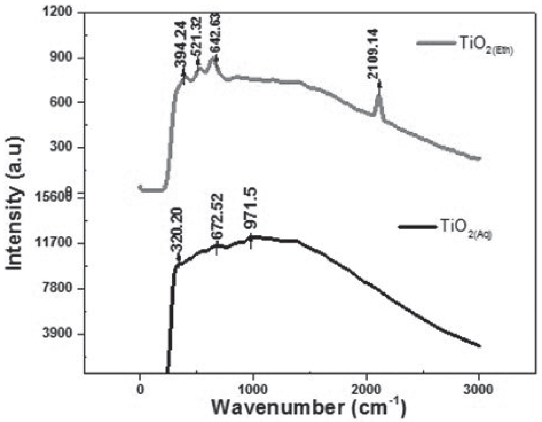
The NPs were subjected to Raman spectroscopy; the obtained spectra are displayed in Fig. 4. The vibrational peaks at 320.20-394.24, 521.32, 642.63-672.52, 971.5 and 2109.14 cm-1 can be assigned to the Raman modes of TiO2 NPs according to literature [39] and are in agreement to the findings of XRD analyses. The TiO2 vibrational modes are due to symmetric, asymmetric, and bending vibration of Ti-O-Ti bond. The peaks at 320.20 and 672.52 cm-1 in TiO2(Aq) and at 394.24, 521.32 and 642.63 cm-1 in TiO2(Et) are owed to A1g and Eg modes of vibrations [39].
SEM (Scanning Electron Microscopy) Analysis
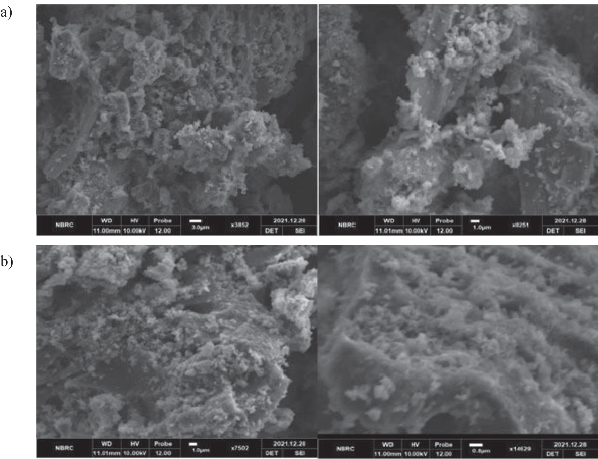
SEM analysis is used to examine the surface morphological characteristics of synthesized NPs. The SEM analysis has shown the round shape and irregular surface morphologies of nanoparticles (Fig. 5(a,b)); the prepared powders consist of a mixture of large grains and fine particles.
Anti-Bacterial Activity
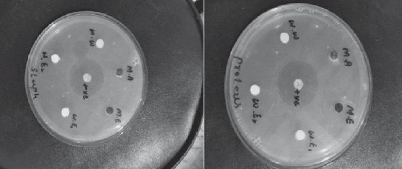
The synthesized NPs were tested for their antibacterial potential against Proteus Mirabbils (Gram-negative) and Staphylococcus aureus (Gram-positive) by disc diffusion method [35]. Ciprofloxacin was used as a positive control. A vernier caliper was used to measure the zones of inhibition in mm. The antibacterial activity data are shown in Table 2.
The investigated TiO2(Aq) and TiO2(Et) NPs have shown comparatively higher inhibition zones (12 and 7 mm) against Gram-negative (P. Mirabilis) as compared to those (6 and 5 mm) against Gram-positive (S. aureus) bacteria. However, both kinds of NPs have shown significantly lower antibacterial potential as compared to the standard drug (Ciprofloxacin).
Hemolytic Activity of TiO2 NPs
The toxic hemolytic effects of the NPs were evaluated on human red blood cells using TritonX-100 and phosphate buffer solution as positive and negative controls, respectively. The investigated TiO2(Aq) and TiO2(Et) have shown negligible hemolytic effects 3.17% and 2.54% as compared to the Triton-X100 (positive control, 89.72%) and phosphate buffer (negative control, 0.43% lysis). The results thus depict the possible safe use of the synthesized TiO2 NPs.
Conclusions
The investigated method for the synthesis of NPs is ecofriendly and cost effective because it involves the plant material as a stabilizing and reducing agent. Titanium dioxide nanoparticles (TiO2(Aq) and TiO2(Et)) were synthesized by treating titanium tetraisopropoxide (TTIP) with the aqueous/ethanolic extracts of Morus nigra leaves. The obtained NPs were characterized by FTIR spectroscopy, X-ray diffraction (XRD) studies, scanning electron microscopy (SEM) and Raman spectroscopy. The crystallite sizes of TiO2(Aq) and TiO2(Et) NPs were found to be 0.17 and 0.076 nm, respectively. FTIR spectroscopic analysis verified the coating of plant material upon the surface of nanoparticles. SEM analysis has shown the round shapes and irregular surface morphologies of nanoparticles. The bio-synthesized TiO2 NPs displayed negligible toxic hemolytic effects and had a little inhibition of bacterial strains Staphylococcus aureus and Proteus Mirabilis with comparatively higher activities against the later.

