

Introduction
Exposure to heavy metals in aquatic environments is a significant environmental concern due to the potential accumulation of these metals in humans through the consumption of aquatic products like fish, posing health risks [1]. Cadmium (Cd2+) and lead (Pb2+) are particularly toxic, even at low exposure levels, especially for aquatic organisms [2]. Fish, being at higher trophic levels, can accumulate substantial amounts of these metals, and their accumulation patterns depend on uptake and elimination rates [3]. Metal exposure in aquatic environments leads to the accumulation of harmful substances in specific tissues of aquatic animals, necessitating research on bioaccumulation in fish tissues in contrast to biotransformation and excretion [4]. The accumulation in fish tissues is influenced by factors such as the type of metal, exposure concentration and duration, water temperature, salinity, hardness, fish species, age, and metabolic activity [5, 6]. Monitoring metal accumulation patterns in fish tissues is a reliable indicator for assessing contamination in aquatic environments [7]. While some metals are essential for fish physiology, excessive accumulation can become toxic, impacting physiological functions and potentially leading to high mortality and loss of indigenous fish species [8].
Changes in blood parameters can provide insights into the overall health and well-being of aquatic organisms [9]. By analyzing these parameters, researchers can diagnose structural and functional alterations in fish exposed to toxicants, contributing to a holistic understanding of the impact of Cd2+ and Pb2+ on fish physiology [10]. Environmental contaminants are often encountered as mixtures, complicating assessments based on individual chemical toxicity [11]. Water quality criteria typically consider individual chemicals in acute and chronic bioassays, but in reality, metals coexist in ambient waters and interact, affecting uptake, bioaccumulation, and toxicity [12].
Cd2+ and Pb2+ exposure leads to metal buildup in animal brains through the blood-brain barrier, impacting the central nervous system and leading to neurological diseases due to cellular dysfunction and cerebral edema [13]. Acetylcholine is a neurotransmitter essential for movement control, memory, and cerebral blood flow in both central and peripheral nervous systems [14]. It is broken down by the enzyme acetylcholinesterase (AChE), a serine protease [15]. Measuring AChE activity serves as a key biomarker for neurotoxicity induced by metals exposure [16], as Cd2+ and Pb2+ toxicity causes neurobehavioral disorders that directly impact AChE activity [17, 18].
The Channa marulius has been selected for the current study due to its importance in local fisheries, often targeted for its economic value and consumed as a food source in many regions [19]. Despite its ecological and economic significance, C. marulius, like many freshwater species, faces threats from habitat degradation, overfishing, and pollution [20]. Consuming fish contaminated with heavy metals poses significant health risks. Cd2+ can lead to kidney damage and bone weakening, while Pb2+ exposure may cause neurological issues, particularly in children, affecting their cognitive development [4, 5]. By examining the individual effects of metals, researchers can discern the unique contribution of each metal to tissue accumulation, hemato-biochemical alterations, and neurotoxicity in fish. Furthermore, investigating the combined effects of Cd2+ and Pb2+ is essential due to the frequent occurrence of environmental contaminants as mixtures. Therefore, the current study evaluates and compares the individual and combined sublethal effects of Cd2+ and Pb2+ on C. marulius tissue accumulation, hemato-biochemical parameters, and neurotoxicity.
Materials and Methods
Test Chemicals and Fish Sampling
Cadmium, represented as CdCl2·H2O in its monohydrate form and a purity of 98%, and lead, in the form of PbCl2 and a purity of 98%, obtained from Merck, Germany. A total of 160 mature catfish (C. marulius) were obtained from Chashma Lake in Mianwali, Punjab, Pakistan. These fish exhibited a mean body length of 26 ± 2.71 cm and a mean body weight of 144 ± 22 g. The selected fish, considered to be in good health, were promptly transported to the laboratory in a 500 L tank equipped with aeration systems. The handling procedures adhered to the guidelines outlined in the animal welfare protocol. Upon arrival at the laboratory, the catfish were given a 14-day acclimatization period in a fiberglass tank of dechlorinated tap water, which was refreshed daily. Throughout this acclimatization phase, the fish were provided with optimal conditions to adjust to their new environment. The daily diet (morning and evening) for the catfish (2% of their body) consisted of a commercial basal fish feed sourced from Aqua Feed Company in Pakistan with a crude protein level of 35%.
Experimental Design
After the acclimatization period, the collected fish were distributed in triplicate into four groups (10 fish/ tank), each treatment with 40 fish. The capacity of each tank was 90 L. Group 1 served as the control and was maintained in dechlorinated water. Meanwhile, Groups 2, 3, and 4 were exposed to 30% of the 96-hour LC50 values for CdCl2 (22.71 mg/L), PbCl2 (16.02 mg/L), and a combination of CdCl2 + PbCl2 (22.71 + 16.02), respectively. This exposure occurred under constant conditions and a 12-hour light/dark cycle, spanning a duration of 40 days. Previous research indicated mean average concentrations of Cd2+ and Pb2+ in Chashma Lake water as 0.32 and 0.73 mg/L, respectively [21]. For this study, a 30% LC50 concentration was considered a critical threshold, potentially leading to adverse effects in fish exposed to Cd2+ and Pb2+. The 96-hour LC50 values for Cd2+ and Pb2+ in C. marulius were reported as 75.70 and 53.42 mg/L, respectively, by Batool et al. [22]. Throughout the study, water quality parameters were diligently monitored using the APHA [23] method. The environmental conditions remained stable for all groups, with a temperature of 26.05 ± 0.04°C, dissolved oxygen (DO) at 5.97 ± 0.17 mg/L, total hardness of 138.9 ± 3.07, pH of 7.13 ± 0.04, total ammonia at 0.22 ± 0.05, nitrite at 0.08 ± 0.001, and nitrate levels at 1.83 ± 0.03 mg/L. To ensure consistent aeration, both the treated and control test media were aerated using a capillary system integrated into the air pump.
Fish Tissue Sampling and Assessment of Biochemical and Metal Contents
After the experimental period, sacrificial procedures were performed on five fish from each group. The gills, liver, kidney, and intestinal tissues were carefully dissected using sterile equipment as outlined by Habib et al. [21]. Subsequently, the liver tissue underwent a wash with phosphate-buffered saline (PBS) at a pH of 7.4 to remove any erythrocytes or clots. To generate a 10% homogenate from the tissue, a precise amount was extracted, finely minced, and then homogenized using ice-cold 0.1 M phosphate buffer with a pH of 7.4. After homogenization, the samples underwent centrifugation (1,500 ×g at 4°C). The resulting supernatants were collected and stored at -80°C for subsequent determination of catalase (CAT) and superoxide dismutase (SOD) levels. Commercially available kits from Biodiagnostics Co. were used for these analyses. Concentrations of Cd2+ in the sampled tissues, including gills, intestine, and kidney, were assessed using a graphite furnace atomic absorption spectrophotometer (AAS, PerkinElmer 3300, USA).
Analysis of Hematological and Biochemical Parameters
Blood samples were obtained from both the control and treated fish groups at the end of the experimental period. The sampling involved randomly selecting five fish specimens from each tank after a fasting period of at least 24 hours. To ensure the fish were unconscious and stress was minimized during handling, a buffered aqueous solution of MS-222 at a concentration of 40 mg/l was employed. Blood was drawn from the caudal vein using a sterile syringe shortly after rendering the fish unconscious. The collected blood samples were transferred into tubes containing the anticoagulant agent EDTA for subsequent analysis. Various blood parameters, such as the red blood cell (RBC) count, were assessed using a Neubauer counting chamber following the method outlined by Habib et al. [24]. The concentration of hemoglobin (Hb) was determined using the cyanomethemoglobin method [25], and the hematocrit (Hct) value was determined according to the procedure outlined by Amouri et al. [26].
Plasma was isolated for further analysis of biochemical parameters by centrifuging the blood at 3000 rpm for 12 minutes. The isolated plasma was stored in a deep freezer. Blood plasma glucose levels were analyzed using an assay kit (Human Diagnostics Worldwide) following the procedure described by Trinder [27]. Cortisol concentration was assessed using a commercially available enzyme-linked immunoassay (ELISA) kit obtained from Alpha Diagnostic International (San Antonio, TX, USA). The total protein content was evaluated calorimetrically, following the method outlined by Henry [28]. Additionally, alanine aminotransferase (ALT) and aspartate aminotransferase (AST) levels were determined using a calorimetric technique, following the procedure outlined by Reitman and Frankel [29].
Neurotransmitter
Gill and liver tissues were collected and used to measure AChE activity. Each type of tissue was homogenized ten times in a 0.1 M PBS solution. The homogenized samples were then centrifuged at 10,000×g for 30 minutes at 4°C. The resulting supernatant was collected for analysis. AChE activity was assessed using the method described by Pretto et al. [30], with the activity reported in terms of nanomoles (nmol) per minute per unit of tissue protein.
Statistical Analysis
Statistical analysis of the data involved employing one-way ANOVA. The results are expressed as mean ± standard error. The Duncan multiple-range test was utilized to assess the differences among the treatment means. Significance was considered at P < 0.05. All statistical analyses were conducted using Prism GraphPad (version 8.1.2).
Results and Discussion
The health and performance of aquatic organisms, particularly fish, are closely intertwined with the quality of their surrounding environment. Heavy metals like Cd2+ and Pb2+ enter aquatic ecosystems through various sources, including human activities, industry, and agriculture. The resulting toxicity from these heavy metals forces fish to adapt by adjusting both their behavioral and physiological activities to cope with encountered oxidative stress [31].
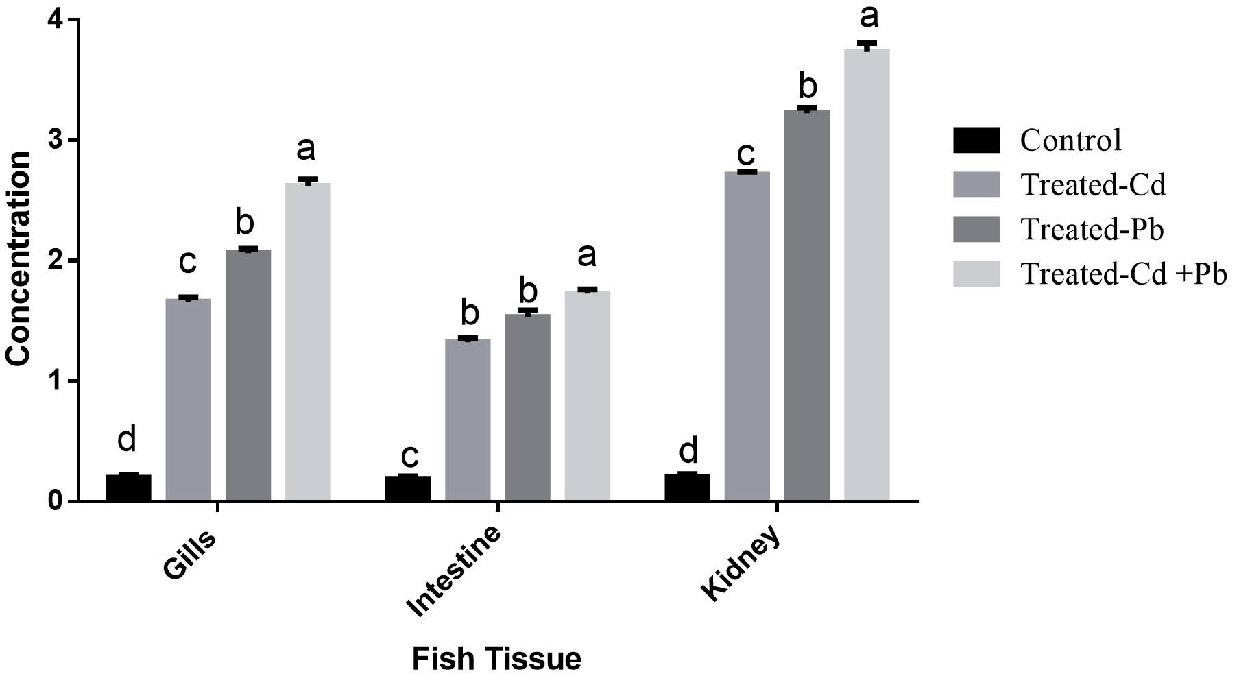
The concentration of Cd2+ and Pb2+, both individually and in combination, in fish tissues is depicted in Fig. 1 after a 40-day exposure period. The results reveal a remarkable trend, indicating a significantly (P < 0.05) higher concentration of these heavy metals in the fish tissues, specifically in the order of kidney > gills > intestine compared to the control group. Comparatively, in both fish gills and kidneys, the group treated with Pb2+ demonstrated a considerably (P < 0.05) higher concentration compared to the group treated with Cd2+ individually. Conversely, in the intestine, no significant difference was observed between the Cd2+-treated group and the Pb2+-treated group. However, the concentration of heavy metals was markedly more pronounced when administered in a mixed form (Cd2+ + Pb2+ treated group) as opposed to their presence in a single form. Similar results were reported in other studies. Tunçsoy and Erdem [32] exposed Oreochromis niloticus to metals (Cd, Zn, and Co) both individually and in a mixture, while Qu et al. [33] found higher concentrations of heavy metals in Carassius auratus tissues when exposed to a combination of metals. Different studies conducted in sublethal concentrations of Pb2+ and Cd2+, such as Al-Balawi et al. [34], investigated the sublethal effects of Pb2+ on Clarias gariepinus, noting higher accumulation in the gills, liver, and kidney, with a lower level found in the muscle. In another study, Malarvizhi et al. [35] reported a higher concentration of Cd2+ in the gills, liver, and kidney of Cirrhinus mrigala after 30 days of exposure. In this study, the increased concentration of Cd2+ and Pb2+ in fish kidneys following sublethal exposure indicates a significant accumulation of these heavy metals within the renal tissues. This elevated concentration highlights the susceptibility of fish kidneys to the absorption and retention of Cd2+ and Pb2+ during sublethal exposure scenarios [36]. The accumulation and harmful impact of metals vary based on the specific species, developmental stage, gender, type of metal, its concentration, duration of exposure, and the physical and chemical attributes of the water [37, 38, 8].
The catalase CAT and SOD activities in fish liver following exposure to heavy metals, both individually and in combination, are illustrated in Fig. 2. The findings indicate that there is no significant difference in fish exposed to Cd2+ and Pb2+ when exposed individually. However, a significantly lower concentration was observed in the combined form compared to both individual exposures and the control group. These results align with the studies of Atli et al. [39], who subjected Oreochromis niloticus to sublethal levels of Cd2+ and Pb2+. Rajeshkumar et al. [40] similarly investigated Cyprinus carpio after exposure to multiple heavy metals (Cr2+, Cd2+, and Pb2+). The reduction in the activities of CAT and SOD in C. marulius after exposure to Cd2+ and Pb2+ can be attributed to the harmful effects of these metals on the antioxidant defense system. Cd2+ and Pb2+ are known to induce oxidative stress by generating reactive oxygen species (ROS) within the organism [41, 42].
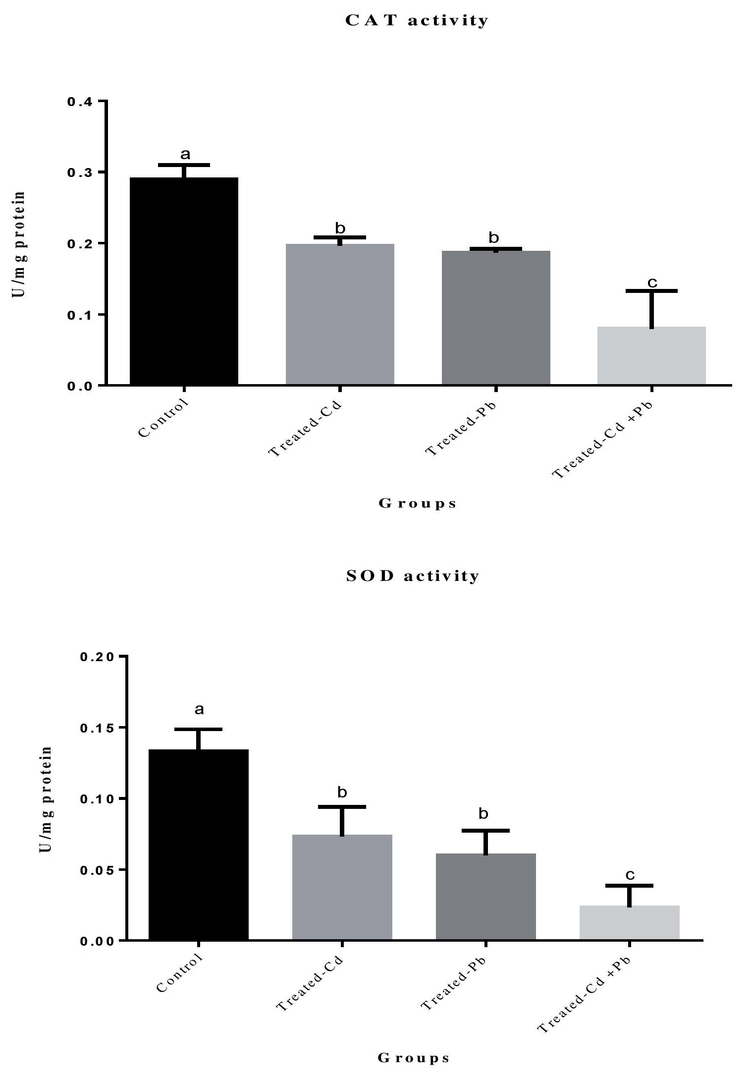
Table 1.
Hemato-biochemical parameters of Channa marulius after exposure to heavy metals individually and in combined form for 40 days.
Hematological and biochemical indicators serve as valuable tools in assessing the health status of fish. They offer early detection capabilities for potential issues, aiding in the effective management of fish populations in both aquaculture and natural environments [43, 44]. Table 1 presents the hemato-biochemical parameters of C. marulius after a 40-day exposure to heavy metals, both individually and in combination. The fish group exposed to Cd2+ and Pb2+ in combined form exhibited significantly (P < 0.05) lower concentrations of RBC count, Hb, and TP compared to those exposed to each heavy metal individually. Hct levels, however, showed no significant differences among all exposed groups, whether individually or in combined form. Similarly, Fazio et al. [5] found reduced RBC, Hb, Hct, and TP levels in Mystus seenghala following sublethal exposure to Cd2+. The toxic effects of heavy metals on fish hematopoietic tissues, responsible for red blood cell production, were evident in our findings. Prolonged exposure to heavy metals can impede the normal functioning of the hematopoietic system, leading to a decline in red blood cell production [45-47].
In terms of cortisol, glucose, AST, and ALT levels, the fish group exposed to heavy metals in combined form demonstrated significantly (P < 0.05) elevated values. Conversely, no significant differences were observed in the Cd2+ and Pb2+ treated groups when exposed individually. Survival rates indicated a higher percentage in the Cd2+-treated group (94.29), followed by the Pb2+-treated group (91.43) when exposed individually. However, in the combined form, the survival rate decreased to 85.71%. The studies, including Fazio et al. [5], demonstrated a substantial increase in glucose, cortisol, AST, and ALT levels in M. seenghala exposed to sublethal levels of Cd2+.
Acetylcholinesterase Inhibition
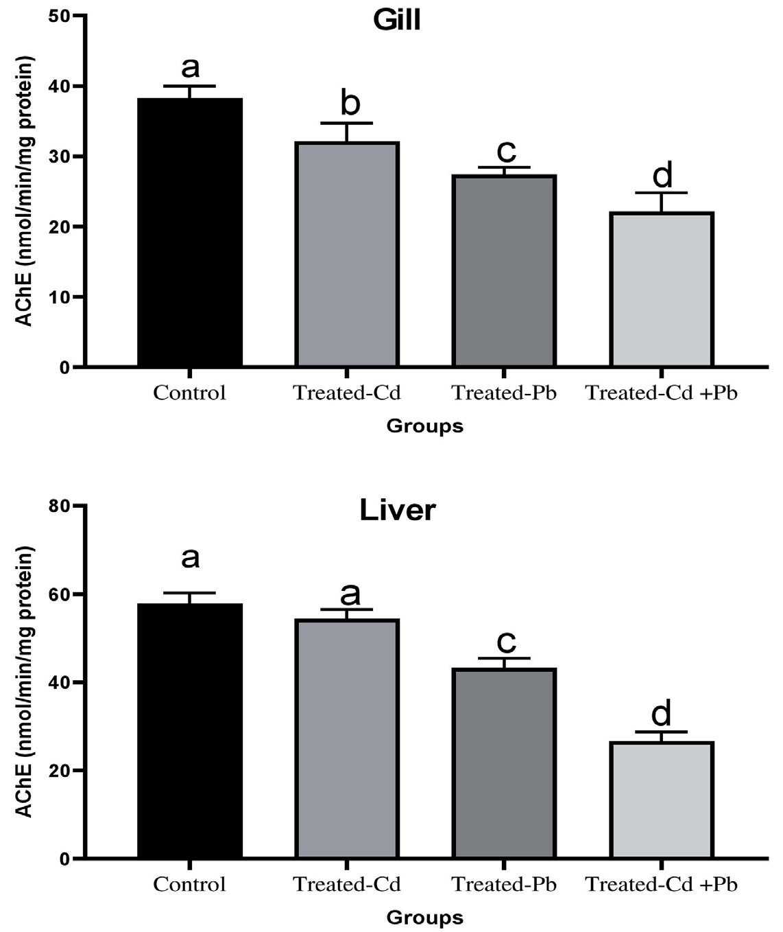
AChE is crucial for cholinergic neurotransmission at neuromuscular junctions and brain synapses. It is produced in the endoplasmic reticulum and then processed in the Golgi apparatus, where it becomes either a membrane-bound enzyme or a secreted molecule [48- 50]. Fig. 3 shows AChE activity in C. marulius exposed to Cd2+ and Pb2+. The results indicate that AChE activity in both gill and liver tissues was significantly affected by exposure to Cd2+ and Pb2+, whether individually or combined. The most substantial decrease in AChE activity was observed in fish exposed to both metals together, followed by those exposed to Pb2+ alone. Interestingly, in liver tissue, there was no significant difference in AChE activity between the control group and the Cd2+-exposed group. However, fish exposed to both Cd2+ and Pb2+ in combination exhibited significantly lower AChE activity. Liu et al. [48] studied the neurotoxicity in zebrafish (Danio rerio) exposed to Pb2+ and As, observing that combined exposure to these metals can lead to abnormal swimming behavior and exacerbate neurotoxicity.
Conclusions
The findings from this study indicate that exposing C. marulius to a combination of metal forms results in a significant accumulation of these metals in fish tissues, causing more pronounced alterations in hematobiochemical parameters compared to exposure to individual metals and control. Remarkably, the kidney exhibited the highest concentration of heavy metals, given its crucial role in filtration, reabsorption, and excretion processes, followed by the gills. The observed changes in hemato-biochemical parameters suggest a clear manifestation of physiological stress, characterized by rapid and transient modifications in specific stress-related indicators. With respect to neurotransmitter activity, Cd2+ and Pb2+ exposure notably reduced AChE activity in C. marulius. These research outcomes offer valuable insights for assessing pollution stress in aquatic environments and their inhabitants. This information is essential for shaping policies and strategies aimed at reducing the release of chemical compounds and heavy metals into freshwater ecosystems. To comprehensively understand the dynamics of aquatic organisms exposed to chemical mixtures in real-world scenarios, further research should explore the depuration potential of these organisms. Investigating the environmental realism of these scenarios will contribute to a more thorough understanding of the long-term impacts of chemical exposure on aquatic ecosystems.

