

Introduction
Mercury can enter the environment through natural sources such as volcanic activity or through human activities such as industrial pollution [1]. Its exposure is a global concern because it is widespread in the environment due to its multiple industrial, domestic, agricultural, and medical usage. Among its various chemical forms, both humans and animals are mainly exposed to mercury chloride (HgCl2), methylmercury, and elemental mercury. HgCl2 is metabolized primarily in the liver [2]. Mercury pollution in water is a serious threat to natural ecosystems. Various methods and technologies are used to remove mercury from the environment. They include phytoremediation, bioremediation, activated carbon adsorption, extractions, and others [3]. Mercury exposure shows symptoms like nausea, irritability, tremors, headache, hypertension, hallucinations, and even death in certain cases [4]. Mercury chloride is a hazardous heavy metal that has been linked to severe liver and kidney damage in both humans and animals [5]. Mercury chloride toxicity is primarily attributed to the production of reactive oxygen species (ROS) and the consequent development of oxidative stress, which results in lipid peroxidation and disruption of cellular antioxidants [6]. Inhaled HgCl2 vapor primarily affects the brain, while mercurous and mercuric salts primarily damage the gastrointestinal lining and kidneys. Methyl mercury, on the other hand, is distributed throughout the body [7]. The mercury concentrations of agricultural soils ranged from 0.42 to 155.00 mg/kg, determined by atomic fluorescence spectrometry of 146 soil samples, and mercury concentrations in local crops (rice, maize, pepper, eggplant, tomato, and bean) all exceeded the Chinese food safety limits [8]. The model predicted that the major emission pathway of mercury to the environment is landfill leachate, which accounted for 99.8% of the total emissions. 83% of mercury in the leachate was estimated to be inorganic form, and the rest 17% was methylmercury [9]. Organisms of aquatic biota are easily exposed to and ingest mercury deposits in the tissues, especially in the gonads, liver, kidney, and gills. Long-term mercury exposure damages the nervous system and causes tremors, spasms, memory loss, hallucinations, severe sadness, increased excitability, delirium, and personality alterations [10]. Furthermore, MDA, which is a crucial byproduct of lipid peroxidation, has frequently been used as a hallmark of oxidative stress in several tissues [11]. Aside from its negative effects on oxidative stress and MDA generation, HgCl2 has also been linked to renal function impairments such as urinary protein excretion, electrolyte imbalance, and renal tubular necrosis [12]. Additionally, HgCl2 has also been demonstrated to cause hepatotoxicity, which is characterized by liver damage such as hepatocyte death, steatosis, and increased liver enzymes [13]. It has been indicated that during mercury therapy, the aspartate transaminase (AST), alanine transaminase (ALT), and alkaline phosphatase (ALP) levels in the serum rose [14]. Mercury’s potential biological effects are being investigated, particularly the relationship between cardiovascular conditions like hypertension, coronary heart disease, and myocardial infarction and mercury toxicity [15]. Likewise, HgCl2-induced brain tissue damage [16]. Furthermore, the high polyunsaturated fatty acid concentration of brain tissues makes them more vulnerable to oxidative damage, which is crucial to necrosis and cell death [17]. On the other hand, mercury is regularly found in the thyroid, particularly in the follicular cells, and with aging, more people have mercury in their follicular cells. Cadmium is another hazardous element that is frequently found in the thyroid. Numerous hazardous metals have corrosive properties that might result in hyperthyroidism, thyroid cancer, and autoimmune thyroiditis. The majority of the potential adverse effects that hazardous metals could cause on the human thyroid are currently speculative [18]. It is capable of causing a wide variety of toxic effects, such as corrosive damage, severe gastrointestinal problems, acute renal failure, circulatory collapse, and ultimately death. Melatonin, a tryptophan metabolite primarily produced by the pineal gland, is incredibly potent and efficient at eliminating oxidative stress [19]. Melatonin has been shown to protect against oxidative stress in a variety of organs, besides the thyroid gland [20] but also, as previously revealed, in many other tissues and organs, such as the kidney [21], heart [22], liver [23], or brain [24]. It has been demonstrated that MEL has been shown to confer cardio protection in several cardiac diseases [25]. On the other hand, MEL has been discovered to be protective against epithelial cell damage, hepatocyte apoptosis, autophagy flux, oxidative stress, and inflammatory signaling [26]. In recent years, there has been an increase in interest in the molecular mechanism behind MEL-induced neuroprotection, which has led to an increase in studies. Studies reveal that anti-inflammatory, antioxidant, and anti-apoptotic mechanisms have positive effects on the recovery of intracerebral hemorrhage, cerebral ischemia-reperfusion injury, spinal cord injury, Alzheimer’s disease, Parkinson’s disease, and meningitis [27]. Given the aforementioned, a detailed investigation into MEL’s impact on HgCl2-induced liver, heart, thyroid, and brain damage in rats is highly recommended. Investigating melatonin’s possible impact on HgCl2-induced oxidative stress, MDA generation, and histological abnormalities in rat kidney, liver, heart, thyroid, and brain dysfunctions is the aim of this study.
Experimental
Animals
Eighteen female albino rats (Rattus norvegicus) (120–180 g), 5 weeks old, were procured from the Salahaddin University-Erbil, Animal House. Rats were employed in the in vivo trial, which required many administrations and handling for 28 days. They were housed in stainless steel cages with good ventilation and a 12-hour light/dark cycle. All rats received standard commercial pelleted food and had unlimited access to tap water.
Experimental design
The experimental animals were put into three groups of six rats each. The following was the experimental design for the in vivo investigation after a 2-week acclimatization period:
For 28 days, rats in Group 1 (control) were provided with unrestricted access to tap water and rat food.
Group 2 (HgCl2): For 28 days, rats were given 4 mg/Kg b.w, (40 mg/L) of HgCl2 in drinking water and a normal diet.
Group 3 (HgCl2 + MEL) rats got Mercury chloride 4 mg/kg body weight (40 mg/L) in ad libitum tap water + MEL (30 mg/kg body weight rats via diet) for 28 days.
Histological Processing
After the withdrawal of blood samples, animals were dissected and the liver, right kidney, heart, cerebrum, and thyroid gland were removed. Their weights were recorded by High-Precision Electronic Balances, then put in formalin 10% fluid for fixation. Preserved tissue samples from fixative solution (formalin 10%) exposed to serial processes began with dehydration, clearing, and impregnation using a series of graded ethanol (Scharlau, Spain) in ascending concentrations and then immersed in xylene (Scharlau, Spain). Finally embedded in paraffin wax (Scharlau, Spain) and cooled. The paraffin sections were cut by a rotary microtome, and after that, samples were stained with hematoxylin and eosin. Finally, photos were taken with a novel digital microscope, and they were analyzed.
Blood Sample Collection
At the end of the experiment, the rats were anesthetized intraperitoneally with ketamine hydrochloride (50 mg/kg) and xylazine (10 mg/kg). Through heart puncture, blood samples were collected and then transferred to a test tube containing gel and clot activator, where they were centrifuged at 1000 g for 15 minutes before being held at -20 C. Then, the extracted liver and kidney were weighed by electronic balance.
Biochemical Analyses
Blood samples were centrifuged at 3000g for 15 minutes to separate serum. The Cobas c411 fully-automated biochemical analyzer was used to measure serum alkaline phosphatase (ALP), alanine aminotransferase (ALT), and aspartate aminotransferases (AST).
Serum Malondialdehyde Determination
Serum MDA was evaluated using a simple and sensitive spectrophotometric approach (Spectronic GENESYS 6 UV – Visible spectrometer, Thermo Electron Corporation) and the thiobarbituric acid (TBA) reagent from Merck KGaA, Germany. One ml of 17.5% trichloroacetic acid (TCA) and 1 ml of 0.66% TBA were mixed, and 1ml of 0.66% TBA was well mixed in a 150 µl serum sample, boiled in boiling water for 15 minutes, followed by 15 minutes of room temperature cooling. The remaining serum protein was precipitated using 1 ml of 70% TCA, which was added, vortexed well, and allowed to stand for 20 minutes at room temperature. After that, the mixture was centrifuged for 15 minutes at 2000 rpm to separate the supernatant, which was then used for spectrophotometric scanning at (532 nm).
Statistical Analysis
The data in Table 1 were analyzed to confirm their distribution according to normality tests (Shapiro-Wilk and Kolmogrove-Smirnove tests). The data are normally distributed, so they were analyzed parametrically by using One-way ANOVA and Tukey tests as a post hoc test using GraphPad Prism Version 8.0.2. The results were presented as mean ± standard error of the mean (SEM). P < 0.05 represents statistically significant.
Table 1.
Effect of melatonin on serum malondialdehyde, alkaline phosphatase, alanine aminotransferase and aspartate aminotransferase of rats treated with HgCl2.
[i] Data are presented as mean ±standard error of mean. The results were analyzed with One-way ANOVA test. MDA; malondialdehyde, ALP; alkaline phosphatase, ALT; alanine aminotransferase, AST; aspartate aminotransferase, *; p˃0.05, #; p˃0.05, ##; p˃0.001, *; indicates the comparison between HgCl2 group and control group, while, #; indicates the comparison between HgCl2+MEL group and HgCl2 group. µmole/L; micromole per liter, IU/L; International Units Per Liter.
Results
Biochemical Analysis
The results of the study showed that rats treated with HgCl2 had significantly (P < 0.05) greater serum MDA levels than the control group, as indicated in Table 1. However, MEL treatment (30 mg/kg body weight rats) for 5 weeks significantly decreased it in comparison to the HgCl2-treated rats. Statistical analysis revealed that liver function test biomarkers were measured like ALP, ALT, and AST. Here, they elevated after HgCl2 treatment, while MEL could ameliorate the result significantly. On the other hand, serum creatinine and uric acid did not change by HgCl2 administration, whereas serum urea was markedly elevated in HgCl2-treated rats and MEL reduced it significantly (p < 0.01).
Microscopic Examination
Liver
The liver sections of the control group had normal arrangements of liver hepatocytes, the central hepatic venule, and the portal tract (Fig. 1A). While, rats treated with HgCl2 showed moderate mixed inflammatory cell infiltration predominantly lymphocytes in the portal tract with interface hepatitis associated with moderate vascular congestion, mild edema, mild focal fibrosis, and mild focal fatty changes (Fig. 1B, C, D, and E). On the other hand, both HgCl2 and MEL treatment showed mild mixed inflammatory cell infiltration predominantly lymphocytes in the portal tract with interface hepatitis associated with mild vascular congestion, mild focal fibrosis, and mild edema (Fig. 1F and G).
Kidney
The kidney section of the control group showed a normal arrangement of glomeruli and kidney tubules (Fig. 2A). Meanwhile, the administration of rats with HgCl2 showed a mild infiltration of mixed inflammatory cells, primarily lymphocytes, together with mild vascular congestion, rare focal fibrosis, and mild focal tubular degenerative alterations (Fig. 2B, C, D, E, F, and G). In contrast, the combination of HgCl2 and MEL administration resulted in a modest infiltration of mixed inflammatory cells, predominately lymphocytes, along with mild vascular congestion, mild edema, and a rare focal mild fibrosis (Fig. 2H).
Heart
The observed sections of the heart in the control group showed a normal arrangement of the heart endocardium and myocardium (Fig. 3A). In the HgCl2 group, the sections showed mild mixed inflammatory cell infiltration predominantly lymphocytes associated with mild vascular congestion, mild edema, rare focal mild fibrosis, and mild focal myocardial degenerative changes (Fig. 3B, C, and D). In contrast, in the HgCl2 and MEL groups, the microscopic examination showed mild mixed inflammatory cells, primarily lymphocytes, infiltration with mild edema, minor vascular congestion, and rare focal mild fibrosis (Fig. 3E).
Cerebrum
The cerebral cortex of the brain of the control group showed normal morphology of cerebral cortex neurons (Fig. 4A and B). However, rats treated with HgCl2 showed mild focal neuronal cell loss (death), mild cerebral edema, mild focal gliosis with reactive changes, and mild focal vascular congestion (Fig. 4C, D and E). In contrast, rare focal neuronal cell loss (death), mild cerebral edema, rare focal gliosis with reactive changes, and mild focal vascular congestion were observed in HgCl2 and MEL groups (Fig. 4F).
Thyroid Gland
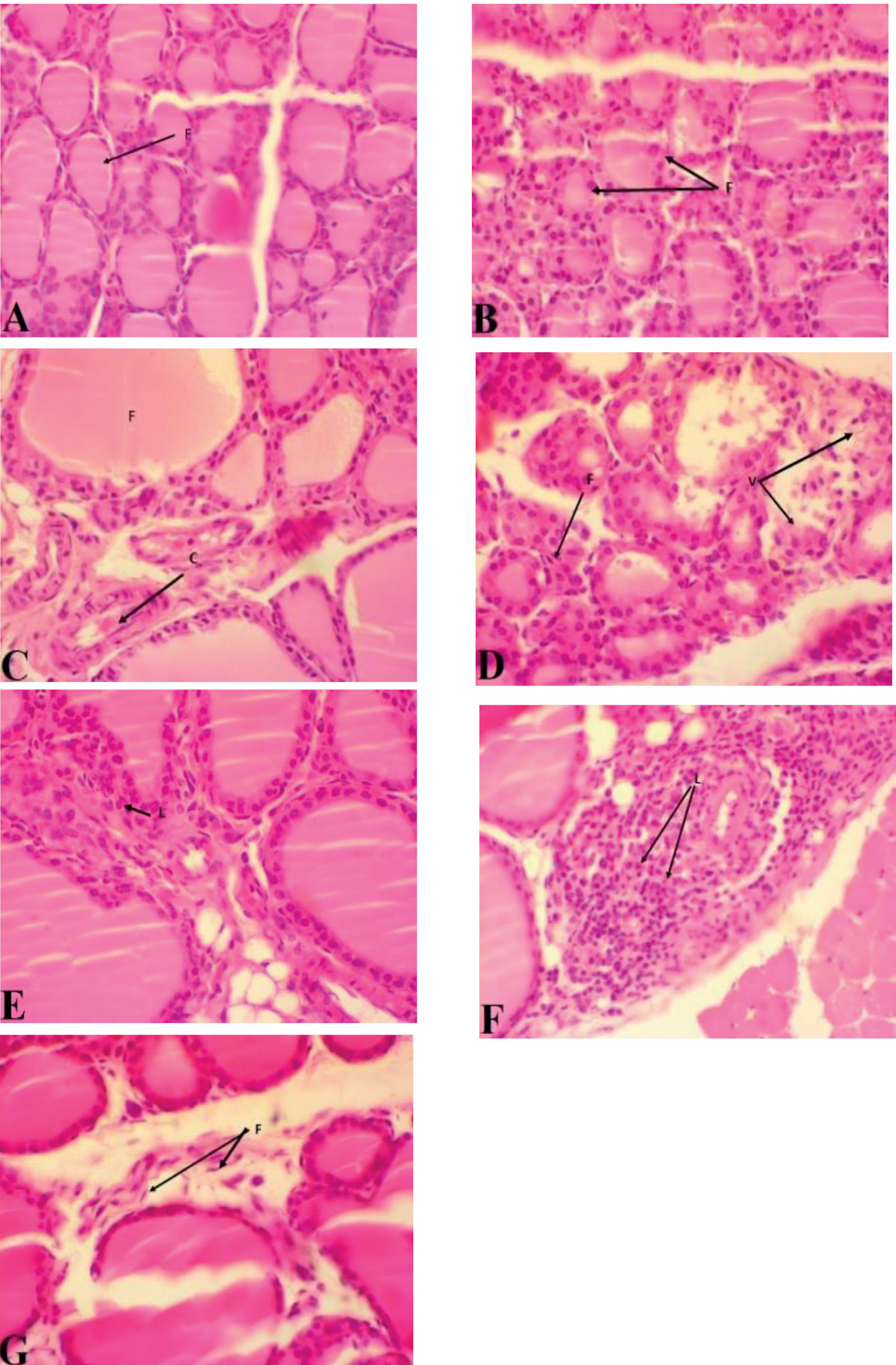
Thyroid gland sections of the control group showed a normal arrangement of thyroid follicles (Fig. 5A). These sections of rats treated with HgCl2 showed rare mixed inflammatory cell infiltration predominantly lymphocytes associated with mild focal vascular congestion, mild edema, rare focal mild fibrosis, rare focal degenerative changes, and focal follicular cellular hyperplasia with cystic dilatation (Fig. 5B, C, D and E). In contrast, rare mixed inflammatory cell infiltration predominantly lymphocytes associated with mild focal vascular congestion, mild edema, and rare focal mild fibrosis were observed in rats treated with HgCl2 and MEL as well (Fig. 5F and G).
Discussion
Free radicals are produced by mercury chloride, which raises oxidative stress, results in nephrotoxicity [5, 28], and accelerates hepatotoxicity [28]. In addition to promoting oxidative stress, HgCl2 treatment starts the synthesis of highly reactive molecules such as ROS [6]. In this investigation, we determined that when, compared to the control group, HgCl2 considerably increased the lipid peroxidation’s end byproduct, such as MDA. Numerous experiments have shown parallel results [29]. Mercury chloride resulted in increased oxidative stress and lipid peroxidation in rat liver tissue, as indicated by increased MDA levels and reduced levels of antioxidants such as catalase and glutathione peroxidase [30]. As a result, the level of lipid peroxidation rises and the activities of antioxidant enzymes decrease. Overall, it is suggested that HgCl2 induces oxidative stress and lipid peroxidation, which increases MDA levels [13]. It has been reported that exposure to mercury can cause a range of health problems, such as neurological disorders, developmental delays, and cardiovascular disease [1]. Our results agree with previous studies indicating that the observed histoarchitectural distortion of the liver in the HgCl2-treated group, such as hepatocellular vacuolation, congestion of the central vein, and sinusoidal dilatation, could be attributed to HgCl2-induced hepatotoxicity [31]. Melatonin is the hormone that is produced in the pineal gland [32], and it has been shown to have various functions in the body [33]. On the other hand, MEL supplementation (30 mg/kg body weight rats) for 5 weeks markedly reduced it as compared with the HgCl2-treated rats. One of the functions of MEL is its ability to decrease MDA, which is a biomarker of oxidative stress [34]. The mechanism of action by which MEL decreases MDA can be explained in three ways [35, 36]. First, tissue injury can lead to an increase in MDA levels in the body [37]. It has been demonstrated that MEL decreases tissue damage by inhibiting the production of free radicals and lowering inflammation [38]. Its antioxidant properties, which assist in scavenging free radicals and inhibit them from causing tissue damage, mediate these effects [39]. For instance, it has been found that MEL causes a reduction in oxidative stress in the liver by eliminating free radicals and lowering lipid peroxidation [40, 41]. Second, free radicals are scavenged by MEL [42]. Free radicals are unstable molecules that can harm tissues and cells [43]. By removing free radicals from the human body and counteracting their effects, melatonin can defend against this damage [44]. Studies have shown that MEL can minimize oxidative stress by scavenging free radicals and decreasing lipid peroxidation in a variety of tissues [45]. Finally, MEL enhances the activity of endogenous antioxidant enzymes [39]. By scavenging free radicals and preventing lipid peroxidation, these enzymes aid in protecting against oxidative stress [46]. Melatonin has been found to boost the activity of vital antioxidant enzymes like catalase, glutathione peroxidase, and superoxide dismutase [47]. The current study explored the impact of HgCl2 treatment on liver function test biomarkers, including ALP, ALT, and AST, as well as MEL’s potential therapeutic effect. Statistical analysis revealed significant elevations in the levels of ALP, ALT, and AST following HgCl2 treatment, indicating potential liver toxicity induced by mercury chloride exposure [30]. Remarkably, MEL administration exhibited a significant ability to mitigate the adverse effects of HgCl2 on liver function. Rats treated with MEL displayed significantly reduced levels of ALP, ALT, and AST compared to those treated solely with HgCl2 [48]. The histopathological examination of liver sections provided valuable insights into the effects of HgCl2 exposure and the potential ameliorating impact of MEL. Hepatocytes, central hepatic venules, and the portal tract were all arranged normally in the liver section of the control group, indicating physiological liver morphology. Conversely, rats treated with HgCl2 displayed a distinct pattern of histological alterations. These alterations included moderate mixed inflammatory cell infiltration, primarily lymphocytes, within the portal tract, accompanied by interface hepatitis. Additionally, moderate vascular congestion, mild edema, mild focal fibrosis, and hepatic tissues showed mild focal fatty alteration. All of these results point to the fact that hepatic inflammation is significantly triggered by HgCl2 exposure, along with vascular congestion and degenerative changes [49]. Interestingly, the histopathological analysis of liver sections from rats treated with both HgCl2 and MEL revealed a distinct pattern. Similar to the HgCl2-treated group, there was mild mixed inflammatory cell infiltration, primarily lymphocytes, in the portal tract, accompanied by interface hepatitis. However, the severity of the observed effects was notably attenuated compared to the group treated solely with HgCl2. Mild vascular congestion, mild focal fibrosis, and mild edema were also evident in this group, indicating that MEL administration had a mitigating effect on the histological alterations induced by HgCl2 exposure [50]. The microscopic examination of kidney sections provided significant insights into the impact of HgCl2 exposure and the potential reparative effect of MEL. The glomeruli and kidney tubules in the kidney sections from the control group were arranged normally, indicating unaltered renal morphology. Conversely, rats treated with HgCl2 displayed distinct histopathological changes in their kidney tissues. These alterations included mild mixed inflammatory cell infiltration, primarily lymphocytes, within the renal tissue. Furthermore, mild vascular congestion and mild edema were observed, along with rare focal instances of mild fibrosis and mild focal tubular degenerative changes. These findings collectively suggest that HgCl2 administration induces mild inflammatory responses in the kidney, along with vascular congestion and degenerative changes [51]. Of particular interest, the histological examination of kidney sections from rats given both HgCl2 and melatonin treatment revealed a distinct pattern. Similar to the group treated with HgCl2 alone, mild mixed inflammatory cell infiltration, primarily lymphocytes, was seen in the renal tissue. However, compared to the group receiving only HgCl2, the intensity of inflammatory reactions was reduced in the combined therapy group. Additionally, mild vascular congestion, mild edema, and rare focal instances of mild fibrosis were observed. Notably, the presence of focal tubular degenerative changes observed in the HgCl2-treated group was absent in the HgCl2 and MEL-treated groups, suggesting a potential reparative effect of MEL [21]. They showed that melatonin reduces kidney damage by inducing mitophagy in diabetic nephropathy. It has been shown that MEL regulation of the kidney’s renin-angiotensin system plays a critical function in reducing the symptoms of chronic renal disease [52]. The microscopic examination of heart sections provided valuable insights into the effects of HgCl2 exposure and the potential mitigating impact of melatonin. Heart sections from the control group displayed a normal arrangement of heart endocardium and myocardium, indicating preserved cardiac morphology. Conversely, heart sections from rats treated with HgCl2 exhibited discernible histopathological alterations. These alterations included mild mixed inflammatory cell infiltration, predominantly lymphocytes, within the cardiac tissue. Additionally, mild vascular congestion, mild edema, rare focal instances of mild fibrosis, and mild focal myocardial degenerative changes were observed. These findings collectively suggest that exposure to HgCl2 results in mild inflammatory responses in the heart, along with vascular congestion, edema, and degenerative changes in the myocardium [53]. They demonstrated that the oxidative tissue damage produced by HgCl2 was manifested in tissue samples by decreased GSH and elevated MDA levels. The thromboplastic activity significantly increased after Hg injection, confirming the cardiotoxic effects of HgCl2. It has been suggested that myofibrils, low dosages of HgCl2, and the energy-producing mitochondria of cardiomyocytes were shown to be negatively affected also [54]. It has been observed that long-term exposure to low doses of mercuric chloride leads to non-specific qualitative and quantitative changes in all structural components of the heart, including ongoing damage to the tissue barrier and dynamic and resorptive insufficiency of the hemo-microcirculatory bed, which causes chronic swelling, diffuse fibrosis, and an increase in cardiac decompensation activities. Surprisingly, a unique pattern was observed upon microscopic analysis of heart sections from rats that had received treatment with both melatonin and HgCl2. Similar to the HgCl2-treated group, mild mixed inflammatory cell infiltration, primarily lymphocytes, was observed in the cardiac tissue. However, the severity of inflammatory responses was notably ameliorated compared to the group treated solely with HgCl2. Additionally, mild vascular congestion and mild edema were observed, along with rare focal instances of mild fibrosis. The presence of focal myocardial degenerative changes observed in the HgCl2-treated group was absent in the HgCl2 and MEL-treated groups, suggesting a potential protective effect of MEL. It has been revealed that melatonin has been proven to have considerable benefits in lowering cardiac pathology and avoiding cardiac muscle death in mouse species in response to ischemia-reperfusion [55]. Furthermore, under some conditions, MEL may prevent cardiac muscle hypertrophy, hence reducing the development of heart failure [56]. Cardiotoxicity is a side effect of several commonly used conventional medicines [57]. Recently, research has revealed that MEL serves as an antioxidant and is efficient in preventing cardiac damage caused by pharmaceutical medicines [58]. However, it has been found that MEL, due to its pleiotropic effects, is a prospective therapeutic candidate [59]. This indoleamine is involved in endocrine physiology and chronobiology regulation [60]. Melatonin is a chemical that has antiaging, antioxidant, antiapoptotic, antiarrhythmic, immunomodulatory, and antiproliferative properties [61]. Melatonin’s antioxidative and anti-inflammatory effects may be responsible for its reported protective effect on heart histology [62]. The ability of MEL to reduce the negative effects of HgCl2 on cardiac tissues may be attributed to its capacity to scavenge reactive oxygen species and modify inflammatory pathways [63]. The microscopic examination of the cerebral cortex provided crucial insights into the effects of HgCl2 exposure and the potential mitigating influence of MEL. Brain sections from the control group displayed normal cerebral cortex neuron morphology, indicative of preserved neural structure. Conversely, rats treated with HgCl2 exhibited distinct histopathological alterations in the cerebral cortex. These alterations encompassed mild focal neuronal cell loss (death), mild cerebral edema, mild focal gliosis characterized by reactive changes, and mild focal vascular congestion. These findings collectively suggest that HgCl2 exposure induces mild neuronal damage, gliosis, cerebral edema, and vascular congestion in the cerebral cortex [64]. They also showed that HgCl2-induced impairment of memory, oxidative stress, and brain damage. Likewise, it has been shown that HgCl2 poisoning impaired mice learning and memory, raised blood and brain mercury levels and elevated interleukin-6, interleukin -1, and tumor necrosis factor levels in serum [49]. Intriguingly, the microscopic examination of brain sections from rats treated with both HgCl2 and MEL unveiled a distinct pattern. Similar to the group treated with HgCl2 alone, rare focal instances of neuronal cell loss (death), mild cerebral edema, and rare focal gliosis with reactive changes were observed in the cerebral cortex. Additionally, mild focal vascular congestion was present. While the exact mechanisms need further investigation, the presence of MEL seemed to confer a protective effect against some of the observed alterations induced by HgCl2 [65]. It has been shown that experimental stroke models have demonstrated the neuroprotective effects of melatonin [66]. The microscopic examination of thyroid gland sections provided valuable insights into the impact of HgCl2 exposure and the potential attenuating effect of MEL. Thyroid gland sections from the control group displayed a normal arrangement of thyroid follicles, indicative of preserved thyroid morphology. Conversely, rats treated with HgCl2 exhibited distinct histopathological changes in the thyroid gland. These changes included rare instances of mixed inflammatory cell infiltration, predominantly lymphocytes, within the thyroid tissue. Additionally, mild focal vascular congestion, mild edema, and rare focal instances of mild fibrosis were observed. Notably, focal follicular cellular hyperplasia with cystic dilatation was also present, suggesting potential thyroid gland dysfunction induced by HgCl2 exposure. Studies have shown that additional hazardous metals, such as cadmium, are frequently found in the thyroid [18]. Hypothyroidism, autoimmune thyroiditis, and thyroid cancer may occur as a result of the corrosive effects of many hazardous metals [67]. Intriguingly, a different pattern was seen when thyroid gland sections from rats treated with both MEL and HgCl2 were examined under a microscope. Similar to the HgCl2- treated group, rare instances of mixed inflammatory cell infiltration, primarily lymphocytes, were observed within the thyroid tissue. Additionally, mild focal vascular congestion, mild edema, and rare instances of mild fibrosis were noted. Importantly, the presence of MEL seemed to have minimal impact on the observed histopathological changes induced by HgCl2, suggesting limited mitigating effects in this context (Iwan et al., 2021). According to this study, MEL may have had a limited impact on thyroid histopathology due to several factors, including the duration of treatment, dose of MEL, and specific mechanisms underlying thyroid responses to mercury exposure. Additional investigations are warranted to elucidate the mechanisms and conditions under which MEL might exert more substantial protective effects on thyroid tissues. Furthermore, it has been found that potassium iodate-induced elevated levels of lipid peroxidation in pig thyroid are decreased by MEL [20].
Fig. 1.
The histological effect of MEL on the liver of rats treated with HgCl2. The control group (A) showed normal arrangement of hepatocytes (black arrow; H) and central hepatic vein (CV). The HgCl2 group (B, C, D, E) showed an increase in the number of lymphocytes (black arrow), central vein congestion (black arrow), and vacuolation inside hepatocytes (black arrow; degeneration) near the central vein and dilated, congestion of central vein (black arrow; CV) and an increased number of lymphocytes (black arrow; L). HgCl2 treated with MEL group (F, G) indicated a reduction in the number of lymphocytes (black arrow; L) surrounding the bile duct (black arrow; BD) and a reduction in the number of lymphocytes (black arrow; L) surrounding the bile duct (black arrow; BD). [Note: A, B, D, E, F, and G (400X), D (100X), and Stain (H and E)].
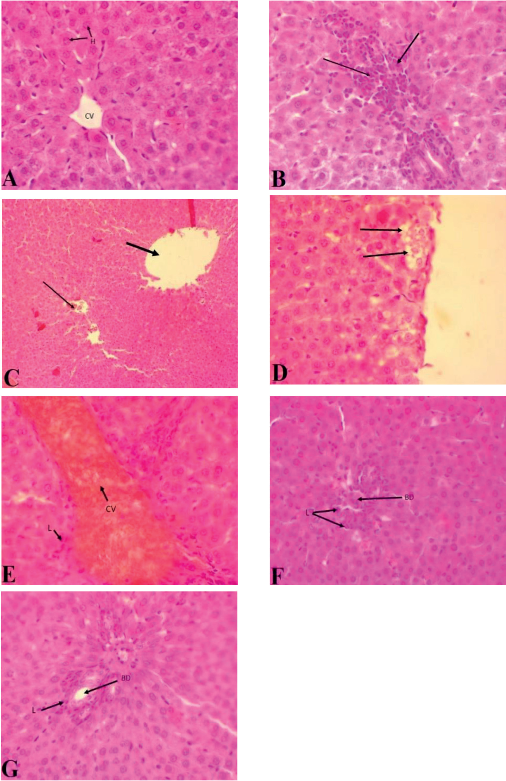
Fig. 2.
The histological effect of MEL on kidneys of rats treated with HgCl2. The control group (A) showed normal arrangement of glomeruli (G) and kidney tubules (T). The HgCl2 group (B, C, D, E, F, G) showed congestion of blood vessels (C), hemorrhage (H), increased number of lymphocytes (black arrow; L), vacuolar degeneration inside tubules (black arrow; V), increased number of lymphocytes (black arrow; L), vacuolar degeneration inside tubules (black arrow; V), increase number of lymphocytes (black arrow; L ), congestion of blood vessels (black arrow; C), hemorrhage (H), increase number of lymphocytes (black arrow; L) around glomerulus, congestion of blood vessel ( black arrow; C ) and mild lymphocytes (black arrow; L). The HgCl2 treated with MEL group (H), showed blood vessel congestion (C), edema found (black arrow; E ), and an increased number of lymphocytes (black arrow; L). [Note; A, B, C, D, E, F, G and H (400x), Stain (H and E)].
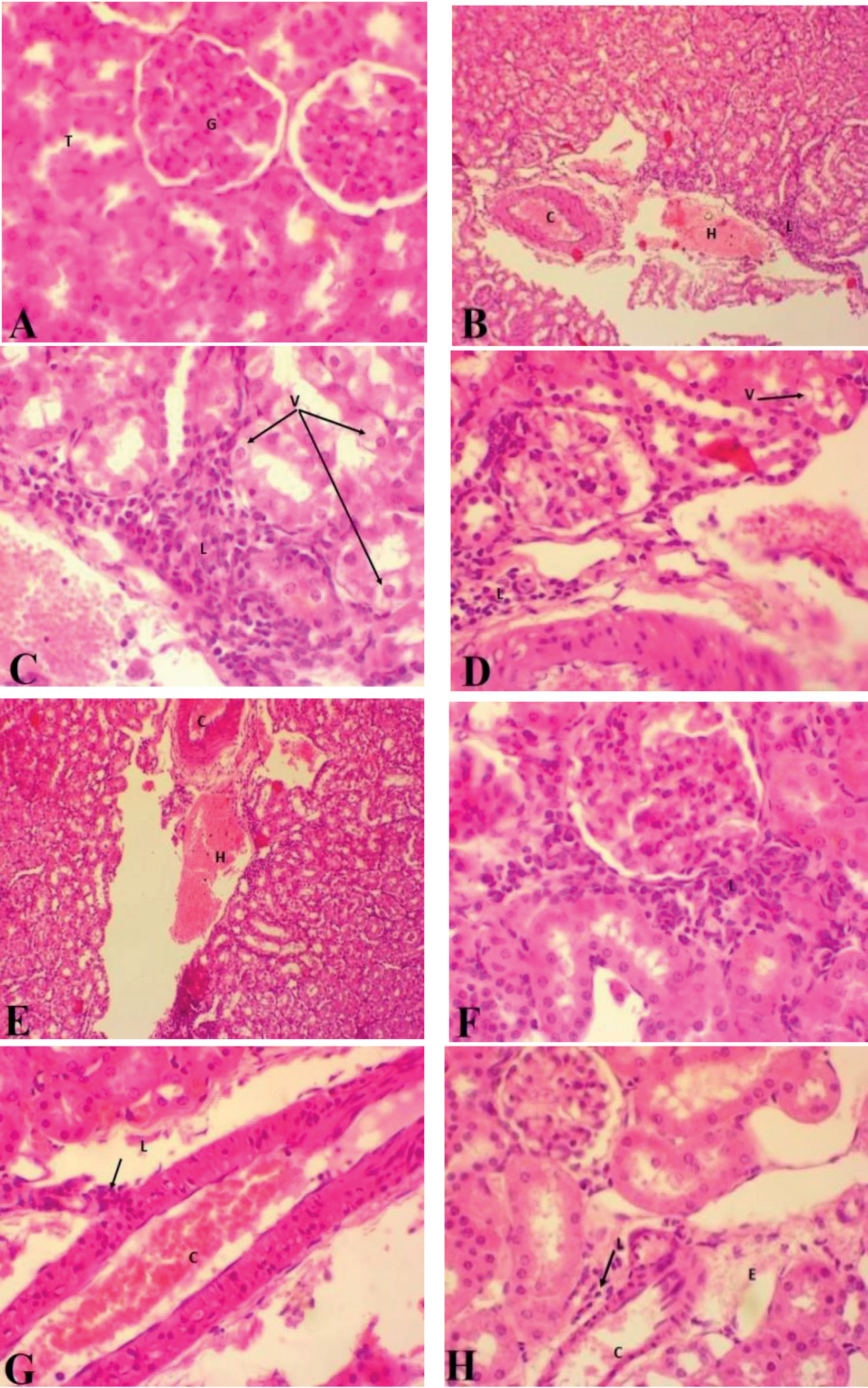
Fig. 3.
The histological effect of MEL on the heart of rats treated with HgCl2. The control group (A) showed a normal arrangement of the myocardium (black arrow). The HgCl2 group (B, C, D) showed hemorrhage (H) lymphocytes (black arrow; L), loss of normal orientation of muscles (black arrow; M), necrosis found (black arrow; N), lymphocytes (black arrow; L) found, loss of normal orientation of muscles (black arrow; M), necrosis found (black arrow; N) and lymphocytes (black arrow; L) found. The HgCl2 treated with MEL group (E) showed a better arrangement of muscle fibers found (M). [Note; A, C, D, E (400X), and B (100X) Stain (H and E)].
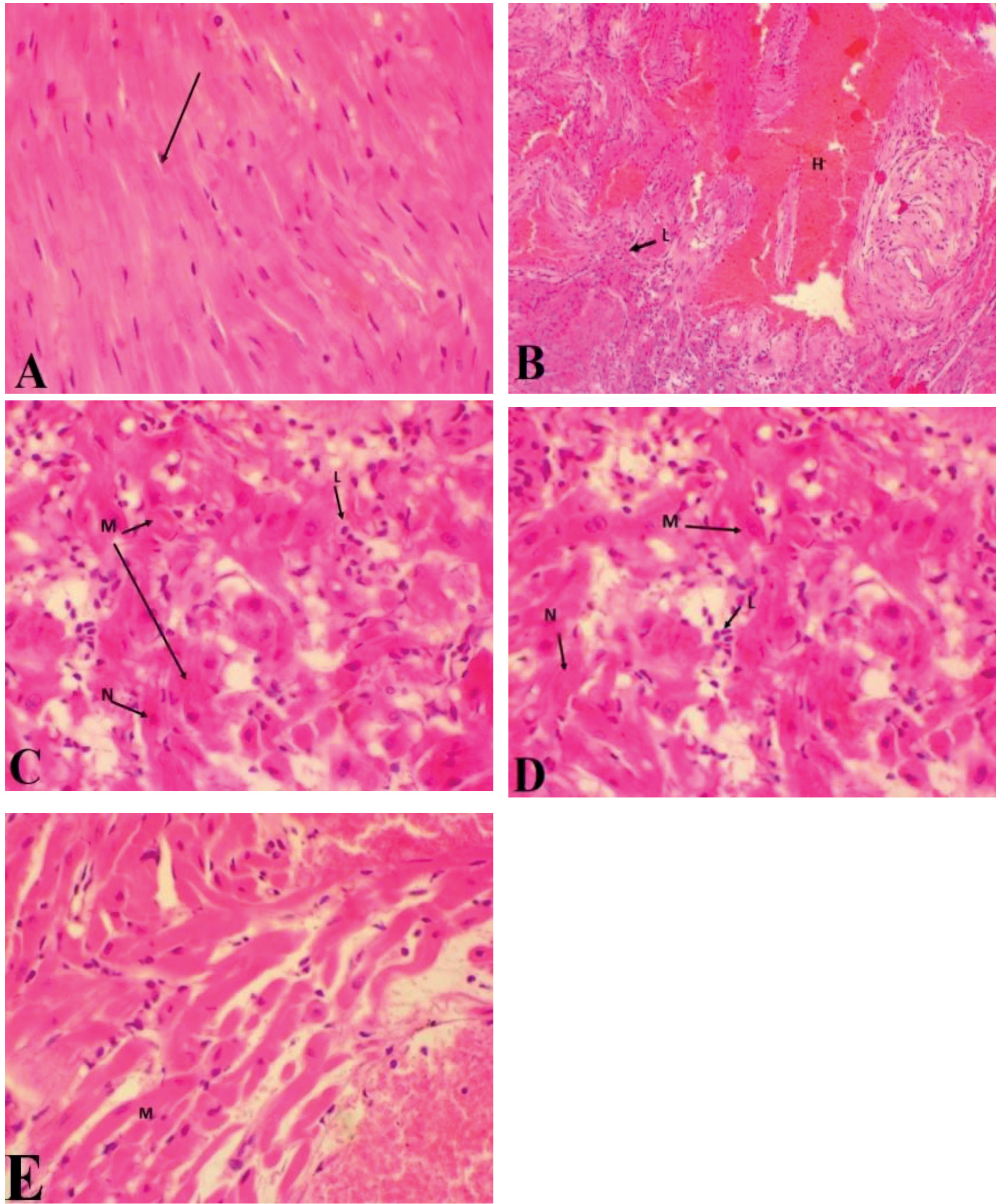
Fig. 4.
The histological effect of MEL on the cerebrum of rats treated with HgCl2. The control group (A, B) showed normal morphology of cerebral cortex neurons (black arow; N) and normal morphology of cerebral cortex neurons (black arow; N). The HgCl2 group (C, D, E) showed mild focal neuronal cell loss, mild focal vascular congestion (black arow; C), mild focal neuronal cell loss, mild cerebral edema (black arow; E), pycnotic cells (black arow; P), necrosis, edema (black arow; E), astrogliosis (black arow; A), congestion of blood vessels and red blood cells found outside blood vessels (hemorrhage) (RBC) with necrosis arrow. The HgCl2 treated with MEL group (F) showed less astrogliosis (black arow; A). [Note: A, B, C, D(100X), and E, F, G, H, I, (400X) Stain (H and E)].
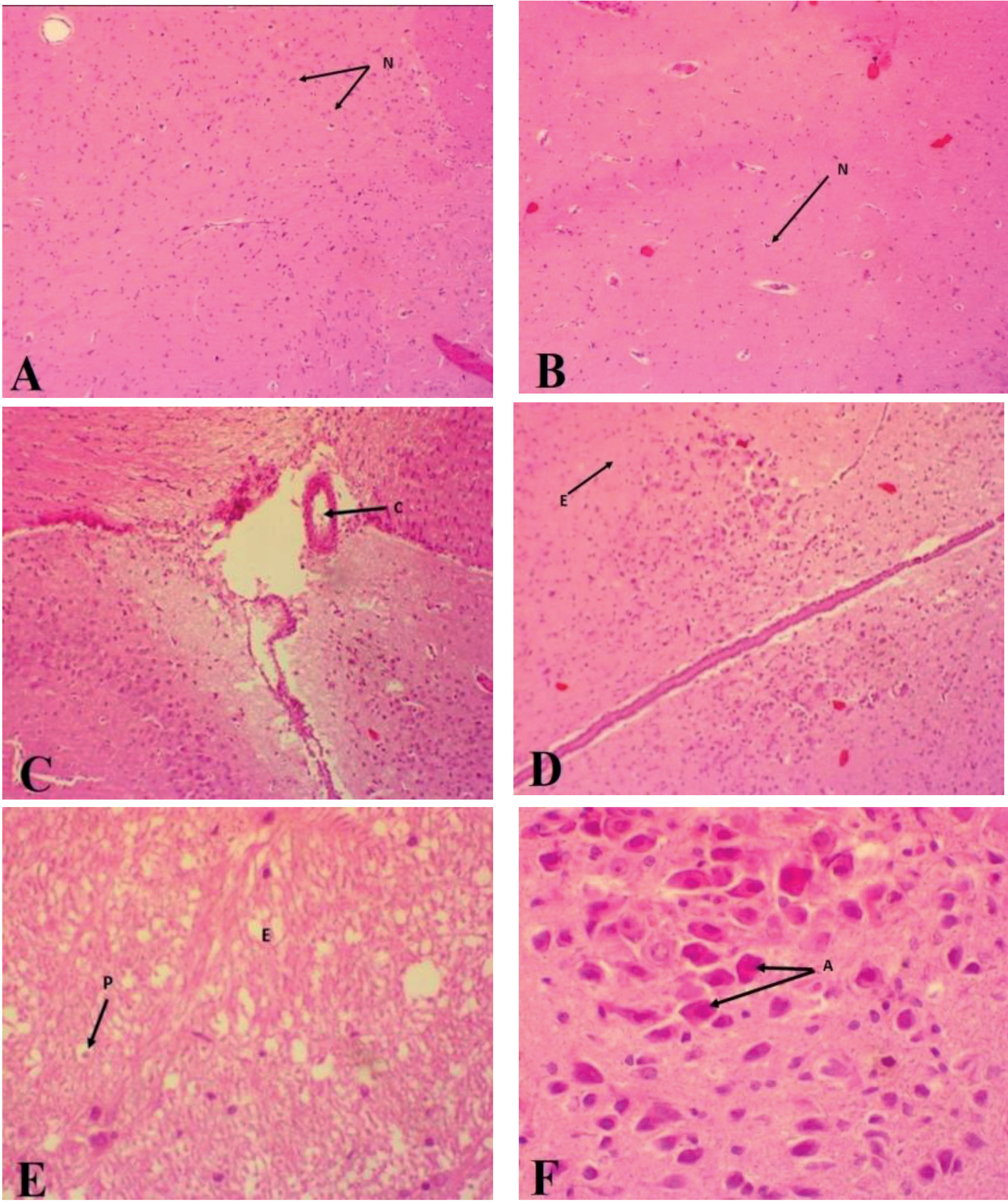
Fig. 5.
The histological effect of MEL on the thyroid gland of rats treated with HgCl2. The control group (A) showed normal arrangement of thyroid follicles (black arow; F). The HgCl2 group (B, C, D, E) showed mild thyroid follicle hyperplasia, mild increase in size (black arow; F), edema, mild blood vessel congestion (black arow; C), mild increase in size (black arow; F), cellular degeneration with vacuolation inside follicular cells degeneration (V), mild increase fibroblast (black arow; F) and mild increase lymphocytes (black arow; L).The HgCl2 treated with MEL group (F, G), showed an increase in the number of lymphocytes (black arow; L) between follicles, edema, and a small amount of fibroblast cells. [Note; A, B, C, D, E, F, G (400X), Stain (H and E)].
Conclusions
In conclusion, the findings of this investigation provide compelling evidence for the adverse effects of HgCl2 exposure on multiple organ systems in rats. HgCl2-induced oxidative stress, marked by elevated oxidative damage markers like MDA, was evident across various organs. These findings align with previous research highlighting HgCl2’s role in generating ROS and initiating oxidative stress. The remarkable potential of MEL as a mitigating agent against HgCl2-induced oxidative damage was underscored. Melatonin’s multifaceted mechanisms contributed to its protective effects. Firstly, its antioxidant properties were evident through the reduction in MDA levels. Melatonin’s ability to neutralize free radicals and reduce inflammation plays a crucial role. Moreover, MEL acted as a scavenger for free radicals, countering their damaging effects on cells and tissues. Furthermore, MEL augmented endogenous antioxidant enzymes, reinforcing the defense against oxidative stress. Regarding specific organ responses, the liver exhibited notable alterations in function, with elevated ALP, ALT, and AST levels post-HgCl2 exposure. Melatonin administration demonstrated its potential to ameliorate these impacts, indicative of hepatoprotective effects. In the kidney, melatonin’s mitigating influence was evident through attenuation of inflammatory responses and fibrosis. Histopathological changes in the heart, cerebral cortex, and thyroid gland following HgCl2 exposure highlighted the damaging effects on these organs. Melatonin supplementation showcased its potential to reduce inflammation, edema, and degeneration in these tissues. The role of MEL in protecting against cardiac damage and its pleiotropic actions in various physiological processes were evident.

