


Introduction
Crude oil is the most prevalent organic pollutant in all ecosystems due to spills during production, storage, transportation, and tank failures. It is classified as hazardous waste because of its cytotoxic, mutagenic, and carcinogenic effects on living organisms. These pollutants, present in contaminated desert soils, can impact both human health and the ecosystem by seeping into nearby subsoil and groundwater [1]. Since many hydrocarbons are water-insoluble, they remain separated in the non-aqueous phase of the liquid [2]. The contamination of hydrocarbons has major negative impacts on ecosystems, such as decreased plant growth and seed germination. Hydrocarbons can coat plant roots, reducing the absorption of water and nutrients. Moreover, these molecules can infiltrate plant tissues, causing damage to cell membranes. This damage can lead to the leakage of cell contents and blockage of intercellular gaps, which lowers the rates of respiration and metabolite transport [3].
According to the analysis of desert soil samples, the amount of lead (total or bioavailable) in soils contaminated with crude oil was three times higher than in uncontaminated soils [4, 5]. A high concentration of copper can decrease bacterial growth and viability, thereby inhibiting the ability of bacterial isolates to degrade hydrocarbons. Overall, high concentrations of heavy metals can increase the toxicity of the culture and decrease bacterial growth [6].
Desert soils with low organic carbon content, sandy texture, and reduced degradation rates generally exhibit poor microbial diversity and proliferation. Higher C ratios and lower internal surface areas are associated with lower abundances of degraders in the sandy component of polluted soils. Traditionally, the fertility of desert soils has been assessed using various physical and chemical characteristics, including aggregate stability, water-holding capacity, soil erosion, carbon fractions, and nutrient content. However, biological parameters such as microbial biomass and basal respiration, along with biochemical characteristics like hydrolases involved in the C, N, and P cycles, are more sensitive indicators closely linked to the microorganisms present in desert soil, offering a more accurate representation of the true degradation state [7].
Currently, chemical and mechanical treatments are employed in decontamination processes; however, these methods are expensive and often inefficient. In response to these challenges, bioremediation has emerged as a promising approach for cleaning oil-contaminated environments [8]. Bioremediation employs living organisms, most often microorganisms, plants, or byproducts produced by living organisms, to degrade, detoxify, or sequester toxic chemicals present in natural waters and soils. Bioremediation can be adapted for use in treating soil, sediment, sludge, water, or even air. Treatments can be either ex situ or in situ. Ex-situ procedures involve removing contaminated materials from a polluted site for treatment in another location, while in situ approaches treat contaminants in place without transferring samples to a treatment facility [9].
According to previous studies, there are several methods for utilizing microorganisms, especially bacteria, to break down environmentally hazardous waste materials such as crude oil. The existence and activity of these microorganisms, which are greatly influenced by factors such as pH, temperature, moisture content, and the composition of the microbial community (including fungi and bacteria), determine the effectiveness of bioremediation [10].
Recently, molecular and biochemical methods have been employed to identify and characterize bacteria and their specific properties at contaminated sites. These techniques serve as complementary tools to classical chemical and physiological analytical methods (such as measuring pollutant concentrations and speciation, redox potential, etc.) to monitor spatial and temporal changes in bacterial community composition and functioning during bioremediation processes. The 16S rRNA gene has become a crucial DNA/gene fragment for identifying bacterial species, supporting comparative and phylogenetic research and classification efforts [11].
Moreover, bacterial isolates can effectively degrade a significant quantity of organic pollutants associated with oil, such as toluene, n-alkanes, aliphatic compounds, monoaromatic compounds, and polycyclic aromatic hydrocarbons (PAHs). However, high-molecular-weight PAHs are challenging to degrade for most genera [12]. A promising new technique for detecting petroleum hydrocarbons in environmental samples is comprehensive gas chromatography (GC). In gas chromatography, compound peaks are arranged along the x-axis and y-axis according to their chemical class and carbon number, respectively. This results in distinct chemical classes for petroleum, including cycloalkanes, alkanes, and one- , two-, and multi-ring aromatics. Additional groupings within each class display homologous series. Gas chromatography has proven to be an effective method for identifying and quantifying specific compounds and classes of compounds for crude oil and refined petroleum products [13].
Bacterial isolates utilize organic contaminants as nutrient sources, energy, and carbon. Acinetobacter, Bacillus, Comamonas, Alcaligenes, Pseudomonas, and Micrococcus are some of the more effective bacterial genera for the initiation of bioremediation [14-19]. However, previous studies indicate a noticeably greater decrease in bacterial numbers and diversity in newly polluted soils compared to older ones, with Pseudomonas being more common than other detected genera [20]. Furthermore, a similar study was conducted to isolate, purify, and identify bacterial strains from the Yanbu coastal region in Saudi Arabia. These strains can consume and break down hazardous substances from unintentional spills, oil leaks, and contamination from petrochemical industrial waste. According to this study, Pseudomonas species, which demonstrated the highest oil-degrading ability among all isolates, could be effectively utilized for bioremediation [21].
The highway between Jordan and Iraq, used for transporting crude oil, has two locations contaminated by oil spills resulting from tanker accidents, potentially impacting local flora. Desert soil samples collected from these sites were dry. This investigation aimed to identify and characterize bacterial isolates from oil-polluted arid soil at these accident locations and evaluate their ability to degrade various hydrocarbons, including crude oil, toluene, naphthalene, and hexane. In a subsequent phase known as ‘In situ bioremediation’, these bacterial isolates could be utilized to remediate crude oil spills and facilitate plant regrowth.
Materials and Methods
Soil Sampling
Five soil samples, weighing 250 g were taken from two oil-contaminated sites in the northeastern region of Jordan at depths of 5, 10, and 15 cm. The soil samples were ground, put in polyethylene bags, and kept at 4ºC until needed. The samples were then sieved through a 2 mm pore size sieve.
Culture Methods
150 g of soil samples were suspended in 300 mm of minimal-salt medium (Stanier’s media) in 500 mm Erlenmeyer flasks supplemented with 400 ppm of crude petroleum oil. The flasks were then shaken at 160 rpm at 37ºC. As controls, samples devoid of crude oil contamination [22].
Enumeration of Bacteria
Using Stanier’s medium as the diluent, a range of serial dilutions (10-5 to 10-18) were utilized to create isolated colonies on tryptic soy agar (TSA) and nutrient agar (N.A). Different media plates were used to isolate and select each colony type [22].
Oil-Degrading Bacteria Isolation Using Different Hydrocarbon Sources
After being cultivated on Staneir’s agar, a minimal salt medium, 400 ppm of hydrocarbon compounds (crude oil, toluene, naphthalene, and hexane) were added, and the bacterial colonies were incubated for ten days at 37ºC.
Morphological Features and Biochemical Characterizations
Each bacterial isolate was characterized morphologically based on gram staining, size, color, and colony characteristics (form and elevation). Biochemical and physiological tests were used to further identify the bacterial isolates [23]. The isolated bacterial colonies were a variety of colors, including yellow, brown, creamy white, and even green. Their shapes were described as circular, filamentous, punctiform, and rhizoid.
Molecular Characterization
Extraction of Genomic DNA
The protocol outlined in the previous study was followed for bacterial DNA extraction. Following an 18-h incubation period at 37ºC, 10 mm of nutrient broth were inoculated with each bacterial isolate. 15 min at 14,000 rpm was used for centrifuging 2 mm of the broth culture. 30 μl 25% w/v SDS, 3 μl Proteinase K (20 mg/ml), and 567 μl TE buffer were used to suspend the pellet. It was then incubated at 65ºC for 30 min. The mixture was incubated with 100 μl of 5 M NaCl and 80 μl of CTAB/NaCl for 10 min at 65ºC. After adding equal amounts of phenol, chloroform, and isoamyl alcohol (25:24:1), the mixture was mixed and centrifuged for 5 min at 14,000 rpm. After a second centrifugation of the mixture for 5 min at 14,000 rpm, the solution’s supernatant was moved to a fresh tube. After adding 1.4 mg/ml of RNase enzyme to the mixture and letting it sit at 37ºC for 35 min, the mixture was cooled with ice, and an equal amount of ice-cold isopropanol was added to precipitate the DNA. After rehydration in 100 μl of TE buffer (pH 7.5), the DNA pellets underwent three rounds of washing with 70% ethanol [24].
Identification of Bacterial Isolates through Sequencing of 16S rDNA
The 16S rDNA target sequence was amplified using the method outlined by a previous study [25]. 25 µl of the master mix (10 mM Tris–HCl pH 8.3, 50 mM KCl, 1.5 mM MgCl2, 200 μM deoxynucleoside triphosphates, and 2.5 U of Taq DNA polymerase) is included in the 50 μl PCR mixture. Additionally, there are 2 μl of the reverse and 2 μl of the forward 16S rDNA universal primer, 5 μl of DNA template, and 16 μl of dH2O. Reaction mixtures were subjected to 35 cycles of PCR reaction (denaturation at 94ºC for 45 sec, annealing at 55ºC for 1 min, and extension at 72ºC for 1 min) in a DNA thermal cycler (Xp cycler, USA, 2008) at 96ºC heat shock for 1 min. Every reaction mixture was kept cold at 4ºC. 5-AGAGTTTGATCCTGGCTCAG-3 using the forward primer sequence (FD1) and 5-GGTTACCTTGTTACGACTT-3 as the reverse primer sequence (RD1) [26]. Finally, with the previous study description, 16S rDNA samples were sent to Macrogen, Inc., Koria, for sequencing [26].
Petroleum Hydrocarbon Determination
The EPA method 3510 was followed to extract soil samples for GC analysis. A 40 mL VOA vial was filled with 10 g of soil. Subsequently, 10 mL of methylene chloride, 15 mL of surrogate working solution (phenylacetylene), and 5 g of anhydrous sodium sulfate were added to the VOA vial. The sealed vial was submerged in a Grant, Inc., Germany, sonic bath for 5 min. After shaking the vial, it was submerged in the sonic bath for five more min. Gas chromatography (Calruse 500 autosampler, Perkin Elmer, USA) was used to quantify the hydrocarbon compounds in the extract. The apparatus included a flame ionization detector (FID) and a capillary column, Rtx-1 (30 m x 053 mm; 0.1 µm film thickness; Silica fused, Philadelphia, Pa., USA). At 290 and 320ºC, respectively, the temperatures of the injection port and detector were maintained constant. After being maintained at 50ºC for 5 min, the oven’s temperature was raised to 320ºC at a rate of 10ºC per min. The air and hydrogen gas flow rates were set at 2 mm per min for the flame ionization detector. The carrier was helium gas. Based on the retention time of the sample, the starting temperature and progress rate were chosen [27].
Heavy Metal Determination
Using a diluted HCl acid extraction method, heavy metals, including lead, zinc, cadmium, copper, ferrous, manganese, and zinc, were extracted from soils [28]. A 100 mL plastic bottle was filled with 200 mg (dry weight) of soil samples, followed by adding 2 mL of HF, 4 mL of 25% HNO3, and 4 mL of 25% HCl. The samples were incubated in a water bath at 70ºC for 2 h after being shaken for 2 min. After adding 50 mL of boric acid, the sample was once again submerged in the water bath for 15 min. Using dH2O, the volume of each sample filtrate was increased to 100 mL, and an atomic absorption spectrophotometer (Analytic Jena, Inc.).
Results and Discussion
Enumeration of Bacterial Colonies
Results presented in Table 1 show the number of bacterial colonies (CFU) on nutrient agar in each soil sample. A range of crude oil concentrations (400, 600, 800, 1000, 1500, and 2000 ppm) were used to enhance the biodegradation ability of the tested bacterial isolates over a period that ranged from 50 days to 300 days. Results indicated that M2A generated the highest CFU at either the least (400 ppm) or the highest (2000 ppm) concentrations of crude oil, where the obtained CFU values were 1.66×10⁷ and 1.87×10¹⁷, respectively. In contrast, the M1A sample showed the least (2.49×10⁵ and 1.25×10¹⁴) CFU at a concentration of 400 ppm and 2000 ppm, respectively. This study compared the number of viable bacterial colonies in samples isolated from oil-contaminated (test samples ranging from 2.49×10⁵ to 1.87×10¹⁷ CFU·g-1 soil) and uncontaminated (negative control; 9.8×10⁴ CFU·g-1 soil) soil. The variations in colony counts observed in the test samples could be attributed to their origins in both ancient and recently polluted locations. The highest toxicity levels of crude oil pollutants and the most detrimental effects on microbial diversity were observed in the old, contaminated areas. [29]. Additionally, Table 1 illustrates how adding various crude oil concentrations (400-2000 ppm) to the bacterial cultures at each site increased bacterial growth in tandem with rising crude oil concentrations.
Growth of Oil-Degrading Bacteria on Minimally Salted Media with a Different Source of Hydrocarbons
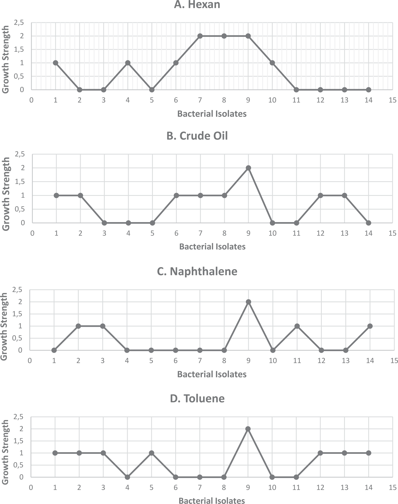
Among the 14 bacterial isolates, 7 were capable of degrading specific n-alkanes, such as hexane; 8 could degrade compounds with a single benzene ring, like toluene; and 5 isolates were capable of degrading compounds with two benzene rings, such as naphthalene. Pseudomonas fluorescens exhibited the highest growth rate on all hydrocarbons, while Micrococcus luteus and Lactobacillus casei appeared with the highest growth rate on hexane (Fig. 1). Conversely, low levels of growth on naphthalene have been observed in other isolates, including Rhodococcus rhodochrous, Bacillus subtilis, Pseudomonas putida, and Serratia liquefaciens. Furthermore, only toluene could be used as the exclusive energy and carbon source by Staphylococcus simulans, Rhodococcus rhodochrous, Bacillus subtilis, Staphylococcus epidermidis, Staphylococcus saprophyticus, and Serratia liquefaciens (Fig 1). According to our findings, the identified bacterial isolates utilize the supplemented hydrocarbons solely as a source of carbon and energy [30].
Table 1.
Growth of bacterial isolates from soil samples at different concentrations of crude oil.
Fig. 1.
Growth rate of oil-degrading bacteria on minimal salt media supplemented with four kinds of hydrocarbons, A) Hexane, B) Crude oil, C) Naphthalene, D) Toluene. II- +1: very low number of scattered colonies, +2: large number of medium-sized colonies, +3: bacterial growth all over the plate. III-These results appeared after triplicate experiments with these isolates.
Biochemical Characterization and Morphological Features of Oil-Degrading Bacteria
The isolated bacterial colonies exhibited a variety of colors, including yellow, brown, creamy white, and even green. Their shapes varied, described as circular, filamentous, punctiform, and rhizoid. In terms of Gram staining, the bacterial isolates included gram-negative bacilli, gram-positive bacilli, and gram-positive cocci (Table 2). Table 2 summarizes biochemical and morphological test results identifying bacterial genera such as Staphylococcus, Pseudomonas, Rhodococcus, Serratia, Bacillus, Acetobacter, Micrococcus, and Lactobacillus. The medium’s temperature and pH significantly influenced the bacterial isolates’ oil degradation capacity. An optimal temperature of 37ºC was identified, facilitating growth across all bacterial species, initiating enzymatic processes, and enhancing hydrocarbon degradation.
Based on their morphological and biochemical traits, the 14 bacterial isolates obtained from culture on a minimal medium enhanced with crude oil were identified (Table 2). The isolates represented a variety of bacterial species. These bacterial isolates exhibited diverse colors, such as yellow, brown, and white, indicating the production of different pigments (Table 2). Recent studies have highlighted that the genus Pseudomonas predominates and demonstrates the highest oil-degrading ability among all isolates in crude oil-contaminated soils. [31, 32]. This study also confirmed that Pseudomonas was the predominant genus isolated from the two contaminated sites. Fig. 1 illustrates that Pseudomonas fluorescens exhibited the highest growth ability across various hydrocarbons, as noted in recent references [33]. The findings of this study suggest potential applications for bioremediation.
On the other hand, according to other research, certain bacterial species are associated with the following: Lactobacillus [34], Rhodococcus [35], Serratia [36], Acetobacter [37], Micrococcus [38], Staphylococcus [39], and Bacillus [40].
Molecular Identification of the Bacterial Isolates
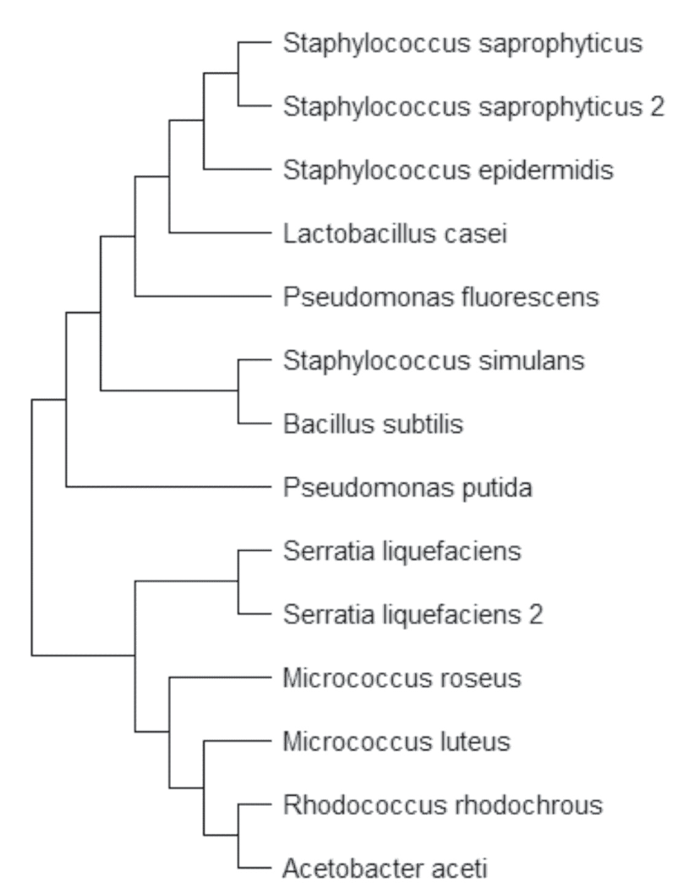
All 14 bacterial isolates had their genomic DNA extracted, and the target sequence of the 16S rDNA was amplified using a DNA thermal cycler (Xp cycler, USA, 2008). The 16S rDNA sequences of all isolates were identified through sequencing (Macrogen, Inc., Korea), and bacterial species were classified (see Table 3). As previously described, the isolated bacterial strains were identified using 16S rDNA sequencing [41]. Table 3 presents the 16S rDNA sequences of the newly isolated species and strains deposited in the NCBI database. Fig. 2 illustrates the phylogenetic tree of oil-degrading bacteria based on their 16S rDNA sequences. PCR-amplified 16S rRNA sequences were used for molecular identification, a sensitive and specific method for detecting microorganisms [41]. Each of the 14 bacterial isolates was molecularly identified, with all isolates producing a PCR product of approximately 1500 bp using universal primer pairs specific to the 16S rDNA gene (Fig. 2). Refer to Table 4 for details on the molecular identification of each bacterial isolate using universal 16S rDNA primers, confirming their species identity.
Petroleum Hydrocarbon Determination
Total petroleum hydrocarbons (TPH) were determined by GC/FID analyses of n-alkanes (C4-C30), monoaromatic hydrocarbons, and polyaromatic hydrocarbons (PAHs). The five oil-contaminated soils showed TPH contents ranging from 6,825 to 107,590 ppm (Fig. 3). Specifically, the M1A soil sample exhibited the highest levels of TPH (107,590 ppm), along with o-xylene and naphthalene. In contrast, the M2B soil sample had the lowest TPH concentration (6,825 ppm). Gas chromatography was utilized to quantify TPH.
The type and concentrations of total petroleum hydrocarbons (ppm) in the examined soil samples were determined using gas chromatography. As depicted in Fig. 3, soil samples M1A and M1C exhibited the highest levels of total petroleum hydrocarbons (107,590 ppm and 27,741 ppm, respectively). Samples M1B, M2A, and M2B showed lower ppm levels. A recent study indicated that soil moisture content in the subsurface layer of crude oil-contaminated sites was higher than that of surface soils. However, the dry surface layer had higher TPH concentrations than the subsurface layer in crude oil-contaminated sites [42].
Table 2.
a) Morphological Features, Biochemical and Physiological characterization of oil degrading bacteria.
Table 2.
b) Morphological Features, Biochemical and Physiological characterization of oil degrading bacteria.
Heavy Metals Determination
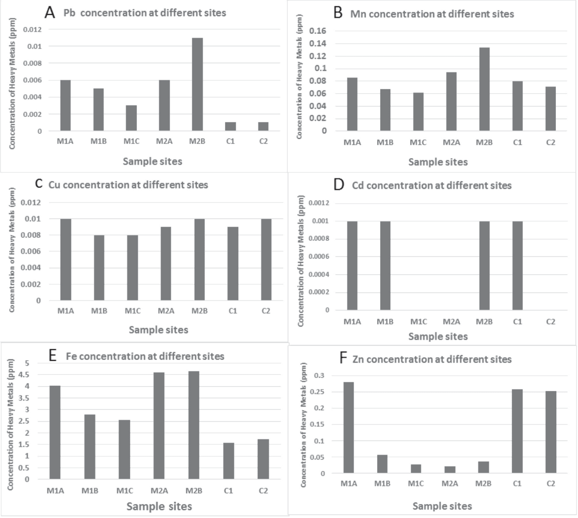
Iron and manganese, traditionally considered significant trace elements with low toxicity, positively affected bacterial isolates. Conversely, metals such as zinc and copper demonstrated toxicity at higher concentrations. Lead and cadmium, also considered significant trace elements, exhibited high toxicity and reduced biological activity. The heavy metal concentrations (ppm) of Cu, Cd, Zn, Pb, and Mn in contaminated soil samples were reported in mg/kg dry soil weight. Fig. 4 illustrates the types and amounts (ppm) of heavy metals in five soil samples collected from two locations within contaminated sites. The results in Fig. 4 indicate that soil sample M2B contained the highest levels of Mn, Cu, Fe, Cd, and Pb. In contrast, soil sample M1C showed the lowest levels of all heavy metals (Cu, Cd, Pb, Fe, Zn, and Mn), while M1A exhibited the highest levels of Zn, Cu, and Fe. M2B had the lowest levels of Zn among the samples. A high concentration of Cu was detected in the M1A sample, leading to high toxicity and reduced hydrocarbon degradation ability in all bacterial isolates. Consequently, the total petroleum hydrocarbon (TPH) concentration remains high with long-term contamination.
Table 3.
Bacterial species identification based on 16s rDNA sequencing data.
Fig. 3.
I-Total petroleum Hydrocarbons in 5 soil samples and the control sample. II- M: soil contaminated by oil, C: soil uncontaminated by crude oil, A: 0-5 cm of depth, B: 5-10 cm of depth, C: 10-15cm of depth and D: 15-20 cm of depth. IIIwww.Restek.com (Retention time of hydrocarbons in the same condition from this website).
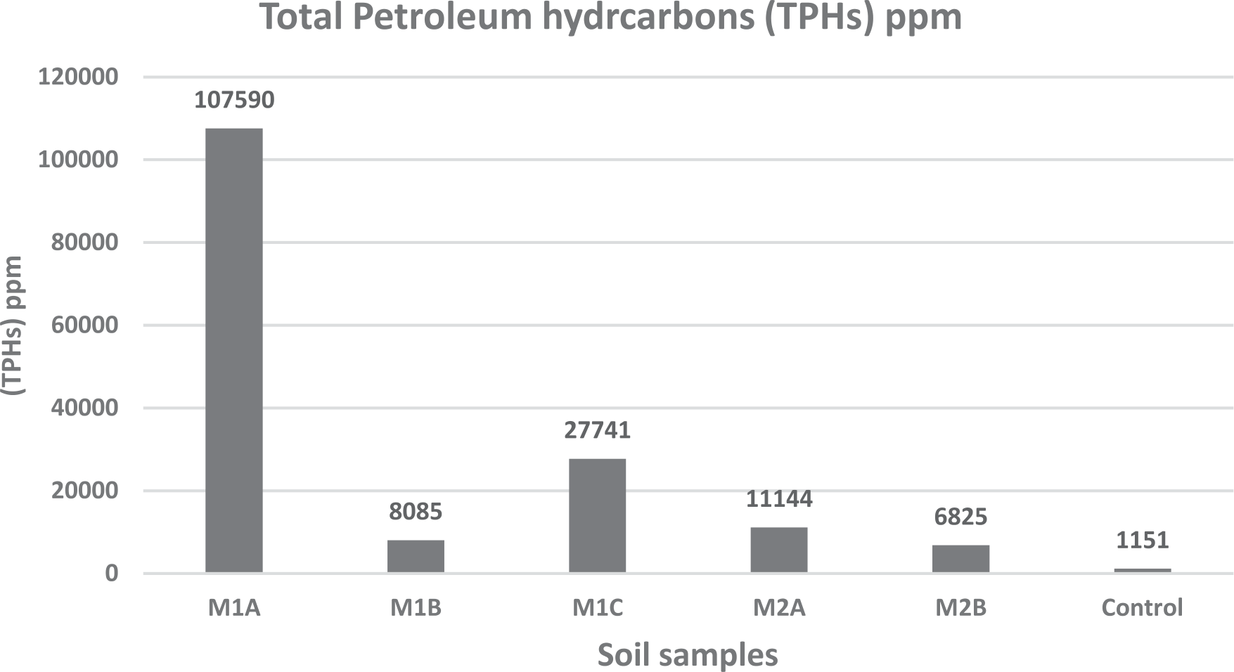
Table 4.
List of the oil degrading bacterial isolates extracted from different soil samples.
Fig. 4.
I- Showed the concentration of many heavy metals (Zn, Cu, Pb, Mn, Cd, Fe) for different sites (M1A, M1B, M1C, M2A, M2B, C1, C2). II- M: soil contaminated by oil, C: soil uncontaminated by crude oil, A: 0-5 cm of depth, B: 5-10 cm of depth, C: 10-15 cm of depth and D: 15-20 cm of depth. III-Reference soils, that were taken from soil uncontaminated by crude oil.
Heavy metal contamination has adversely affected the viability of crude oil biodegradation in oil-contaminated soil. Certain heavy metals can be detrimental to microorganisms, inhibiting their growth. Copper toxicity, for instance, primarily affects microorganisms through interactions with nucleic acids, altering enzyme active sites, oxidizing membrane constituents, and potentially generating harmful hydroxyl free radicals [43].
Flame atomic absorption spectroscopy was employed to analyze heavy metals, which influenced bacterial growth and subsequently affected crude oil degradation. The highest Cu, Fe, Mn, and Pb concentrations were observed in M2 soil samples. Consequently, these samples exhibited the lowest isolate counts, likely due to growth inhibition. Cadmium, lead, cobalt, copper, and zinc were among the metals detected most frequently. These metals exhibited increased toxicity in the following order: lead<zinc<copper<cadmium [44].
Conclusions
This study aimed to identify oil-degrading bacteria from contaminated soil near the Jordanian–Iraqi border. All isolates have been designated in the NCBI database and were identified through molecular detection and biochemical analysis of bacterial species. The optimum temperature for the isolates’ oil degradation activity was found to be 37ºC. Pseudomonas fluorescens exhibited the highest growth across all selective hydrocarbons used in this study. High copper concentrations were found at the accident site, resulting in high toxicity and reduced hydrocarbon degradation ability among all bacterial isolates. As a result, total petroleum hydrocarbon (TPH) concentrations remain high despite the presence of aged crude oil spills at the site. The species identified in this study could be utilized for both in situ and ex-situ cleanup of contaminated sites, such as oil-contaminated desert soil. Isolated bacteria have demonstrated the ability to degrade oil, making them suitable for remediation efforts to clean up oil-contaminated soil.

