

Introduction
The presence of active pharmaceutical compounds (PACs) in wastewater poses significant risks to both environmental and human health. With global pharmaceutical consumption exceeding 1,000 tons daily across veterinary and human medicine, the scale of pharmaceutical discharge is rapidly escalating. By 2020, global consumption was projected to reach approximately 4.5 trillion pharmaceutical doses per day [1]. Among these, ciprofloxacin (CIP), a widely used second-generation fluoroquinolone antibiotic, is commonly prescribed for urinary and respiratory tract infections [2]. Following administration, a substantial proportion of CIP is excreted unmetabolized, approximately 20– 35% through feces and 40–50% via urine, resulting in its persistence in aquatic environments.
CIP contamination is particularly concerning due to its role in promoting antibiotic resistance. In aquatic ecosystems, residual CIP contributes to the proliferation of antibiotic-resistant genes (ARGs) and antibiotic-resistant bacteria (ARB), posing a major public health threat [3]. The global impact of ARGs is profound, with estimates attributing the deaths of 25,000 newborns in the EU, 23,000 in the US, and 58,000 in India to ARG-related infections each year [4]. Projections indicate that, without intervention, ARB-related mortality could reach 10 million annually by 2050 [5]. Given these alarming trends, the effective removal of CIP from wastewater is imperative. However, conventional wastewater treatment plants are often inadequate for eliminating this compound [6].
To address this issue, a range of advanced oxidation processes and adsorption techniques have been explored for CIP removal [7, 8]. Although these methods demonstrate high efficacy, their operational complexity and high costs limit scalability and widespread application [9, 10]. Consequently, there is growing interest in developing cost-effective, environmentally sustainable alternatives [11, 12]. Among these, biologically based treatment systems have emerged as promising candidates, offering lower operational costs and reduced environmental impact compared to conventional physical or chemical methods [13].
Microalgae-based treatment systems have demonstrated potential for the sustainable removal of PACs from wastewater [14]. Several studies have reported the capacity of microalgal consortia to effectively degrade CIP under controlled conditions. For instance, Zambrano et al. achieved a 78% CIP removal efficiency using microalgal consortia cultivated in synthetic wastewater [15], while Ricky et al. reported removal efficiencies ranging from 59% to 84% in hospital effluents. Notably, a maximum removal efficiency of 65% was achieved in systems utilizing synthetic wastewater [16]. These findings underscore the promise of microalgae as a sustainable, nature-based solution for pharmaceutical wastewater remediation.
This study explores the potential of indigenous, acclimated industrial microalgal consortia for the simultaneous bioremediation of ciprofloxacin (CIP) and nitrate from pharmaceutical wastewater. By utilizing locally sourced microalgal strains within naturally adapted consortia, the approach aims to provide a cost-effective and sustainable alternative for tertiary treatment in pharmaceutical effluent management.
To evaluate the performance of the microalgal consortia, three distinct cultivation systems were employed: (i) distilled water (DW), serving as a control with minimal nutrient background; (ii) BG-11 medium, a standardized nutrient-rich medium commonly used for algal growth; and (iii) untreated pharmaceutical wastewater (WW), representing a complex, real-world effluent matrix. The comparative analysis of these systems allowed for the assessment of both bioremediation efficiency and adaptive capacity under varying environmental and nutrient conditions.
Furthermore, the role of nitrate as a competing substrate in the BG-11 medium was investigated to determine its potential influence on CIP removal dynamics. By assessing nitrate-CIP interactions, the study aims to elucidate the nutrient-priority mechanisms that may affect the uptake, transformation, or degradation of pharmaceutical contaminants by microalgae.
Material and Methods
Microalgae Consortium
Microalgal consortia were isolated from pharmaceutical wastewater and cultured in 250 mL conical flasks containing non-sterilized BG-11 medium. The cultures were maintained under continuous illumination at a light intensity of 5,400 lux, with a room temperature of 25°C and an aeration rate of 1.25 L/min to ensure adequate gas exchange.
Pharmaceutical Wastewater Sampling and Characterization
Pharmaceutical wastewater samples were collected from an active pharmaceutical manufacturing facility located in Karachi, Pakistan. Upon collection, the samples were immediately pretreated to remove coarse particulate matter through sedimentation and filtration processes. The pretreated wastewater was then stored at 4 °C to prevent microbial degradation and maintain chemical stability prior to use in experimental procedures [17, 18].
Compositional analysis of the pretreated wastewater revealed the presence of elevated levels of inorganic nutrients and organic pollutants [19-21]. Specifically, the wastewater contained nitrate at a concentration of 18.83 mg/L, phosphate at 61.6 mg/L, and exhibited a notably high chemical oxygen demand (COD) of 26,550 mg/L, indicative of a substantial organic load (Table 1). The initial pH of the wastewater was measured at 7.8, falling within the slightly alkaline range.
For experimental consistency and to simulate realistic operational conditions, the wastewater was further diluted to achieve target concentrations of COD at 2,000 mg/L, nitrate at 1 mg/L, and phosphate at 3.4 mg/L. These values were selected based on preliminary trials to optimize micro-algal growth and pollutant removal efficiency without inducing inhibitory effects. As a control, distilled water (DW), devoid of any nutrients, was used to assess the baseline performance of the microalgal consortium in a nutrient-free environment.
Experimental Setup and Photobioreactor Operation
Following the attainment of the stationary growth phase in preliminary cultures, varying volumes of the industrial native microalgal consortia were harvested from conical flasks. Fig. 1 shows the experiment setup with operation. Biomass separation was achieved via centrifugation at 3,000 rpm for 5 minutes. The resulting microalgal pellets were subsequently washed three times with sterile distilled water to eliminate residual media components and potential contaminants.
Subsequent batch experiments were conducted in 0.5 L culture photobioreactors (PBRs) under controlled laboratory conditions. Each PBR was maintained at ambient temperatures ranging from 25 °C to 30 °C, supplied with a continuous aeration rate of 1.25 L/min, and exposed to constant illumination at an intensity of 5,400 lux to support photosynthetic activity.
Three different media types were used to evaluate the performance of the microalgal consortia: distilled water (DW), BG-11 medium, and pretreated pharmaceutical wastewater (WW). Each PBR was filled with a working volume of 300 mL and inoculated with microalgal biomass to achieve an initial cell density of 0.5 g/L. To assess the influence of non-biological factors on ciprofloxacin (CIP) removal, a control reactor without microalgal inoculation was also established under identical conditions.
All experimental treatments were performed in duplicate to ensure reproducibility and statistical reliability of the results [22-24].
CIP Spiking and Removal Experiments
CIP with a purity of 99% was procured from Sigma-Aldrich (USA) for use in this study. To evaluate the CIP removal efficiency of the microalgal consortia, batch experiments were conducted over 8 days using PBRs with a working volume of 300 mL. Each reactor was inoculated with microalgal biomass at an initial concentration of 0.5 g/L and operated under three distinct media conditions: DW, BG-11 medium, and untreated PWW.
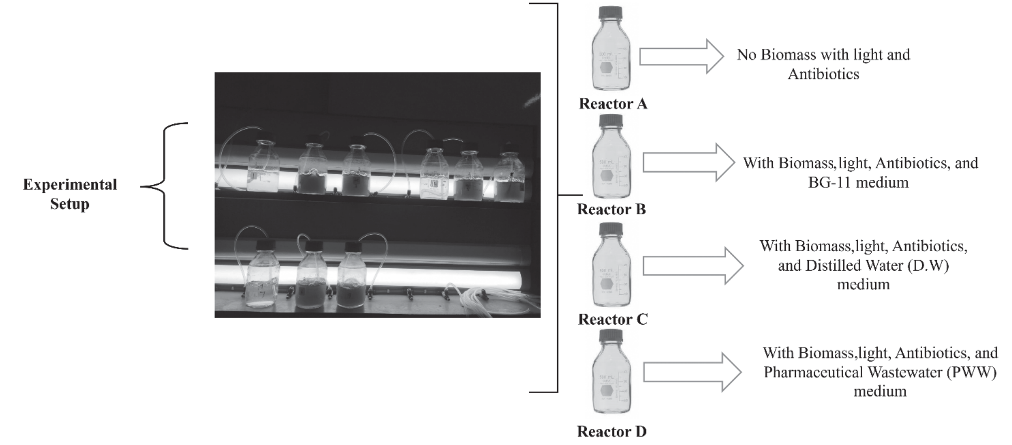
Table 2.
Optimal conditions CIP removal under different optimal conditions by using different types of microalgae.
A control reactor, spiked with CIP but devoid of microalgae, was also maintained for 8 days to account for abiotic degradation and sorption effects. Based on recent environmental monitoring data from Pakistan, indicating CIP concentrations in wastewater ranging from 0.4 to 1.81 µg/L [25]. A higher concentration of 3 mg/L was selected for this study to ensure measurable removal dynamics and simulate a high-contamination scenario relevant to industrial effluent.
All experimental runs were performed simultaneously under standardized and optimized cultivation conditions, as detailed in the Table 2. To support biomass suspension and gas exchange, each PBR received a continuous aeration rate via an air pump at a flow rate of 1.5 L/min throughout the experiment.
Analytical Methods
Analysis of CIP
During the 8-day cultivation period, 1 mL of microalgal culture was collected daily from each photobioreactor to monitor the temporal removal of ciprofloxacin (CIP). The collected samples were subjected to centrifugation at 15,000 rpm for 15 minutes to separate the microalgal biomass. The resulting supernatant was then passed through a 0.45 µm membrane filter to eliminate any residual particulate matter prior to chemical analysis.
CIP concentrations in the filtrate were quantified using high-performance liquid chromatography (HPLC) equipped with a Shim-pack VP-ODS LC-20A system. Separation was achieved using a C18 reverse-phase column (5 µm, 150 mm length × 4.6 mm internal diameter) maintained at a temperature of 35 °C. A 10 µL aliquot of each filtered sample was injected into the HPLC system [26].
The mobile phase consisted of a mixture of acetonitrile and HPLC water in a volumetric ratio of 20:80 (v/v), adjusted to pH 3.0 using 5% (v/v) phosphoric acid to enhance separation efficiency. The mobile phase was delivered at a constant flow rate of 1.0 mL/min. Detection of CIP was performed by measuring the UV absorbance of the eluent at 274 nm [27]. All reagents and solvents used were of analytical grade to ensure accuracy and reproducibility [28].
Determination of Nitrate
Nitrate concentrations in the aqueous phase were measured using the colorimetric brucine method, as recommended by the United States Environmental Protection Agency (EPA) [29]. This technique is widely used due to its sensitivity and specificity for nitrate ions (NO₃⁻) in water samples. In this method, nitrate reacts with brucine in an acidic medium to produce a yellow-colored complex, the intensity of which is directly proportional to the nitrate concentration and is measured spectrophotometrically at an appropriate wavelength.
To evaluate the effectiveness of the microalgal consortia in removing nitrate from the different media, the nitrate removal efficiency was calculated using the equation given below :
Where:
NR = Nutrient removal efficiency (%)
Cin = Influent nutrient concentration (mg/L)
Cout = Effluent nutrient concentration (mg/L)
Determination of Dry Cell Weight and Chlorophyll Cell
To evaluate the growth dynamics of the microalgal consortia, the dry cell weight (DCW) and chlorophyll content, including chlorophyll-a (Chl-a) and chlorophyll-b (Chl-b), were quantified throughout the experimental period.
Dry Cell Weight (DCW)
For biomass quantification, a 10 mL aliquot of culture was collected from each photobioreactor. The samples were filtered through pre-weighed Whatman membrane filters (pore size 0.45 µm) that had been pre-dried at 60 °C to ensure moisture removal [30-32]. After filtration, the retained biomass on each filter was dried in a laboratory oven at 60 °C for 24 hours to achieve a constant weight [33]. The dry cell weight was calculated using the equation given below [34].
Where:
Cb = Biomass concentration (g/L)
Wt = Final weight of filter + biomass (g)
Wf = Weight of clean filter (g)
V = Volume of sample filtered (mL)
Chlorophyll-a and Chlorophyll-b Determination
To extract chlorophyll pigments, 1 mL of algal culture was centrifuged to pellet the cells. The pellet was then incubated with methanol at 60 °C for 5 minutes to extract Chl-a and Chl-b, followed by a second centrifugation for 5 minutes to remove cellular debris. The supernatant containing the chlorophyll pigments was analyzed spectrophotometrically.
The optical densities (OD) were measured at 652 nm and 665 nm using a UV–Visible spectrophotometer. The concentrations of Chl-a and Chl-b were then calculated using equations (3) and (4).
Where:
A652 and A665 represent the absorbance values at 652nm and 665 nm, respectively.
Results and Discussion
pH Variation During Cultivation
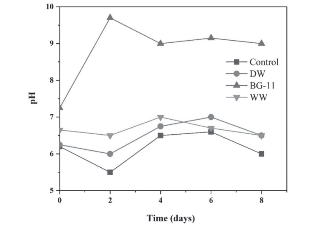
The temporal variation in pH across different reactors is presented in Fig. 2. Notably, the pH in the reactor supplemented with BG-11 medium exhibited a sharp increase from an initial value of 7.25 to a peak of 9.7 within the first two days of cultivation. This alkaline shift is attributed to vigorous microalgal growth, as supported by biomass accumulation shown in Fig. 3a, and is primarily driven by photosynthetic activity and nutrient assimilation.
In contrast, reactors containing distilled water (DW), pharmaceutical wastewater (WW), and the control (CIP-only, no microalgae) displayed a gradual decline in pH. Specifically, the pH decreased from initial values of 6.2, 6.25, and 6.65 to final values of 5.5, 6.0, and 6.5, respectively. The observed acidification in these reactors can be linked to limited photosynthetic activity and the accumulation of acidic metabolic byproducts in the absence or underperformance of active nutrient uptake.
The pronounced increase in pH within the BG- 11 reactor during early cultivation can be attributed to microalgae-mediated photosynthesis, during which bicarbonate ions are consumed and hydroxide ions (OH⁻) are released because of oxygen evolution. This process drives the carbonate equilibrium toward increased alkalinity, thereby making it more evident. [35, 36]. After the initial spike, the pH in the BG-11 reactor stabilized and remained relatively constant for the remainder of the 8-day experimental period, indicating a steady-state growth phase and balanced metabolic activity.
In contrast, no significant changes in pH were observed in the other reactors after the second day, suggesting either low microalgal activity or the establishment of a non-optimal environment for photosynthetic pH regulation.
Change in DCW and Chlorophyll Content
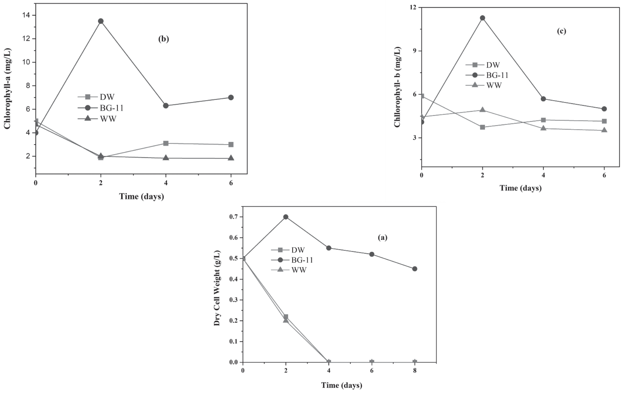
Fig. 3a shows the variations in the DCW of microalgae under various reactor conditions. Throughout the trial, there was a significant fluctuation in microalgal growth. Due to ideal nutrient availability and conducive photosynthetic conditions, DCW increased by 40% in the BG-11 reactor. However, DCW in the DW and WW reactors had dropped by 50% by the second day, most likely due to nutrient limitations.
Fig. 3b and 3c show that the chlorophyll contents increased in the BG-11 medium [37], reaching 13.5 mg/L for Chl-a and 11.282 mg/L for Chl-b. However, on the second day of cultivation, the concentrations of Chl-a and Chl-b decreased in the DW and WW reactors. These patterns align with the observed shifts in DCW, as shown in Fig. 3a.
Removal of CIP
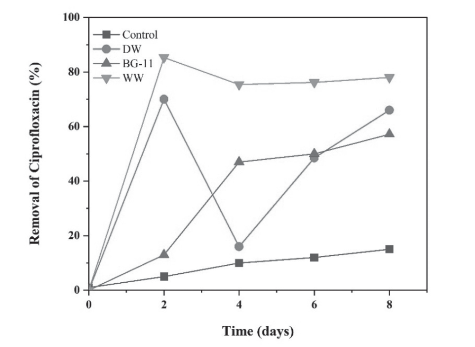
The CIP removal efficiency observed under different reactor conditions is presented in Fig. 4. During the initial two days of treatment, the reactor containing pharmaceutical wastewater (WW) exhibited the highest initial CIP removal efficiency, achieving 85.3%. Simultaneously, the microalgal consortium cultivated in DW also demonstrated considerable removal capacity, attaining 70% CIP elimination within the same period. In contrast, the reactor supplemented with BG-11 medium displayed markedly lower initial removal efficiency, with only 13% of CIP removed after two days.
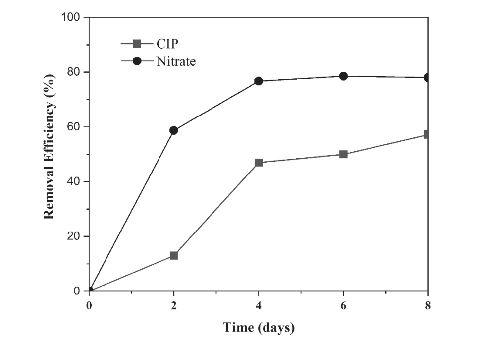
The reduced CIP removal in the BG-11 reactor may be attributed to two potential factors: (i) the presence of nitrate occupying binding sites within the microalgal biomass, thereby limiting available sites for CIP adsorption, or (ii) the preferential uptake of nitrate as a primary nutrient for microalgal growth (Fig. 5), which could delay the absorption and biotransformation of CIP.
Following the initial two days, a decline in CIP removal efficiency was observed in the WW reactor, with the final removal rate stabilizing at 78% by the end of the 8-day cultivation period. Meanwhile, the DW reactor exhibited a transient decrease in CIP removal efficiency to 15%, followed by a recovery phase that culminated in 66% removal by Day 8. Notably, the BG-11 reactor showed a progressive increase in CIP removal after Day 2, ultimately achieving 57% removal by the end of the experiment. This improvement may be associated with a decline in nitrate concentrations over time, thereby reducing competition for adsorption and metabolic processing sites (Fig. 4).
The control reactor, which represented abiotic conditions (no microalgae), exhibited only 15% CIP removal throughout the experimental duration, emphasizing the essential role of biotic mechanisms, particularly microalgal activity, in enhancing CIP elimination across all treatment systems.
These findings are consistent with previous studies, 78% CIP removal efficiency using a microalgal consortium cultivated in synthetic wastewater [38]. After eight days of incubation in untreated pharmaceutical wastewater, our native microalgal consortium demonstrated strong bioremediation capability, achieving a CIP removal efficiency of 85.3%. These results highlight the adaptive potential of indigenous microalgal consortia to effectively degrade or adsorb pharmaceutical contaminants under real wastewater conditions without the need for synthetic enrichment.
Influence of Nitrate on CIP Removal
The impact of nitrate presence on ciprofloxacin (CIP) removal is illustrated in Fig. 5. In the BG-11 medium reactor, nitrate uptake increased sharply to 58.7% within the first two days of cultivation, while CIP removal remained relatively low at 13%. This inverse relationship suggests a potential competitive interaction, wherein nitrate availability may suppress CIP removal by occupying active uptake or adsorption sites on the microalgal biomass, or by being preferentially utilized as a primary nutrient source.
Between Days 2 and 4, a shift was observed. CIP removal increased significantly to 34%, coinciding with a marked reduction in nitrate uptake, which slowed to 18%. This shift implies a possible change in nutrient prioritization or a delayed onset of CIP metabolism once nitrate levels declined. However, following Day 4, a concurrent decrease in dry cell weight (DCW), as shown in Fig. 3a, suggests potential toxicity effects associated with CIP accumulation or stress-related inhibition of cellular growth and function.
Similar inhibitory outcomes have been observed, with only 10% CIP removal over a 10-day retention period in a high-rate algal pond (HRAP) using a mixed culture derived from a wastewater stabilization pond, treating residential wastewater spiked with 2 mg/L CIP. Their findings, like the present study, indicate that CIP toxicity and nutrient competition can significantly influence the efficiency of microalgal-based bioremediation systems [39]. Limited light penetration and high nitrate concentrations, which can function as scavenge reactive species and hinder CIP removal, are the reasons for low CIP removal rates in HRAP systems [40].
Mechanism of Ciprofloxacin and Nitrate Removal by Indigenous Industrial Microalgal Consortia
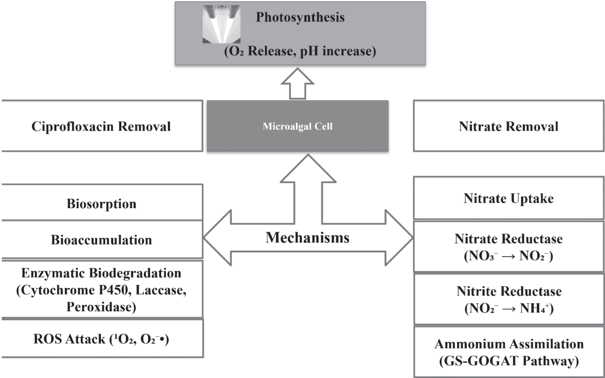
The removal of ciprofloxacin (CIP) and nitrate by indigenous industrial microalgal consortia occurs through a combination of biological, physicochemical, and enzymatic processes that reflect the adaptability of microalgae to complex environments such as pharmaceutical wastewater. These micro-algal consortia, isolated from high-strength pharmaceutical effluents, have shown substantial capacity to mitigate both organic micropollutants and inorganic nutrients, making them highly effective for bioremediation. The mechanism is further discussed below, and Fig. 6 illustrates the mechanism of CIP and nitrate removal.
Ciprofloxacin Removal Mechanism
The degradation of ciprofloxacin by microalgae involves a multi-step process, beginning with biosorption, where CIP molecules adhere to the surface of the microalgal cells. This is facilitated by the presence of functional groups on the algal cell walls, such as hydroxyl, carboxyl, and amine groups, which can interact with CIP through electrostatic attraction, hydrogen bonding, and van der Waals forces. This initial phase is particularly important in the early stages of treatment, where rapid binding of CIP to the cell surface occurs before any intracellular activity begins.
Following biosorption, CIP is subject to bioaccumulation, where the antibiotic is transported into the algal cells. This internalization likely occurs through passive diffusion or active transport mechanisms and leads to the concentration of the compound within intracellular compartments. Once inside the cell, biodegradation processes are initiated. Enzymes such as cytochrome P450 monooxygenases, peroxidases, and laccases may facilitate the oxidative transformation of CIP into intermediate products. These processes involve structural modifications, such as hydroxylation, dealkylation, or aromatic ring cleavage, reducing the toxicity and persistence of the parent compound.
A particularly important factor in CIP degradation is the generation of reactive oxygen species (ROS) through photosynthesis under light exposure. Microalgae, during photosynthetic activity, produce singlet oxygen and superoxide radicals, which can directly attack and break down complex molecules like CIP. This photosensitized degradation enhances the overall breakdown of CIP, especially in illuminated culture systems such as photobioreactors. The observed high removal efficiencies, up to 85.3% in reactors containing real pharmaceutical wastewater, underscore the synergy between these removal pathways.
Nitrate Removal Mechanism
Nitrate removal in microalgal systems is primarily driven by assimilatory uptake, a metabolic process wherein nitrate ions (NO₃⁻) are actively absorbed by the cells through specific nitrate transporters embedded in the plasma membrane. Once internalized, nitrate is enzymatically reduced to nitrite (NO₂⁻) by nitrate reductase and subsequently to ammonium (NH₄⁺) by nitrite reductase. The resulting ammonium is then assimilated into amino acids via the glutamine synthetase-glutamate synthase (GS-GOGAT) pathway, supporting protein synthesis and cellular growth.
In reactors with nutrient-rich BG-11 medium, a significant increase in dry cell weight (DCW) and chlorophyll content was observed, indicating robust growth due to efficient nitrate assimilation. However, the high concentration of nitrate in BG-11 initially appeared to inhibit CIP uptake, likely due to a competitive interaction at the cellular level. This competition may be the result of nitrate occupying key transport pathways or metabolic energy being preferentially directed toward nitrate assimilation over the energy-intensive degradation of CIP.
Moreover, photosynthetic activity by microalgae contributes to a rise in pH, as bicarbonate ions are consumed, and hydroxide ions are released during carbon fixation. The elevation in pH can indirectly support nitrate uptake and may also alter the speciation of other compounds in solutions, affecting their bioavailability. Interestingly, as nitrate levels declined over time in the BG-11 reactors, CIP removal increased, suggesting that reduced nitrate stress allowed the consortium to redirect metabolic resources toward organic pollutant degradation.
Synergistic Behavior and Environmental Implications
The combination of biosorption, intracellular biodegradation, ROS-mediated breakdown, and nutrient-driven growth creates a robust and flexible system for tackling both pharmaceutical residues and nutrient pollution. In particular, the high CIP removal efficiency in real pharmaceutical wastewater indicates that microalgal consortia can function under harsh and complex wastewater conditions, including high COD and nitrate content.
This study demonstrated that microalgal consortia are not only capable of adapting to different cultivation media but can also dynamically shift their metabolic priorities based on nutrient availability. The interaction between nitrate and CIP uptake observed in the BG-11 system exemplifies the need to understand and balance nutrient conditions for optimized bioremediation performance. Importantly, the minimal removal of CIP in the abiotic control reactors (only 15%) reinforces the conclusion that the observed degradation was predominantly biological, dependent on the metabolic activity of the microalgal consortium.
Conclusions
This study demonstrated the effectiveness of indigenous industrial microalgal consortia for the simultaneous removal of ciprofloxacin (CIP) and nitrate from pharmaceutical wastewater. The native consortia achieved up to 85.3% CIP removal in untreated effluent, confirming their adaptability to complex wastewater conditions. CIP removal was driven by a combination of biosorption, intracellular accumulation, enzymatic biodegradation, and reactive oxygen species (ROS)-mediated oxidation. Nitrate was primarily removed through assimilatory uptake and enzymatic reduction, supporting microalgal growth and metabolic activity. Notably, high nitrate levels initially limited CIP removal in BG-11 medium, indicating competition between nutrient assimilation and pollutant degradation. As nitrate levels decreased, CIP removal efficiency increased, suggesting a dynamic metabolic shift. The low CIP removal observed in abiotic controls confirmed the central role of biological processes. These findings highlight the potential of native microalgal consortia as a low-cost, sustainable approach for tertiary treatment of pharmaceutical wastewater. Their ability to respond to varying nutrient conditions and degrade both organic and inorganic contaminants underscores their suitability for integration into advanced wastewater treatment systems.