


Introduction
Plants are an indispensable part of landscaping. they usually withstand the stress factors alone [1]. These stress factors affect all characteristics of the plants [2-4]. Actually, Climate has direct impact to change all properties of plants [1]. Some plants have taken strategies to adapt to such difficult environmental conditions (high temperature, UV radiation, pollution) They perform many functions locally where they exist [5]. For the halophytes plants, they usually exhibit high concentrations of reactive oxygen species (ROS), which cause cell damage and sometimes plant death. Throughout history, the halophytes plants have shown specific strategies to acclimate, morphologically and physiologically, to harsh conditions. they have developed defense mechanisms, against oxidative stress by producing bioactive metabolites [6]. Therefore, they are equipped with a powerful antioxidant system including enzymes (superoxide dismutase, catalase, etc.) and non-enzymatic components (secondary metabolites) [7]. These secondary metabolites are distributed in different plant parts and have considerable roles, including contributing to eliminating the accumulation of reactive oxygen species (ROS) at toxic level during abiotic stresses [8]. Previous studies have examined the value of halophytes as sources of Phenolic extracts that have large biological properties such as antimicrobials, anticancer, anti-inflammatory and antioxidant effects [9]. In Tunisia, several halophytes have are the center of attention of scientists interested in plants’ phytochemistry, particularly in identifying individual compounds for modern drug development [10]. In this context, we focus research on Reaumuria that is a genus of flowering plants in the family Tamaricaceae, including 12-21 species. The generic distribution center is in North Africa, Anatolia, Sicily, the Middle East, the Caucasus, Pakistan, northern China, Mongolia, Tibet, and Central Asia. They tend to be perennial xerophytes and halophytes shrubs or subshrubs. Reamuria vermiculata is one of the most important species belonging to this genus. It grows in many gypsum and salt areas. It can also be observed in the embankment, rockery, and even on the coast and is native to Asia, and Europe, particularly in Italy; south of Sicily. It is a ubiquitous plant in North-West Africa (Morocco, Tunisia, Algeria and Libya). Reaumuria vermiculata L. (Tamaricaceae) is a xero-halophytic shrub distributed in different bioclimatic stages with rainfall varying from 50 to 400 mm [11]. It is widely known in tropical and subtropical regions [11]. Indeed, temperatures between 10ºC and 30ºC seem favorable to the germination of this species. Reaumuria vermiculata is utilized in folk medicine to prevent many diseases due to the multiple positive effects on human health induced by many chemical compounds. Recent research has demonstrated that this species has anticancer activity against liver (Huh-7), colorectal (HCT-116), breast (MCF-7) and prostate (PC-3) tumor cell lines [12]. Furthermore, Manel et al. reported that the R vermiculata plant possesses antioxidant, anti-inflammatory and cytotoxic activities against A-549 lung carcinoma cells [13]. To the best of our knowledge, few studies have assessed the phytochemical profile of the aerial part of Reamuria vermiculata. Nevertheless, there are no reports on the nutritional value, antioxidant and biological characterization of roots, stem and leaves of Reamuria vermiculata. Therefore, in this work, a study of the mineral composition, a phytochemical investigation as well as an evaluation of biological activities (antioxidant and antibacterial) was carried out on the different organs (leaves stem and roots) of Reamuria vermiculata collected from the regions of southern Tunisia (Medenine).
Material and Methods
Plant material
Reaumuria vermiculata plants were collected from the region of Medenine (south of Tunisia, 33º21′17″ North 10º30′19″ East), in March 2020. The plant was taxonomically identified by the botanists of Dryland Farming and Oasis Cropping Laboratory (Institute of Arid Lands, Route El-Jorf, 4119 Medenine, Tunisia). This plant are identified and collected from local farmers by Prof. Ali Ferchichi, A voucher specimen was deposited at the herbarium ex-situ in the experimental field of Dry Land Farming and Oasis Cropping Laboratory (Arid Lands Institute of Medenine, Tunisia) under the number of LACCORV-100. After harvest, different parts of the plant were detached as leaves, stems and roots and kept at -20ºC until phytochemical analyses. For mineral analysis, an oven drying (80ºC, 72 h) of 100 g from each part was carried out. The dried samples were then ground to powder.
Sample Preparation
Phenolic compounds were extracted at room temperature using two different solvents: water and ethanol. Ten grams of each dried organ were grounded and mixed with 100 mL of each solvent. The mixture was continuously agitated in the dark for 6 h and filtered by a Whatmann paper (3 mm). The extract obtained was centrifuged at 25ºC for 20 min. Dried extracts were stored at 4ºC in the dark until further analysis.
Mineral Contents
One g of powdered plant material was ignited and incinerated in the muffle furnace at 530ºC for 5 h. Then, the resulting ash was then dissolved using 5 mL of hydrochloric acid (20%). The dissolved solution was adjusted with distilled water into a volumetric flask of 50 mL Each mineral element was analyzed individually by an atomic absorption spectrometer (Shimadzu A 6800, Kyoto, Japan) [14].
Preliminary Phytochemical Screening
Through qualitative reactions, the phytochemical tests detect different families of existing compounds in Reaumuria vermiculata. The detection of these chemical compounds was based on the visual observation of color change or formation of a precipitate after additing of specific reagents.
Estimation of saponins
According to [15], saponins content in Reaumuria vermiculata was estimated by dissolving 5 mg of extract in 10 mL of hot distilled water (50ºC). The formation of stable foam demonstrates the existence of saponins.
Estimation of Flavonoids
The flavonoid detection reaction consists of placing 5 mL of the extract in a test tube, to which 1 mL of concentrated hydrochloric acid and some fragments of magnesium are added. After three minutes, a pink or red color indicates the presence of flavonoid [16].
Estimation of Glycosides
Add, 1 mL of acetic acid, 1 mL of concentrated sulfuric acid and then 3 drops of 2% FeCl3 to 1 mL of the extract solution in a test tube. A blue-green color or a brown ring indicates the presence of cardiac glycosides [17].
Estimation of Steroids and Triterpenes
In this technique, 5 mL of the extract is dissolved in chloroform, with 1 mL of acetic anhydride and then without shaking adding 0.5 mL of concentrated H2SO4 is added to the bottom of the tube. The formation of a brownish-red ring at the contact area of the two liquids reveals the presence of triterpenes, while the dark green turn of the supernatant layer (aqueous phase) shows the presence of steroids in the extract [18].
Estimation of Alkaloids
Two methods are determined to illustrate the appearance of the alkaloids:
Dra-gendorff’s test: 2 mL 1% HCl and 2 mL MeOH were homogenized with 5 mg of the extracts along the side of the test tube, and then 500 μl Dragen-dorff’s reagent was added to the mixture. The formation of an orange or orange-reddish-brown precipitate confirms the test as positive [18] .
Mayer’s test: included a drop or two of Mayer’s reagent with 1 mg/mL of the extract. The formation of a white or creamy precipitate shows the presence of alkaloids[18] .
Estimation of Polyphenols
10 mg of each extract dissolved in 1 mL of distilled water and then a few drops of ferric chloride 2% is added. A bluish-black color indicates the presence of polyphenols [19].
Determination of Phenolic Compounds
Quantification of Total Polyphenols
The quantitative study of the total polyphenols of the different extracts were quantified using the method described by [20]. A total of 100 mL of sample was dissolved with 750 mL of Folin-Ciocalteu reagent and 750 mL of saturated sodium carbonate solution. After 90 min, recorded the absorbance was at 765 nm with a UV-vis spectrometer. A control is prepared in parallel, under the same conditions, by substituting distilled water for the extract; then, the entire sample is incubated for 30 minutes at 25ºC and the absorbance is read at 765 nm. The determination of total polyphenols content was performed using the calibration line (y = ax + b) obtained by the reading of the optical density as a function of the concentrations of the gallic acid. The polyphenol content is expressed in mg equivalent of gallic acid per gram of dry matter (mg EGA/g DM).
Quantification of Flavonoids
The total flavonoid contents of the samples were determined using the aluminum chloride colorimetric method [21] with slight modifications. One mL of each extract dilution or quercetin (used as standard) blended with 3 mL of distilled water. After manual stirring, 0.3 mL of sodium nitrate (5%) is added and the combination is agitated well. After 5 min, 0.2 mL of aluminum chloride was added to the mixture; then, the mixtures were incubated for half an hour in the dark at ambient temperature; 0.5 ml of sodium hydroxide (1M) was then added. After that, the optical density is determined at 510 nm. The absorbance measurements obtained are then compared to a recorded standard calibration curve to determine the flavonoid concentration in the sample. The total flavonoid content (TFC) of each sample was expressed as mg quercetin equivalent (CE)/g dry weight of plant material (mg CE/g DW). All samples were run in triplicate.
Quantification of Tanins
The total tannin content (TTC) was determined using the vanillin method as described by [22]. A volume of 0.2 mL of each extract was mixed with 1 mL of vanillin reagent (8% HCl (v/v), methanol at 37% (v/v) and 4% vanillin in methanol (m/v)). The associations were first incubated at room temperature, in the dark for 20 min, before measuring the absorbance at 500 nm using a UV spectrophotometer (Perkin Elmer). The results were expressed as mg CE/g. All of the steps performed in triplicate for analysis.
Evaluation of Antioxidant Activity
This study, evaluated the antioxidant power of R vermiculata using three bioassays including DPPH, ABTS, and β-carotene bleaching. Then, this mixture was incubated for 30 min, at room temperature and in the dark, and the absorbance reading was taken using a spectrophotometer at 517 nm.
DPPH Free Radical Scavenging Activity
The ability of the extracts to scavenge the DPPH free radical was used according to [23]. Add 100 µL of different concentrations of extracts (from 1 to 0.007 mg/mL) to 750 µL of 0.004% of DPPH. The mixture was then incubated at room temperature in the dark for 30 min, and the absorbance value was measured at 517nm with a spectrophotometer (Jasco V-530) and compared to the control. The percentage of inhibition (I%) was calculated using the following equation:
Where A0 is the absorbance of the control reaction and A1 is the absorbance of the tested extract sample. The IC50 is equivalent to 50% of the inhibition of DPPH.
ABTS Radical Scavenging Activity
The ABTS radical scavenging activity of extracts was determined according to a method described by [24], with some modification 900 μl of the solution of ABTS was added to 100 μl of the extracts dissolved in ethanol. After 20 min of incubation at room temperature and in the dark, the absorbance of the mixture was measured at 734 nm using a spectrophotometer. The antioxidant activity was calculated using the following equation:
WhereA0 and A1 have the same meaning as in Eq. (1)
β-carotene/Linoleic Acid Bleaching Assay
The β-carotene bleaching inhibition of the extracts was determined using the protocol by Santos and their collaborators [25]. In this analysis, antioxidant capacity is determined by measuring the inhibition of the organic compounds and the conjugated diene hydroperoxides arising from linoleic acid oxidation. 50 ml of distilled water was mixed with 2 mL of β-carotene solution, 20 μl of linoleic acid and 200 μl of Tween-20. Then, 5 mL of this resulting solution was added to 500 μl of extracts and incubated in a water bath at 50ºC for 60 min. The absorption of this reaction was detected at 470 nm.
Determination of Antibacterial Activity
In this work, the R vermiculata extracts was evaluated against three Gram-negative bacteria, including Escherichia coli ATCC 35218, Salmonella typhimurium ATCC 14080, Pseudomonas aeruginosa ATCC 27853, and three Gram-positive bacteria, including Staphylococcus aureus ATCC 25923, Listeria monocytogenes ATCC 19115 and Bacillus subtilis ATCC 6633.
Determination of MIC and MBC
The Minimum Inhibitory Concentration (MIC) and Minimum Bactericide Concentration (MBC) Assays were performed by microplate method and followed a protocol previously employed by [26]. Shortly, samples of decreasing concentration were prepared, inoculated with bacterial inoculum, and incubated at 37ºC for 24 h. After incubation, wells are considered to have MICs corresponding to lower concentrations and no visible bacterial growth. The MBC was assumed as the highest dilution (lowest concentration) of the sample, which showed clear fluid with no development of turbidity and without visible growth of microorganisms).
Statistical Analysis
For all plant parameters, all samples were conducted in three replications. Data are shown as mean±sd. A one-way analysis of variance (ANOVA) using the post hoc analyses with Duncan’s test was conducted to test any significant differences at p<0.05. Multivariate analysis (Principal compounds Analysis and Heatmaps) were performed using the Pearson correlations approach.
Results and Discussion
Qualitative Phytochemical Analysis
Preliminary phytochemical testing of the plant of bioactive compounds helps find chemical constituents in plant material that can guide their quantitative estimation and confirm their medicinal value as a source of pharmacologically active chemicals [27]. This study exhibited that pharmacologically active compounds such as tannins, terpenoids, flavonoids, saponins, quinones, and coumarins were shown in leaves, stems and roots with varying intensities on different solvent extracts of R vermiculata (Table 1). Contrariwise, sterols, cardiac glycoside, and alkaloids were absent in all plant parts (Table 1). After this preliminary study, we found that the roots of the plant contained more active compounds than those present in the stems and leaves when water and ethanol were used as the extractants. However, the presence and absence of such classes of secondary metabolites can be various physiological and biosynthetic reactions taking place inside the plant. Several studies noted that flavonoids, terpenoids, saponins, quinones had been reported to possess a wide variety of biological activities among which are antimicrobial, anti-inflammatory, antiangiogenic, analgesic, antiallergic effects, cytostatic and antioxidant, antiviral, anticarcinogenic, anticancer as well as antidiarrheal properties [28]. The phytocompounds in R vermiculata suggest that this plant is a potential source of chemotherapeutic compounds.
Table 1.
. Results of preliminary phytochemical screening of aqueous and ethanol extracts from the leaves, stems and roots of Reamuria vermiculata.
| Terpenes | Saponins | Tannins | Flavonoids | Quinones | Coumarins | Alcaloïde | Cardiac Glycosides | Sterols | |
|---|---|---|---|---|---|---|---|---|---|
| WL | + | + | + | + | + | + | - | - | - |
| WS | + | + | + | + | + | + | - | - | - |
| WR | ++ | ++ | ++ | ++ | ++ | ++ | - | - | - |
| EL | + | + | + | + | + | + | - | - | - |
| ES | + | + | + | + | + | + | - | - | - |
| ER | ++ | ++ | ++ | ++ | ++ | ++ | - | - | - |
Total Polyphenol TPC, Flavonoid TFC and Condensed Tannin Contents CTC Analysis
Total polyphenols, flavonoids and condensed tannins contents of different R.vermiculata extracts (from leaves, stems and roots) were presented in Fig. 1. The range of phytochemicals varied between leaves, stems and roots, and depended on whether water or ethanol was used as the extractant. Results showed a high significant differences between solvents used (p<0.001) for TPC, TFC and CTC. The highest polyphenol contents were obtained in aqueous extracts of roots 1153.29 mg GAE/g dr followed by ethanolic extract 943.31mg GAE/g dr compared to the stems and leaves. Similarly, the roots had the highest total flavonoid content compared to the stems and leaves. However, the aqueous extracts had a higher amount of flavonoid in the roots (308.16 mg CE/g DW) followed by the stem and leaves of the same extract (160.27 mg CE/g DW and 72.79 mg CE/g DW respectively). Considering, condensed tannins, the aqueous extracts have the highest quantity of roots at 31.84 mg CE/g DW, stems at 13.54 mg CE/g DW and leaves at 3.9 mg CE/g DW. In contrast, ethanol extracts present the lowest condensed tannins at 3.34, 6.44 and 9 mg CE/g DW from roots, stems and leaves respectively. These results indicated the influence of the extraction solvent on the total content of phenolic compounds extracted. However, a study has been described concerning the polyphenolic content in R vermiculata aerial part collected from Gabes, Tunisia It measured that the highest quantity of phenol (58.19 mg gallic acid/g), was obtained in methanol extract. Similarly, the best yields of flavonoid were recorded in methanol extract (18.98 mg CE/g). Considering total tannins contents, the best yields in R. Vermiculata shoots were achieved in dichloromethane extract (27.98 mg CE/g). Furthermore, our results agreed with [29] which showed that total phenolic compounds in Mesembryanthemum edule depend on organs. Roots and stems are defined by a high production of these metabolites with values ranging between 989 and 364 mg GAE g−1DW. According to these results, it can be deduced that more phenolic compounds from R vermiculata organs are easily extracted using water; this agrees with [30], which noted in their research that water is a universal solvent for polyphenol extraction, nontoxic for human consumption, cheap and usually easy to exploit and also ethanol is a good solvent for the extraction of food phytochemicals. Recent reports have shown that this difference in active compounds in Reamuria vermiculata can be explicated by the differences in genotype, plant part used, environmental conditions (climatic, seasonal, geographical), and solvent used in the study [31]. The TPC of Reamumia vermiculata significantly correlated with TFC (r = 0.3691) and TTC (r = 0.521). Other significant relationships are shown with TFC and TTC (r = 0.9785) (Table 3).
Fig. 1.
Total polyphenols (A), total flavonoids (B), and Condensed tannins (C) contents in ethanolic and aqueous extracts from different plant organs. ***: p<0.001 ; Highly Significant differences revealed by two-way ANOVA; different lowercase (a-c) and Uppercase (A-C) letters denote significant differences between the different plant organs in the ethanolic and aqueous extracts respectively, according to Duncan’s multirange comparison of means test (p<0.05).
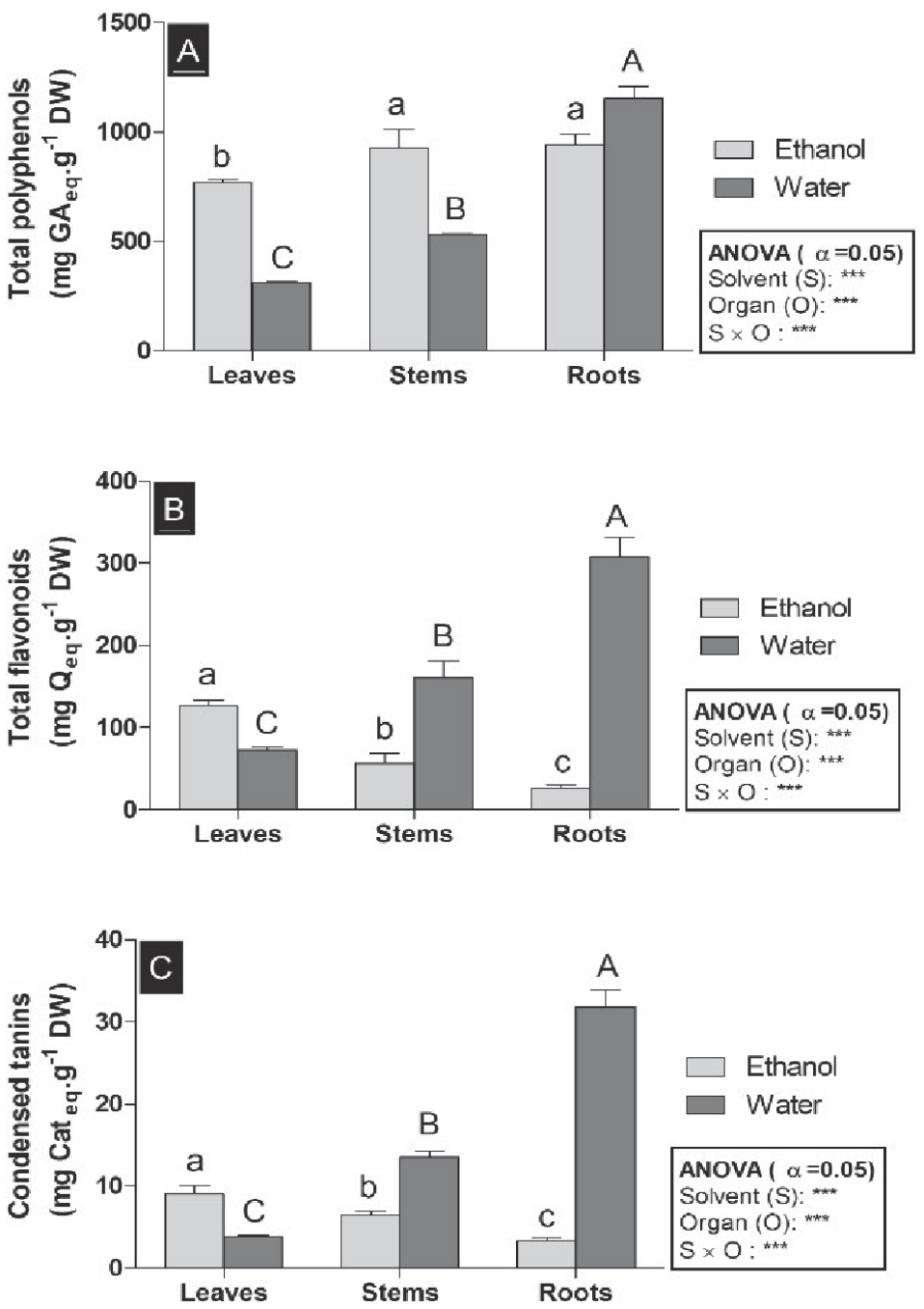
Table 2.
Mineral contents (mg.g-1 DW) extracted from different plant organs.
Determination of Mineral Content
Minerals are vital elements necessary for a plant to complete its life cycle. Seven elements were determined in the R vermiculata plant: the macroelements (Ca, K, Mg, and Na), and the microelements (Fe, Zn, Mn). Our results indicate that Sodium Na, were the most abundant mineral in different organs. Moreover, mineral concentrations were observed.in higher quantities in leaves compared to the roots and stems. The statistical study showed a very high significant difference p<0.001 in the Na. A highly significant difference p<0.01 in Ca and Mg, a significant difference p<0.05 in k and Zn and no significant difference in Mn. As explained in [32], the distribution of Na and Ca in various parts of a plant helps to maintain the hydration of photosynthetic tissue. Calcium, is an important structural mineral, plays a critical function in enzyme activation [33]. Additionally, sodium is the major cation that controls the volume of plasma and the acid-base equilibrium of cells [34]. These concentrations are higher in R vermiculata leaves 1173.8, 656.5 mg/100g and 185.6 for Na, Ca and K respectively. According to the literature, no published study was available about the leaves of this species. However, a study reported by [35] had been described concerning the R. vermiculata plant. It measured that our contents were lower than those obtained for Na (7607 mg/100 g), K (288 mg/100 g) and Ca (918 mg/100 g). However, compared with the halophyte Salicornia ramosissima. They found contents equal to 17.4 mg/100g for Na, 1.1 mg/100 g for k and 271.43 mg/100 g for Ca [36]. These results provide screening minerals elements that are important to treat various diseases.
Antioxidant Activity
The antioxidant activity of R vermiculata extracts was evaluated by the anti-free radical DPPH test and compared to that of the synthetic antioxidant, vitamin E (l‟alpha-tocophérol). The percentages of inhibition of the various extracts are shown in Fig. 2. Statistically, it should be noted that the lowest IC50 value indicates the strongest activity against free radicals. The results obtained showed that the aqueous extracts of roots were found to exhibit the greatest scavenger activity at 0.04 mg/ml, which was followed by the ethanolic extracts of roots and leaves (0.05 mg/ml, 0.07 mg/ml respectively). In general, these extracts are effective scavengers, which were not very far from used standards (l‟alpha-tocophérol) (vitamin E) at 0.06 mg/ml. The statistical study showed that there is a high positive correlation existed between DPPH and total phenolic content(r = 0.7634), and a small correlation with flavonoids (r = 0.3487) and with condensed tannins(r = 0.4429) (Table 3).
For the ABTS radical scavenging activity, the aqueous fraction of roots has an excellent high capacity to scavenge the radical ABTS with an average IC50 value of 0.002 mg/mL followed by the ethanol fraction of roots and leaves (0.003 mg/ml, 0.005 mg/ml) (Fig. 2).
The correlation between ABTS IC50 and total phenols, flavonoids, and condensed tannins shows that they were significantly correlated (r≥0.5531). This allows us to infer that the capacity of the trapped radical ABTS is mainly due to the 87.82% of polyphenols, 55.31% of flavonoids, and 66.56% of condensed tannins (Table 3). To evaluate the capability of R vermiculata fractions to inhibit lipid peroxidation, we performed the beta-carotene bleaching method. The results showed that the aqueous extract of roots exhibited an interesting antioxidant activity (IC50= 0.06 mg/mL).
Furthermore, a correlation is established between β-Carotene IC50 and the levels of total polyphenols, flavonoids, and condensed tannins with a coefficient correlation r.≥0.7669 (Table 3). This enables us to deduce that this capacity is due to the 81.64% of polyphenols, 76.69% of flavonoids, and 85.12% of condensed tannins.
As a result, we can deduce that Phenolic are powerful antioxidants because of their ability to slow or stop oxidative processes by chelating transition metals that are involved in the initiation of free radical reactions initiated [37]. These results probably explain that antioxidant activities depend on organs, in good agreement with previous results by [38] showed a considerable variability of antiradical response in seeds (0.46 μg mL−1) compared to fruits of Nephelium lappaceum (1.46 μg mL−1). Furthermore, [29] showed that antioxidant activity in the halophyte Mesembryanthemum edule was a function of organs. Data showed that independently of the solvent used in that work, the roots extracts have the highest total antioxidant activity (over 280 mg GAE g−1DR) and the most important iron-reducing capacity (over 189 μg mL−1).
PCA Analysis
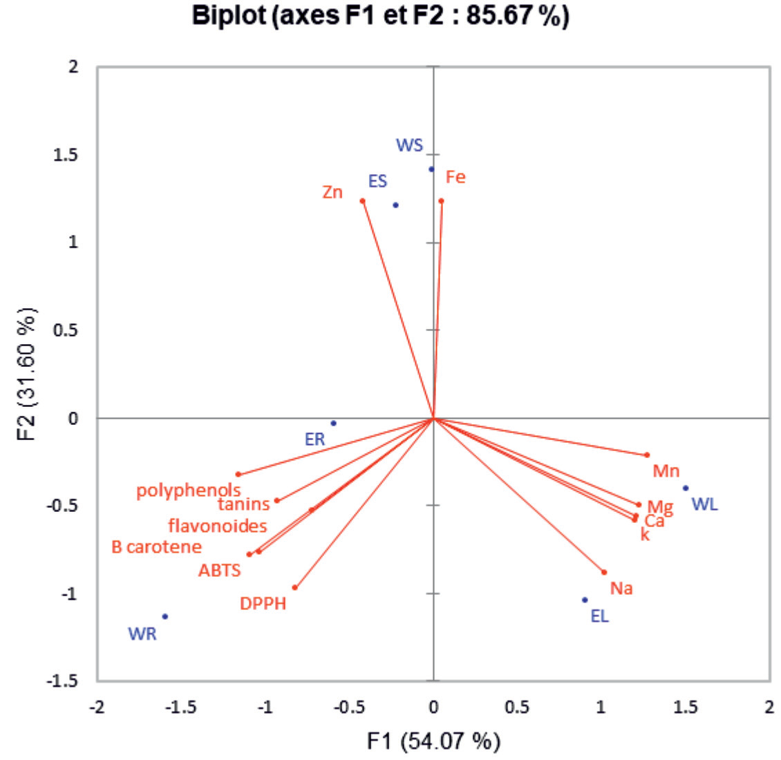
PCA is performed to better understand the relationship between chemical components (TPC, TFC, and CTC) and the antioxidant activity of R vermiculata. The first two axes (F1 and F2) accounted for 85.67% of the data variability. The major components (F1 and F2) accounted for 54.07 and 31.60% of the total variance of the data, respectively (Fig. 3). Simultaneously, the ACP figures express the extent to which the main components (F1 and F2) are correlated with the variables, as well as the correlations between the DPPH ABTS, β-carotene activities and the chemical composition (TPC, TFC, and CTC). The F1 axis showed a high positive correlation with TPC (r = 0.728) TTC (r = 0.4670), β-carotene (r = 0.5825) and ABTS (r = 0.6468). Respectively, In addition, the second axis F2 was highly correlated with the anti-DPPH activity showing an r values equal to 0.5113. However, the TFC was well correlated with the F3 (r = 0.5451) (Table 4).
Fig. 2.
DPPH (A), β-carotene (B), and ABTS (C) antioxidant activities determined for the ethanolic and aqueous extracts from different plant organs. ***: p<0.001; Highly Significant differences revealed by two-way ANOVA; different lowercase (a-c) and Uppercase (A-C) letters denote significant differences between the different plant organs in the ethanolic and aqueous extracts respectively, according to Duncan’s multirange comparison of means test (p<0.05).
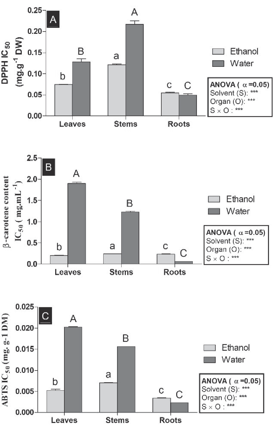
Fig. 3.
Principal components analysis “loading plot”, total phenolic content TPC, total flavonoids content TFC, condensed tannins content CTC and biological activities (DPPH: ABTS β-carotene antioxidant activity of R vermiculata organs extracts.
The difference in clustering (Fig. 4) indicates the variation of concentration minerals (Mn, Cu, K, Mg, Ca, Na, Zn, and Fe), total polyphenols (PT), total flavonoids (FT), condensed tannins, and antioxidant activities (DPPH, ABTS, and b carotene) in different parts in Reamuria. The green color boxes in the clove presents the high density of these compounds and compare our distribution with the combination names, which are presented on the horizontal axis. To distinguish between clusters: the first and second clusters were represented individually by EL and WL that were correlated with high levels of minerals; In contrast, the third cluster gathered the combination of WR with high antioxidant activities and phenolic contents. The combination ES and WS were correlated with the mineral Fe and Zn composed the fifth and sixth clusters, according to our results R vermiculata altered the accumulation of secondary metabolites in significant quantities in roots, which causes powerful antioxidant activities (Fig. 4).
Determination of Antimicrobial Activities
Table 5 summarises the results of antibacterial activity of the leaf, stem and roots of different R vermiculata extracts with various degrees of inhibition, evaluated as MIC (The minimal inhibition concentrations) and MBC (the minimal bactericidal concentrations) against different strains of Gram-negative (Escherichia coli, Pseudomonas aeroginosa and Salmonella typhi) and Gram-positive (Staphylococcus aureus, Bacillus subtilis and Listeria monocytogenes) (Table 5). A high value of MIC can be a sign of low efficacy or that the organisms can expose resistance to the bioactive compounds. (Table 5). The ethanol extract had a significant inhibitory effect on Gram+ and Gram-bacterial strains. In all plant parts, the root extracts showed better activity than the leaf and stem extracts against Listeria monocytogenes with MIC = 0.0781 mg/ml staphylococcus aureus and Bacillus subtilis with MIC = 0.3125 mg/ml. However, Salmonella typhi was more resistant with MIC of 1.250 mg/ml. For Gram-negative bacteria, none of the two extracts has great antibacterial activity against Pseudomonas aeroginosa, Escherichia coli and Salmonella typhi. The Gram-positive bacteria were more sensitive to the antimicrobial properties of Reamuria vermiculata than the Gram-negative ones. In the literature, there are no studies dealing with the antibacterial activity of the R vermiculata extracts. However, many researchers have shown interesting antibacterial activities of the Tamaricaceae family plants. Indeed [39] notified that the ethanol extract of Tamarix Tetragyna showed significant Activity against Staphylococcus aureus (MIC = 0.00125mg/mL) and Escherichia coli (MIC = 0.005 mg/mL). In addition [40] published that the methanol extract of Tamarix exhibited an antibacterial effect with a MIC of 0.146 mg/mL against Listeria monocytogenes, Bacillus subtilis, Staphylococcus aureus and Escherichia coli, In the same way, Halophytes renowned for their bioactive compounds associated with high biological proprieties. For example, diverse extracts of Limoniastrum guyonianum, known for their high antioxidant capacity, have a significant inhibitory effect on bacterial growth against different strains of Gram-negative (Escherichia coli, Pseudomonas aeroginosa and Salmonella typhi) and Gram-positive (Staphylococcus aureus, Enterococcus faecalis).
Fig. 4.
. Heatmap and hierarchical clustering for phenolic contents, antioxidant activities, and chemical composition of Tunisian halophyte Reamuria vermiculata.
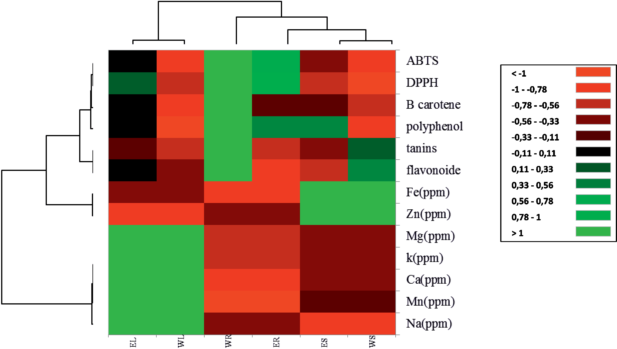
Table5.
Minimal inhibition concentrations (MICs) and minimal bactericidal concentrations (MBCs) values of selected extracts of R. vermiculata against bacteria.
Conclusions
Interestingly, based on the findings discussed for the first time in this report, R vermiculata organs could be employed as a readily available source of bioactive compounds. Mineral analysis of R vermiculata showed that leaves highlighted the highest amount. The roots also contain a high level of polyphenol, flavonoid and condensed tannins that give the plant important antioxidant and antimicrobial properties. Furthermore it was demonstrated that each organ extract was selective for one or different biological activity, as summarized in the principal component analysis PCA biplot. Our results indicate that this edible halophyte possesses a high phenolic content and can be considered an important source of antioxidants and antimicrobial effects compared to other medicinal plants. These findings give scientific evidence of the traditional medicine benefits and provide a promising potential for the isolation of natural antioxidant agent from R vermiculata and identifying the unknown compounds to establish their pharmacological properties.

